Preferential localization of IgG memory B cells adjacent to contracted germinal centers Yuichi Aibaa, Kohei Kometania, Megumi Hamadatea, Saya Moriyamaa,b, Asako Sakaue-Sawanoc, Michio Tomurad, Hervé Luchee, Hans Jörg Fehlinge, Rafael Casellasf, Osami Kanagawad, Atsushi Miyawakic, and Tomohiro Kurosakia,b,g,1 a Laboratory for Lymphocyte Differentiation, RIKEN Research Center for Allergy and Immunology, Yokohama, Kanagawa 230-0045, Japan; bLaboratory for Lymphocyte Differentiation, Graduate School of Frontier Biosciences, Osaka University, Suita, Osaka 565-0871, Japan; cLaboratory for Cell Function and Dynamics, RIKEN Brain Science Institute, Wako, Saitama 351-0198, Japan; dLaboratory for Autoimmune Regulation, RIKEN Research Center for Allergy and Immunology, Yokohama, Kanagawa 230-0045, Japan; eInstitute of Immunology, University Clinics Ulm, 89081 Ulm, Germany; fGenomics and Immunity, National Institute of Arthritis and Musculoskeletal and Skin Diseases, and Center of Cancer Research, National Cancer Institute, National Institutes of Health, Bethesda, MD 20892; and gLaboratory of Lymphocyte Differentiation, World Premier International Immunology Frontier Research Center, Osaka University, Suita, Osaka 565-0871, Japan
Edited* by Jeffrey V. Ravetch, The Rockefeller University, New York, NY, and approved May 21, 2010 (received for review April 21, 2010)
It has long been presumed that after leaving the germinal centers (GCs), memory B cells colonize the marginal zone or join the recirculating pool. Here we demonstrate the preferential localization of nitrophenol-chicken γ-globulin-induced CD38+IgG1+ memory B cells adjacent to contracted GCs in the spleen. The memory B cells in this region proliferated after secondary immunization, a response that was abolished by depletion of CD4+ T cells. We also found that these IgG1+ memory B cells could present antigen on their surface, and that this activity was required for their activation. These results implicate this peri-GC region as an important site for survival and reactivation of memory B cells. immunoglobulin
| immunological memory
A
ppropriate interactions between antigen-specific B and T lymphocytes are essential for humoral immune responses to T-dependent antigens (1, 2). After their initial exposure to antigen, antigen-binding IgM+ B cells migrate from random locations within the B-cell follicles to the border between the follicles and the T-cell–rich areas, where cognate interactions with antigenspecific CD4 T cells occur and subsequent B-cell proliferation is induced (3, 4). Then, shortly after the appearance of extrafollicular foci of antibody (Ab)-secreting plasma cells, clusters of isotype-switched cells such as IgG+ B cells, which can be detected by staining with peanut agglutinin and the GL7 mAb, appear in germinal centers (GCs) within the areas occupied by follicular dendritic cells (5–7). Because the Ig variable-region genes of IgG+ long-lived memory B cells contain somatic mutations, and because these mutations occur primarily in GCs, it is thought that IgG+ memory B cells are mainly derived from the GC. Despite the importance of IgG+ memory B cells in long-term humoral memory (8, 9), their sites of residency and activation remain elusive, in part, because of technical difficulties associated with in situ detection of the rare IgG+ memory B cells specific for a given antigen. Thus, to circumvent this problem, a transgenic mouse line harboring an IgM-type B-cell antigen receptor (BCR) has been used; these studies showed that longlived IgM+ memory B cells reside not just in the marginal zone (MZ), as had been thought, but also in splenic follicles (10). However, given the finding that antigen-experienced IgM B cells and switched IgG2a B cells differentially localize during primary immune responses (11), extrapolation of the above scenario to residency sites of IgG+ memory B cells in physiological settings needs to be done with great caution. Here we focus on where IgG+ memory B cells reside and how these memory B cells are activated upon secondary antigen challenge. Results CD38+IgG1+ Memory B Cells Localize Around GCs. The Ab response to
the hapten nitrophenol (NP) has been characterized extensively 12192–12197 | PNAS | July 6, 2010 | vol. 107 | no. 27
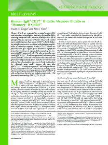
(12, 13). Thus, to determine where IgG-type memory B cells reside, we used this model system. The response to NP in C57BL/6 (B6) mice is dominated by Abs composed of the VH186.2 heavy chain and an Igλ light chain (12, 13). Consistent with these reports, by flow cytometry analysis, ∼60–80% of NP-specific IgG1+ B cells expressed λ light chains on day 60 after alum-precipitated NPconjugated chicken γ-globulin (NP-CGG) immunization (Fig. S1A). We then used immunohistochemical analyses to determine the localization of IgG1+λ+ B cells, which are mixtures of memory and GC B cells as described below, in the spleen on day 30 and day 60 postimmunization. As shown in Fig. S1B, IgG1+ cells were located in several areas including the red pulps and follicles, but the λ-expressing IgG1+ cells were found mainly as clusters residing in the centers of follicles both at day 30 and day 60. Although we expended considerable effort attempting to detect NP-reactive cells in sections using NP-labeled fluorochromes, in our hands the data obtained using this approach were unconvincing. Previous studies have demonstrated that CD38, used in conjunction with GL7, is a good marker for distinguishing between memory and GC B cells: The former are CD38+IgG1+ and the latter are CD38−IgG1+GL7+ (14, 15). Therefore, to localize IgG1-type memory B cells, we stained sections of spleen from NPCGG-immunized mice with anti-CD38, -IgG1, and -GL7 Abs. On day 30, a small number of IgG1+CD38+ B cells were detected in the follicles adjacent to clusters of CD38−GL7+ GC B cells (Fig. 1A). On day 60, these IgG1+CD38+ B cells were still observed near the clusters of CD38−GL7+ GC B cells; the GC B cells were still present, despite a significant reduction in their numbers compared with day 30 by both flow cytometry and immunohistology (Fig. 1B). Results using PNA, another widely used marker for GC B cells, were similar to those with the GL7 mAb (Fig. S2). Moreover, consistent with the above results, ∼50–70% of IgG1+ cells clustering near the GCs on day 60 were λ+ (Fig. 1A). Together, these observations suggest that the majority of IgG1+ memory B cells localized near the contracted GCs on day 60 were specific for NP. IgM- and IgG-type Memory B Cells Localize in Distinct Areas of the Spleen. Several previous reports have described a different lo-
calization of memory B cells from the one we describe above, that is, scattered in follicles or within the marginal zones (10, 16).
Author contributions: Y.A. and T.K. designed research; Y.A., K.K., M.H., and S.M. performed research; A.S.-S., M.T., H.L., H.J.F., R.C., O.K., and A.M. contributed new reagents/analytic tools; Y.A. and K.K. analyzed data; and Y.A. and T.K. wrote the paper. The authors declare no conflict of interest. *This Direct Submission article had a prearranged editor. 1
To whom correspondence should be addressed. E-mail:
[email protected].
This article contains supporting information online at www.pnas.org/lookup/suppl/doi:10. 1073/pnas.1005443107/-/DCSupplemental.
www.pnas.org/cgi/doi/10.1073/pnas.1005443107
NP-CGG + Alum, Day 60
None, Day 30
IgG1
CD38
CD45.1 B220
NP-binding
A CD45.1
A
B220
IgMa
GL7
IgMa
CD38, IgMa, GL7 IgMa
NP-CGG + Alum, Day 60 NP-binding
B220
B220
None
Fucci-red
Because previous reports mainly focused on memory B cells expressing IgM, we next examined the possibility that IgM- and IgG-type memory B cells might differentially localize in the spleen. For this purpose, we used mice that have a heavy-chain locus targeted with the B1-8hi IgH (Igha) gene. Because the light chains in B1-8hi mice are not fixed, only 3–5% of their B cells express λ and bind NP (17). F1 offspring of CD45.1 and CD45.2 congenic mice (CD45.1-CD45.2 F1) were adoptively transferred with B cells from B1-8hi IgH knock-in mice (CD45.2) and immunized with alum-precipitated NP-CGG. In this experimental setting, almost all donor-derived B cells present on day 30 after immunization can be considered as antigen-experienced, because of the following two lines of evidence. First, on day 30, more than 90% of the transferred B cells bound NP, in contrast to about 5% of naïve B cells (Fig. S3). Second, more than 90% of transferred B cells labeled with carboxyfluorescein diacetate succinimidyl ester (CFSE) before transfer became CFSE-negative by day 30 after immunization, indicating that they had undergone extensive proliferation (Fig. S3). As shown in Fig. 2A, two distinct subpopulations of donor-derived B cells were observed in immunized mice on day 60. The largest subpopulation was CD38+GL7−, Aiba et al.
IgG1
IgG1, Fucci-red
10 IgMa+ cells
IgG1+Fucci-red + cells
8
8
6
6
4
4 2 Red Pulp
Fo. Scat.
Fo. Close to GC
0
Red Pulp
Fo. Scat.
Fo. Close to GC
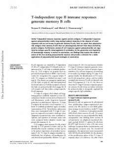
Fig. 2. IgM- and IgG-type memory B cells differentially localize in the spleen. (A) B cells from B1-8hi IgH knock-in mice were adoptively transferred to CD45.1-CD45.2 F1 mice. The mice were immunized with alum-precipitated NP-CGG or left untreated. After 30 days, unimmunized mice were killed and analyzed for the presence of donor-derived, B220+CD45.1− cells by flow cytometric analysis (Upper Left; None, day 30). Immunized mice were killed at 60 days after immunization (NP-CGG + Alum, day 60). Spleens were subjected to flow cytometric (Upper) or immunohistological (Lower) analyses. [Scale bars, 300 μm (Left), 20 μm (Right).] (B) B cells purified from double-transgenic mice (B1-8hi IgH knock-in and Fucci-red) were adoptively transferred to CD45.1CD45.2 F1 mice. The mice were immunized with alum-precipitated NP-CGG or left untreated. After 60 days, spleens from unimmunized (None) or immunized (NP-CGG + Alum, day 60) mice were subjected to flow cytometric (Upper) and immunohistological (Lower) analyses. The panels of histological analysis represent the same region in the same follicle in different sections. [Scale bars, 300 μm (Left), 50 μm (Center), 100 μm (Right).] (C) Quantification of IgMa+ cells in A and IgG1+Fucci-red+ cells in B are shown. Average number ± SD from three mice are shown. Cells in the red pulp were probably plasma cells, as judged by low expression of CD38. Fo. Scat., IgG1+ cells scattered among follicles.
the phenotype of memory B cells, and most of these cells expressed IgM. As expected, those IgM+ cells could bind NP, indicating that NP-reactive IgM-type memory B cells are generated in this experimental system. Histological analysis revealed that the IgM-type memory B cells detected with anti-IgMa mAb were mainly scattered in follicles (Fig. 2A and Fig. 2C Left). We next attempted to determine the localization of IgG1-type memory B cells in the same adoptive transfer experiment as described above. However, despite tremendous efforts, the antiallotypic IgG1a mAb did not work well, especially for histological analysis. Thus, we tried another approach to detect donor-derived IgG1-type memory B cells. We used a transgenic mouse expressing a cell-cycle-sensitive probe, fluorescent indicator for cell-cycle progression (Fucci), in which cells become reversibly fluorescent depending on their cell-cycle status; they are red in the G1, but not S/G2/M, phases (Fucci-red) (18), therefore being suitable for labeling resting cells such as memory B cells. CD45.1-CD45.2 F1 mice were adoptively transferred with B cells from Fucci-red PNAS | July 6, 2010 | vol. 107 | no. 27 | 12193
IMMUNOLOGY
2 0
Fig. 1. Detection of CD38+IgG1+ memory B cells adjacent to contracted GCs. (A) B6 mice were immunized with alum-precipitated NP-CGG. After 30 and 60 days, spleens were harvested and subjected to flow cytometric (Upper) and immunohistological (Lower) analyses. B220+ gated populations are shown in the upper panels. A fluorescence-activated cell sorting (FACS) profile of an unimmunized B6 mouse (day 0) is also shown. White arrows in the first and second rows of the second section indicate cells stained with both anti-CD38 and -IgG1 Abs, and in the third row indicate cells that are IgG1+λ+. [Scale bars, 150 μm (Left), 50 μm (Center).] (B) Quantification of the absolute number of memory (CD38+IgG1+) and GC (CD38−IgG1+) B cells by flow cytometric (Left) and immunohistological (Right) analyses.
CD38, Fucci-red
10
N. of cells/mm2
B
IgG1
Fucci-red
CD38, Fucci -red, GL7
C
CD38, IgMa
CD38
B
CD38
ular dendritic cells are located within and/or surrounding GCs, where they can capture antigen–IgG complexes by virtue of Fcγ (19) and complement (20) receptors during immune responses. Because the above assay relies on anti-IgG1 staining, one concern is that this method might not be detecting B cells but instead cells that had passively acquired secreted IgG1 Abs. To eliminate this possibility, we used a genetic approach in which the Cre recombinase gene is expressed under the control of the Aicda (activationinduced cytidine deaminase; AID) promoter and red fluorescent protein (RFP) is only expressed upon Cre-mediated deletion of a floxed neomycin gene (AID-cre/RFP-ROSA) (Fig. S4) (21, 22). In these mice, the progeny of AID-expressing cells, including memory B cells, are permanently RFP+ (Fig. S5A). Localization of the red fluorescent cells was determined by immunohistological analysis (Fig. S5B). Consistent with the above immunohistological analysis using anti-IgG1 Ab, RFP+CD38+ B cells were also located near the GL7+ GC B cells (Fig. S5B Left). Most of these RFP+CD38+ B cells were IgG1+ (Fig. S5B Right). Based on these findings, we conclude that most IgG1 memory B cells in the spleen are found in clusters near the contracted GCs. Location of the CD38+IgG1+ Memory B Cells That Proliferate Following Antigen Rechallenge. To next examine where memory
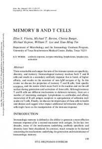
B cells are activated upon secondary antigen challenge (NP-CGG without alum), we used another strain of Fucci transgenic mice, in which cells become reversibly fluorescent (green) in the S/G2/M, but not G1, phases (Fucci-green) (18). As expected, flow cytometric analysis indicated that almost all of the CD38+IgG1+NP+ memory B cells on day 60 after primary challenge were in the resting state. However, by 2 days after secondary challenge, many of them had entered the cell cycle and become green (Fig. 3A). In situ studies were performed to identify the location of the green cells in the spleen (Fig. 3B). Before secondary challenge, a few green B cells were detectable, consistent with the flow cytometric data (Fig. 3A). Two days after secondary challenge, many IgG1 B cells entered the S/G2/M phase. Among these green cells, about two-thirds of them were CD38+ memory cells and the remaining were CD38− GC B cells. These results suggest that IgG1 memory B cells near the contracted GCs, together with the GC B cells, start to proliferate upon secondary challenge. CD4+ T Cells Reside Close to IgG1+ Memory B Cells in the Follicles.
Considering the recent evidence that some T cells, particularly follicular helper T cells (TFH), are localized inside or surrounding GCs during primary humoral responses (23–25), it seemed possible that helper T cells for activating memory B cells might also reside near the contracted GCs. If so, in contrast to the requirement for migration of naïve B and naïve T cells toward the T-B border area for their initial cognate interactions, such active migration might 12194 | www.pnas.org/cgi/doi/10.1073/pnas.1005443107
Prime alone
Fucci-green
IgG1
Prime + Challenge, Day 2
IgG1
IgG1
Fucci-green
CD38
Fucci-green IgG1 CD38
Fucci-green IgG1 CD38
25
120 100
P < 0.01
80 60 40 20 0
20.9
CD38
NP-binding
CD38
NP-binding
55.7
Prime alone Fucci-green
CD38
C
0.7
Fucci-green
IgG1
B
Prime + Challenge, Day 2
88.8
P P+C CD38+ IgG1+ NP-binding
P P+C CD38+ IgG1+ NP-binding FG-
P P+C CD38+ IgG1+ NP-binding FG+
N. of cells/mm 2
Detection of Memory B Cells Based on the AID-Cre–Mediated Expression of Red Fluorescent Protein. In addition to B cells, follic-
A
N. of cells/106 splenocytes
transgenic B1-8hi IgH knock-in mice, and immunized with NPCGG precipitated in alum. As shown in Fig. 2B, donor-derived Fucci-red-labeled cells were observed in mice on day 60 after immunization. Flow cytometric analysis revealed that IgG1+ cells were present in the Fucci-red-labeled cell population. Because we gated out Fucci-red-negative cells, probably including GC B cells, most of these IgG1+ cells expressed CD38 and were able to bind NP, suggesting that NP-specific IgG1-type memory B cells were labeled with Fucci-red. Localization of Fucci-red-labeled cells was examined by immunohistological analysis (Fig. 2B Lower). Clustering of Fucci-red-labeled CD38+ cells was observed near GL7+CD38− GC cells, in addition to cells scattered in follicles. Staining the sections with anti-IgG1 Ab revealed that IgG1+ cells were predominantly located in clusters near GCs (Fig. 2 B and C Right). Together, these data demonstrate differential localization of IgM- and IgG1-type memory B cells.
20 P < 0.05
15 10 5 0
P P+C CD38+ IgG1+
P P+C CD38+ IgG1+ FG-
P P+C CD38+ IgG1+ FG+
Fig. 3. Activation of CD38+IgG1+ cells after antigen rechallenge. Fucci transgenic mice were immunized with alum-precipitated NP-CGG. After 60 days, mice were injected with NP-CGG without adjuvant (Prime + Challenged, day 2) or left untreated (Prime alone) and were killed 2 days later. Spleens were subjected to flow cytometric (A) and histological analysis (B). Percentages of CD38+Fucci− and CD38+Fucci+ cells among NP-binding IgG1+ cells are indicated in the FACS profiles (A). White arrows in the right panel of B indicate the Fucci probe-positive cells stained with anti-CD38 and -IgG1 Abs. (Scale bars, 50 μm.) (C) Quantification of CD38+IgG1+ cells as either negative (FG−) or positive (FG+) for the Fucci probe was performed as described in Fig. 1B. P values were calculated with a two-tailed Student’s t test. P, primed alone; P+C, primed and rechallenged.
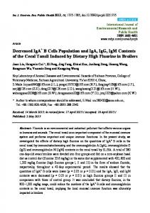
not necessarily be required for activating memory B cells. This possibility was tested by immunohistological analysis of spleen sections on day 60 after primary immunization. As shown in Fig. 4A, CD4+ T cells were found near the region where CD38+IgG1+ and CD38−IgG1+ B cells were localized in the follicles. To further examine whether these CD4+ T cells express TFH markers, we used anti-PD-1 mAb (26) and found that some, but not all, CD4+ T cells in the follicles on day 60 after primary immunization expressed PD-1 (Fig. 4B). Cognate Interaction of IgG1+ Memory B Cells with CD4+ T Cells Is Required for Their Activation. The presence of T cells near the
IgG1 memory B cells prompted us to examine the functional requirement for such helper T cells in humoral memory responses. To address this question, B6 or Fucci-green transgenic mice that had been immunized with alum-precipitated NP-CGG were treated with anti-CD4 mAb and control Abs before secondary challenge (NP-CGG without alum). The initial proliferation of the memory B cells, as judged by the expression of the Fucci-green probe (Fig. 4C), as well as the production of secondary anti-NP Abs (Fig. 4D) were almost completely abolished by the anti-CD4 treatment. Having demonstrated the importance of CD4+ T cells for activating IgG1 memory B cells, we wished to address whether cognate interactions between the B and T cells are required. Before addressing this question, we examined whether the IgG1 memory B cells are capable of presenting antigen. To do this, NP-CGG-primed B6 mice were boosted with an NP-conjugated fusion protein composed of GFP and amino acids 46–74 of the Aiba et al.
A
C
CD38
IgG1
CD4
CD38 IgG1 CD4
B
PD-1
IgG1
CD4
PD-1 IgG1 CD4
Anti-CD4 mAb treated
Control Ig treated Prime alone Prime + Challenge 1.0
Prime alone
Prime + Challenge
0.9
1.8
CD38
19.3
P < 0.05
10
20 10 0
P P+C Control Ig 103
P P+C Anti-CD4 mAb
8 6 4 2 0
P P+C Control Ig
P P+C Anti-CD4 mAb
P < 0.05 Control Rat Ig treated Anti-CD4 mAb treated
102
10
1 P
P+C
+
Fig. 4. Depletion of CD4 cells prevents activation of memory B cells and abolishes the secondary Ab response. B6 mice were immunized with alumprecipitated NP-CGG. On day 60, spleens harvested from the mice were subjected to immunohistological analysis. (A) A section stained with anti-CD38, -IgG1, and -CD4 mAbs. (Scale bar, 50 μm.) (B) A section stained with anti-PD-1, -IgG1, and -CD4 Abs. White arrows in B indicate cells stained with both anti-CD4 and -PD-1 mAbs. (Scale bar, 50 μm.) Fucci-green transgenic (C ) or B6 (D) mice were immunized with alum-precipitated NP-CGG. After 60 days, mice were treated daily with anti-CD4 mAb or control rat Ab before rechallenge. Three days after the first Ab treatment, mice were rechallenged with NP-CGG without adjuvant (Prime + Challenge) or left untreated (Prime alone). (C ) Spleens from Fucci-green transgenic mice were harvested 2 days after rechallenge and subjected to flow cytometric and histological analyses. Percentages of CD38+Fucci+ cells among NP-binding IgG1+ cells are indicated in the FACS profiles. (D) Anti-NP IgG1 titers in sera from immunized B6 mice were measured by ELISA 7 days after rechallenge. P, primed alone; P+C, primed and rechallenged. P values were calculated with a two-tailed Student’s t test.
I-Edα MHC II subunit (NP-EαGFP) as a secondary challenge antigen. In this setting, antigen presentation can be monitored with the Y-Ae mAb, which is specific for pEα:I-Ab complexes on the surface of antigen-presenting cells (27, 28). As demonstrated in Fig. 5A, CD38+IgG1+ memory B cells were able to present exogenous antigens in the context of MHC class II molecules. In these experiments, IgM+ and IgD+ cells were not depleted before flow cytometric analysis for technical reasons, and thus the large number of NP-binding IgG1− cells is probably IgM+ B cells. Next, to test the requirement for antigen presentation by memory B cells in their activation, sorted CD38+IgG1+NP+ memory B cells were transferred together with CGG-primed CD4+ T cells Aiba et al.
into Rag1−/− mice. The memory B cells were able to mount a secondary Ab response when the recipient mice were administered NP-CGG, but not NP-chicken ovalbumin (NP-OVA) (Fig. 5B). The simplest explanation for these results is that IgG1+NP+ memory B cells efficiently differentiated only when expression of an NP-specific IgG1 BCR allowed efficient uptake of CGG and presentation of CGG peptides to the CGG-primed T cells. NP conjugated with a different carrier, OVA, would be unable to elicit the requisite T-cell help in this system. This idea was substantiated by our findings using MHC class II−/− Rag1−/− mice as recipients, an experimental setting in which MHC class II is only expressed on the transferred cells. As shown in Fig. 5C, transfer of memory B cells together with primed T cells into the MHC class II−/− Rag1−/− mice PNAS | July 6, 2010 | vol. 107 | no. 27 | 12195
IMMUNOLOGY
30
Titer of anti-NPhi IgG1 in sera (Arbitrary Units)
D
P < 0.05
40 N. of cells/mm 2
N. of cells/106 splenocytes
Fucci-green
A
Prime alone
CD38
NP-binding
3.3
Y-Ae
IgG1 Prime + NP-EaGFP
CD38
NP-binding
53.1
IgG1
N. of AFCs/107 splenocytes
B
Y-Ae
106
104
102
1 Rechallenged with
C
None
NP-CGG
NP-OVA
N. of AFCs/107 splenocytes
106
104
102
1 Recipient genotype
MHC II Rag1
Anti-MHC class II Fab
+/+ -/-
-/-/-
-/-/-
-
-
+
Fig. 5. Cognate interaction with antigen-specific T cells is required for the response of IgG-type memory B cells. (A) B6 mice were immunized with alumprecipitated NP-CGG. After 60 days, mice were injected i.p. with NP-EαGFP (Prime + NP-EαGFP) or left untreated (Prime alone). After 24 h, splenocytes were analyzed by flow cytometry with Y-Ae mAb for estimating antigen presentation in the context of MHC class II. Percentages of CD38+Y-Ae+ cells among NP-binding IgG1+ cells are indicated in FACS profiles. (B and C) Memory B cells and CGG-primed T cells were purified from B6 mice immunized with alumprecipitated NP-CGG. (B) Cells were transferred to Rag1−/− mice. One day later, the mice were left untreated (None) or rechallenged with NP-CGG or NP-OVA without adjuvant. (C) Cells were transferred to Rag−/− or Rag−/− MHC class II−/− mice. After 1 day, mice were immunized with NP-CGG without adjuvant. On day 0 and day 3 after cell transfer, some mice were injected with 200 μg of Fab fragments of anti-MHC class II mAb. In both B and C, splenocytes were prepared and subjected to enzyme-linked immunospot (ELISPOT) assays for measuring the number of antibody-forming cells (AFCs) 7 days after cell transfer.
could elicit the secondary NP response, whereas administration of Fab fragments of anti-MHC class II mAb blocked this response, demonstrating the importance of MHC class II expression on memory B cells. Discussion Previous work has shown that NP-specific memory B cells appear in the MZ area shortly after primary immunization in rats, and 12196 | www.pnas.org/cgi/doi/10.1073/pnas.1005443107
then emerge in both the follicle and the MZ at later time points (16). Although the study using rats did not distinguish whether these memory B cells were of the IgM or class-switched type, given that the same localization sites was observed when NPspecific IgM transgenic mouse models were immunized (10) it is most likely that the NP-specific memory B cells observed in the rat system were of the IgM type. This possibility is further supported by our observations that antigen-experienced IgM+ cells are scattered in the follicles on day 60 after primary immunization (Fig. 2 A and C). In contrast to the localization of IgM-type memory B cells, we have shown here that IgG1-type memory B cells (IgG1+CD38+) are mainly located near the contracted GC-like structures still present on day 60 after primary immunization (NP-CGG with alum). Our histochemical resolution did not suffice to conclude whether the IgG1+CD38+ memory B cells are localized near the GC light or dark zone. The presence of these GC-like structures on day 60, albeit much smaller than on day 30, is quite consistent with a recent report demonstrating that GC-like structures persist for up to 8 months after being challenged with sheep red blood cells (SRBCs) twice (29). Because SRBCs induce a very potent polyclonal B-cell response, the persistence of GC-like structures for longer periods in the case of SRBCs probably reflects the fact that a steady-state level of newly activated B-cell clones is high, thereby being continuously recruited into the GC fractions. Together with our data, it now seems clear that GClike structures can persist longer than previously appreciated, and that the duration of such structures is dependent, at least partly, on the nature of the immunogen and adjuvants. By using lymphocytes harboring the Fucci cell-cycle tracker, we have demonstrated here that IgG1+CD38+ memory B cells near GCs, in addition to the IgG1+CD38− (GC B cells from our criteria), begin to proliferate upon secondary challenging on day 60. As discussed above, in the NP-CGG immunization protocol, some IgG1+CD38− GC B cells still remain at day 60 and their proliferation appears to be enhanced upon secondary challenge. Because recent reports have suggested that IgG-type memory B cells undergo differentiation into plasma cells rather than entering into GC pools (29, 30), we favor the idea that the precursors of proliferating IgG1+CD38− B cells on day 2 after secondary challenge are GC, but not memory, B cells. However, at present, we cannot completely exclude the possibility that IgG1+CD38+ memory B cells differentiate into IgG1+CD38− cells, or vice versa, during the 2 days after secondary challenge. Assuming that such preferential localization and activation of memory B cells near the contracted GCs also occur in draining lymph nodes, our observations would explain the previous findings that the ipsilateral lymph node transfers a significantly higher humoral memory response than does the contralateral node at all intervals after a primary challenge (31). Proliferation of IgG1+CD38+ memory B cells and their subsequent differentiation into plasma cells are likely to require cognate interactions between B and T cells. This notion is supported by the following three lines of evidence: (i) the requirement for T cells for the proliferation and differentiation of IgG1 memory B cells; (ii) IgG1+CD38+ memory B cells are able to present antigens; and (iii) the requirement for MHC class II on B cells for differentiation of IgG1+CD38+ memory B cells. Given that CD4+ T cells exist in close proximity to IgG1 memory B cells near the contracted GCs on day 60, we speculate that some of these CD4+ T cells are long-lived follicular helper memory T cells and are responsible for activation of IgG1 memory B cells. If so, we would propose that this close proximity of memory B cells to memory T cells can explain, at least partly, the more rapid kinetics of memory responses because, during primary responses, movement of antigen-specific B cells and antigen-specific T cells toward the T-B border area is required. Aiba et al.
1. Catron DM, Itano AA, Pape KA, Mueller DL, Jenkins MK (2004) Visualizing the first 50 hr of the primary immune response to a soluble antigen. Immunity 21:341–347. 2. Allen CD, Okada T, Cyster JG (2007) Germinal-center organization and cellular dynamics. Immunity 27:190–202. 3. Garside P, et al. (1998) Visualization of specific B and T lymphocyte interactions in the lymph node. Science 281:96–99. 4. Okada T, et al. (2005) Antigen-engaged B cells undergo chemotaxis toward the T zone and form motile conjugates with helper T cells. PLoS Biol 3:e150. 5. Allen CD, Okada T, Tang HL, Cyster JG (2007) Imaging of germinal center selection events during affinity maturation. Science 315:528–531. 6. Hauser AE, et al. (2007) Definition of germinal-center B cell migration in vivo reveals predominant intrazonal circulation patterns. Immunity 26:655–667. 7. Schwickert TA, et al. (2007) In vivo imaging of germinal centres reveals a dynamic open structure. Nature 446:83–87. 8. Radbruch A, et al. (2006) Competence and competition: The challenge of becoming a long-lived plasma cell. Nat Rev Immunol 6:741–750. 9. Tarlinton D (2006) B-cell memory: Are subsets necessary? Nat Rev Immunol 6: 785–790. 10. Anderson SM, Tomayko MM, Ahuja A, Haberman AM, Shlomchik MJ (2007) New markers for murine memory B cells that define mutated and unmutated subsets. J Exp Med 204:2103–2114. 11. Pape KA, et al. (2003) Visualization of the genesis and fate of isotype-switched B cells during a primary immune response. J Exp Med 197:1677–1687. 12. Cumano A, Rajewsky K (1985) Structure of primary anti-(4-hydroxy-3-nitrophenyl) acetyl (NP) antibodies in normal and idiotypically suppressed C57BL/6 mice. Eur J Immunol 15:512–520. 13. Bothwell AL, et al. (1981) Heavy chain variable region contribution to the NPb family of antibodies: Somatic mutation evident in a γ 2a variable region. Cell 24:625–637. 14. Takahashi Y, Ohta H, Takemori T (2001) Fas is required for clonal selection in germinal centers and the subsequent establishment of the memory B cell repertoire. Immunity 14:181–192. 15. Ridderstad A, Tarlinton DM (1998) Kinetics of establishing the memory B cell population as revealed by CD38 expression. J Immunol 160:4688–4695. 16. Liu YJ, Oldfield S, MacLennan IC (1988) Memory B cells in T cell-dependent antibody responses colonize the splenic marginal zones. Eur J Immunol 18:355–362. 17. Shih TA, Roederer M, Nussenzweig MC (2002) Role of antigen receptor affinity in T cell-independent antibody responses in vivo. Nat Immunol 3:399–406. 18. Sakaue-Sawano A, et al. (2008) Visualizing spatiotemporal dynamics of multicellular cell-cycle progression. Cell 132:487–498. 19. Ravetch JV, Bolland S (2001) IgG Fc receptors. Annu Rev Immunol 19:275–290.
Aiba et al.
is possible that there are functional differences between GCdependent and -independent IgG memory B cells. Methods Detailed descriptions of all materials and methods are provided in SI Methods. Mice, Immunization, and Treatment with Anti-CD4 Monoclonal Antibody. C57BL/6 mice were purchased from CLEA Japan. B1-8hi IgH knock-in mice were described in ref. 17. AID-cre mice and RFP-ROSA mice have been described previously (21, 22). MHC class II-deficient mice (36) were crossed with Rag1−/− mice to obtain Rag1−/− MHC class II−/− mice. Fucci-red and -green transgenic mice were established as described in a previous report (18). Lines of transgenic mice were screened for the expression of the Fucci probe in peripheral blood leukocytes. For primary immunization, mice were injected i.p. with 100 μg of NP-CGG in 200 μL of alum (Thermo Scientific) according to the manufacturer’s instructions. For secondary immunization, mice were injected i.p. with 50 μg of NP-CGG or EαGFP-NP in PBS without any adjuvant. For in vivo depletion of CD4+ cells, mice were injected i.p. with 200 μg of anti-CD4 mAb (clone; YTS191.1) every day for 3 dsya before rechallenge with NP-CGG. All of the protocols for animal experiments were approved by the RIKEN Animal Research Committee. ACKNOWLEDGMENTS. We thank Dr. K. A. Pape and Dr. M. K. Jenkins (Department of Microbiology, Center for Immunology, University of Minnesota Medical School, Minneapolis) for providing us with the EαGFP construct, Dr. T. Okada for discussions, and Dr. P. D. Burrows for critical reading of our manuscript. This work was supported by grants to Y.A. and T.K. from the Ministry of Education, Culture, Sports, Science, and Technology in Japan and by a grant to T.K. from Japan Science and Technology Agency, Core Research of Evolutional and Technology.
20. Carroll MC (1998) The role of complement and complement receptors in induction and regulation of immunity. Annu Rev Immunol 16:545–568. 21. Luche H, Weber O, Nageswara Rao T, Blum C, Fehling HJ (2007) Faithful activation of an extra-bright red fluorescent protein in “knock-in” Cre-reporter mice ideally suited for lineage tracing studies. Eur J Immunol 37:43–53. 22. Crouch EE, et al. (2007) Regulation of AID expression in the immune response. J Exp Med 204:1145–1156. 23. Kim CH, et al. (2001) Subspecialization of CXCR5+ T cells: B helper activity is focused in a germinal center-localized subset of CXCR5+ T cells. J Exp Med 193:1373–1381. 24. Breitfeld D, et al. (2000) Follicular B helper T cells express CXC chemokine receptor 5, localize to B cell follicles, and support immunoglobulin production. J Exp Med 192: 1545–1552. 25. Vinuesa CG, Tangye SG, Moser B, Mackay CR (2005) Follicular B helper T cells in antibody responses and autoimmunity. Nat Rev Immunol 5:853–865. 26. Haynes NM, et al. (2007) Role of CXCR5 and CCR7 in follicular Th cell positioning and appearance of a programmed cell death gene-1high germinal center-associated subpopulation. J Immunol 179:5099–5108. 27. Pape KA, Catron DM, Itano AA, Jenkins MK (2007) The humoral immune response is initiated in lymph nodes by B cells that acquire soluble antigen directly in the follicles. Immunity 26:491–502. 28. Itano AA, et al. (2003) Distinct dendritic cell populations sequentially present antigen to CD4 T cells and stimulate different aspects of cell-mediated immunity. Immunity 19:47–57. 29. Dogan I, et al. (2009) Multiple layers of B cell memory with different effector functions. Nat Immunol 10:1292–1299. 30. Benson MJ, et al. (2009) Distinction of the memory B cell response to cognate antigen versus bystander inflammatory signals. J Exp Med 206:2013–2025. 31. Baine Y, Thorbecke GJ (1982) Induction and persistence of local B cell memory in mice. J Immunol 128:639–643. 32. Martin SW, Goodnow CC (2002) Burst-enhancing role of the IgG membrane tail as a molecular determinant of memory. Nat Immunol 3:182–188. 33. Kaisho T, Schwenk F, Rajewsky K (1997) The roles of γ 1 heavy chain membrane expression and cytoplasmic tail in IgG1 responses. Science 276:412–415. 34. Toyama H, et al. (2002) Memory B cells without somatic hypermutation are generated from Bcl6-deficient B cells. Immunity 17:329–339. 35. Weller S, et al. (2001) CD40-CD40L independent Ig gene hypermutation suggests a second B cell diversification pathway in humans. Proc Natl Acad Sci USA 98: 1166–1170. 36. Madsen L, et al. (1999) Mice lacking all conventional MHC class II genes. Proc Natl Acad Sci USA 96:10338–10343.
PNAS | July 6, 2010 | vol. 107 | no. 27 | 12197
IMMUNOLOGY
In addition to this spatial advantage, intrinsic properties of IgG-type memory B cells likely contribute to their more rapid response as demonstrated in previous studies. For example, IgG1type memory B cells are more apt to differentiate into plasma cells than IgM-type memory and/or naïve cells (29, 30). At the molecular level, the cytoplasmic domain of the IgM-type BCR is almost nonexistent, whereas the IgG-type BCR contains an extended cytoplasmic domain which has been suggested to generate unique signals for conferring a memory phenotype (32, 33). Although our study has focused on IgG-type memory B cells, we do not argue that antigen-specific IgM memory B cells are dispensable for T-dependent memory IgG responses. In regard to their contribution, it has been recently proposed that memory IgM B cells, by virtue of their rapid mobilization in GCs and switching to IgG after antigen rechallenge, ensure replenishment of the memory pool, probably including both IgM and IgG types (29). Our data, together with recent work (29), strongly suggest the importance of persisting GC-like structures for normal humoral memory responses. However, previous studies involving genetic deficiency of CD40 and Bcl6 in human and mouse, respectively (34, 35), demonstrated that in the absence of GCs, an unmutated but functional memory B-cell compartment could develop, implying that there may exist at least one, if not multiple, GCindependent pathways of memory B-cell development. However, the IgG memory B cells that develop in the absence of Bcl6 appear not to give rise to long-lived plasma cells (34). Thus, it