RESEARCH ARTICLE
Prenatal Stress Down-Regulates Reelin Expression by Methylation of Its Promoter and Induces Adult Behavioral Impairments in Rats Ismael Palacios-García1,4‡, Ariel Lara-Vásquez1‡, Juan F. Montiel2, Gabriela F. Díaz-Véliz3, Hugo Sepúlveda5, Elías Utreras4, Martín Montecino5, Christian González-Billault4*, Francisco Aboitiz1* 1 Departamento de Psiquiatría, Escuela de Medicina, and Centro Interdisciplinario de Neurociencia, Pontificia Universidad Católica de Chile, Santiago, Chile, 2 Centro de Investigación Biomédica, Facultad de Medicina, Universidad Diego Portales, Santiago, Chile, 3 Programa de Farmacología Molecular y Clínica, Instituto de Ciencias Biomédicas, Universidad de Chile, Santiago, Chile, 4 Laboratory of Cell and Neuronal Dynamics (Cenedyn), Department of Biology, Faculty of Sciences, Universidad de Chile, Santiago, Chile, 5 Center for Biomedical Research, Faculty of Biological Sciences and Faculty of Medicine, and Fondo de Áreas Prioritarias (FONDAP) “Center for Genome Regulation”, Universidad Andrés Bello, Santiago, Chile OPEN ACCESS Citation: Palacios-García I, Lara-Vásquez A, Montiel JF, Díaz-Véliz GF, Sepúlveda H, Utreras E, et al. (2015) Prenatal Stress Down-Regulates Reelin Expression by Methylation of Its Promoter and Induces Adult Behavioral Impairments in Rats. PLoS ONE 10(2): e0117680. doi:10.1371/journal. pone.0117680 Academic Editor: Pascale CHAVIS, INSERM U901, FRANCE Received: October 21, 2013 Accepted: December 30, 2014 Published: February 13, 2015 Copyright: © 2015 Palacios-García et al. This is an open access article distributed under the terms of the Creative Commons Attribution License, which permits unrestricted use, distribution, and reproduction in any medium, provided the original author and source are credited. Funding: This work has been supported by the Millennium Center for the Neuroscience of Memory, Chile, (NC10-001-F), developed with funds from the program for Innovation for Competitivity of the Ministry for Economics, Fomentation and Tourism, Chile (to FA). Also supported by ACT114 (to CG-B) and FONDAP 15090007 (to MM). JFM is a recipient of Becas Chile postdoctoral fellowship. The funders had no role in study design, data collection and analysis, decision to publish, or preparation of the manuscript.
‡ These authors contributed equally to this work. *
[email protected] (CGB);
[email protected] (FA)
Abstract Prenatal stress causes predisposition to cognitive and emotional disturbances and is a risk factor towards the development of neuropsychiatric conditions like depression, bipolar disorders and schizophrenia. The extracellular protein Reelin, expressed by Cajal-Retzius cells during cortical development, plays critical roles on cortical lamination and synaptic maturation, and its deregulation has been associated with maladaptive conditions. In the present study, we address the effect of prenatal restraint stress (PNS) upon Reelin expression and signaling in pregnant rats during the last 10 days of pregnancy. Animals from one group, including control and PNS exposed fetuses, were sacrificed and analyzed using immunohistochemical, biochemical, cell biology and molecular biology approaches. We scored changes in the expression of Reelin, its signaling pathway and in the methylation of its promoter. A second group included control and PNS exposed animals maintained until young adulthood for behavioral studies. Using the optical dissector, we show decreased numbers of Reelin-positive neurons in cortical layer I of PNS exposed animals. In addition, neurons from PNS exposed animals display decreased Reelin expression that is paralleled by changes in components of the Reelin-signaling cascade, both in vivo and in vitro. Furthermore, PNS induced changes in the DNA methylation levels of the Reelin promoter in culture and in histological samples. PNS adult rats display excessive spontaneous locomotor activity, high anxiety levels and problems of learning and memory consolidation. No significant visuo-spatial memory impairment was detected on the Morris water maze. These results highlight the effects of prenatal stress on the Cajal-Retzius neuronal population, and the persistence of behavioral consequences using this treatment in adults, thereby
PLOS ONE | DOI:10.1371/journal.pone.0117680 February 13, 2015
1 / 24
Cellular and Behavioral Effects of Prenatal Stress
Competing Interests: The authors have declared that no competing interests exist.
supporting a relevant role of PNS in the genesis of neuropsychiatric diseases. We also propose an in vitro model that can yield new insights on the molecular mechanisms behind the effects of prenatal stress.
Introduction Stress in early life has been proposed as a main cause of neuropsychiatric diseases such as depression, schizophrenia and bipolar disorders [1–3]. Stress-related problems may have profound effects on adult brain functions which are dependent on the correct structure and operation of neuronal networks. More specifically, prenatal development is a period particularly susceptible to stress. Indeed, application of stress protocols to pregnant animals increases the release of stress-related hormones such as cortisol, which in turn induces higher than normal cortisol concentrations in embryos, which may affect the expression of other factors and in general, development and growth [4–7]. It is very likely that the effect of stress during pregnancy lasts postnatally, even through adulthood, predisposing subjects to cognitive and emotional disturbances. Animal models of prenatal stress usually include exogenous administration of glucocorticoids and restraint of pregnant dams. Regardless of the chosen method to induce prenatal stress, newborn and adult rodents display several abnormalities such as decreased number and complexity of dendritic spines in the hippocampus [8], decreased expression of mineralocorticoid receptors [9], and global changes in DNA methylation including gene promoters [10]. In adulthood, prenatal stress has been associated with anxious behavior [11, 12], decrease in synaptic plasticity [13], and impairments in learning and memory [12, 14, 15], all of which are conditions that support etiological relationships between prenatal stress and some neuropsychiatric diseases. Some of the prenatal effects of stress are likely to involve cytoskeletal reorganization [16]. However, there are very few studies linking prenatal stress with molecules implicated in cytoskeletal modulation, including one report of a decrease in the levels of phosphorylated GSK3β, a protein kinase linked to cytoskeleton dynamics, due to maternal stress during pregnancy [17]. A critical protein in regulating brain development through cytoskeletal modifications is the extracellular matrix glycoprotein Reelin [18–20]. During development, Reelin is secreted by Cajal-Retzius (CR) neurons located principally in the cortex and the hippocampus [21], and functions via activation of a signaling cascade that involves effector proteins like mDab1 and kinases like GSK3β and Cdk5 [22, 23]. Reelin regulates the migration of neurons in laminar structures of the brain, including the hippocampus, cerebral cortex and cerebellum [20] among other regions [24], and has been proposed to play a major role in the origin and structuring of the mammalian neocortex [25, 26]. In the postnatal brain, Reelin has been identified in synaptic boutons (especially in GABAergic hippocampal neurons) and has been implicated in neuronal plasticity [27–29]. Furthermore, human post-mortem studies reveal that bipolar, schizophrenic and depressive patients display low levels of Reelin suggesting a potential role for Reelin in the etiopathology of these diseases [30–32]. Finally, down-regulation of Reelin through DNA methylation at its promoter sequence is found in stressed adult rats and in schizophrenic patients [33, 34]. Together, all this evidence places this protein and the epigenetic regulation of its expression as a likely target for the development of neuropsychiatric pathology [35, 36]. In this report, we analyzed the effects of restraint stress in pregnant rat females on the expression of Reelin and its signaling pathway in the cerebral cortex of the offspring. Moreover,
PLOS ONE | DOI:10.1371/journal.pone.0117680 February 13, 2015
2 / 24
Cellular and Behavioral Effects of Prenatal Stress
anxious behaviors, memory and learning of young, prenatally-stressed rats were analyzed, thereby assessing the postnatal behavioral effects of this manipulation that are consistent with a potential correlation between reelin expression and behavioral impairments. The implications of our findings provide an important contribution to the study of the etiology of neuropsychiatric conditions.
Materials and Methods Animals and restraint stress protocol Pregnant Sprague-Dawley rats (n = 14) were housed in groups of five, under a 12-hour light/ dark cycle (lights on at 7:00 A.M.) with ad-libitum access to food and water in a temperaturecontrolled room. Pregnant rats were randomly assigned to 2 different groups: control (n = 7) and stress (n = 7). Additionally there were no differences in reproduction activity in PNS and control dams (control (n = 10) and stress (n = 11)). Efforts were made to minimize the number of animals used and their suffering. All procedures were in accordance with the guidelines of the National Commission of Scientific Research and Technology of Chile (CONICYT) and approved by the animal care committee of the Pontificia Universidad Católica de Chile. Restraint stress was performed between the embryonic days 11 and 20, as described by Vyas et al. [37]. The prenatal restraint stress (PNS) group of pregnant rats suffered restraint once a day for 2 hours (from 12 P.M. to 2 P.M.). The dimensions of the rodent restraint cages were: length 18 cm, width 6 cm and height 6 cm. The cage enables complete restraint of animal subjects without obstructing normal breathing; furthermore, they may urinate and defecate without being in contact with their waste. Unstressed controls were first handled and then left undisturbed in their home cage. The weight gain was measured from day 10 until day 20. Fig. 1A outlines all procedures presented as a time-line.
Tissue preparation At gestation day 20, we randomly chose 4 pregnant dams from each group for histological and biochemical analysis. Remaining rats were maintained until they gave birth, and their offspring were kept for future behavioral tests. The chosen pregnant rats (Control = 4, PNS = 4) were euthanized by an overdose of sodium pentobarbital, and perfused transcardially with 0.9% saline. At the time of perfusion, adrenal glands and fetuses were removed. Adrenal glands were weighed. E20 fetal brains were hemisected and random brain hemispheres were fixed overnight in 4% paraformaldehyde and cut in coronal sections using a vibratome (Leyca VT 1000S) (Control = 30, PNS = 30). Sections of 70 μm were obtained and maintained in phosphatebuffered saline (PBS), pH 7.4 at 4°C until processing for immunohistochemistry. The remaining unfixed brain hemispheres were maintained at -80°C and randomly used for protein extraction or cortical neuron culture.
Immunohistochemistry For Reelin and NeuN immunohistochemistry, frontal, parietal and retrosplenial brain sections of each fetus were selected (Fig. 1B). Endogenous peroxidase of free-floating sections was inactivated twice for 15 min at room temperature (RT) with H2O2 at 6% in PBS. After 3 washes with PBS, sections were immersed in blocking solution (IHC Select Detection Systems, Millipore) for 2 hours at RT, and then incubated with the monoclonal anti-Reelin (1:1000, Millipore) and anti-NeuN (1:100, Millipore) as primary antibody, over 2 nights at 4°C. After the incubation and the corresponding washes with PBS, brain sections were incubated for 1 hour at RT with the biotynilated anti-mouse secondary antibody (IHC Select Detection Systems,
PLOS ONE | DOI:10.1371/journal.pone.0117680 February 13, 2015
3 / 24
Cellular and Behavioral Effects of Prenatal Stress
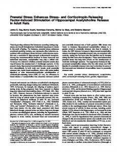
Fig 1. Experimental design and reference map. A, Experimental protocol of prenatal restraint stress. B, Map of neuronal quantification. The frontal (A), parietal (B), and caudal (C) cortical levels were subdivided in a medial (red square; 1), dorsal (blue square; 2) and dorsolateral (green square; 3) tiers. E = embryonic days; P = postnatal days. doi:10.1371/journal.pone.0117680.g001
Millipore), then and after subsequent washes with PBS, sections were incubated with Streptavidin-HPR (IHC Select Detection Systems, Millipore) for 1 hour at RT. After washing, the staining was revealed using diaminobenzidine (1:400, IHC Select Detection Systems, Millipore). Finally, sections were cleaned in distilled water and mounted.
Stereological quantification Reelin and NeuN immunoreactive neurons were quantified in 9 different cortical regions distributed along the anteroposterior and mediolateral axes of the rat brain (Fig. 1B)[38]. Observations were made at three different rostrocaudal levels (frontal, parietal and retrosplenial) in coronal sections. In each of these levels, we took samples from medial, dorsal and dorsolateral tiers (Fig. 1B). More specifically, at the frontal level we took samples from the medial tier, including the prospective of the adult anterior precentral and cingulate areas of the prefrontal cortex, from the dorsal tier including the early location of the motor cortex and from the dorsolateral tier including early somatosensory cortex (Fig. 1B, section A). At the parietal level, the cortex was subdivided into a medial tier including the future cingulate and motor cortex, and dorsal and dorsolateral tiers, both including early somatosensory cortex (Fig. 1B, section B). More posteriorly, the caudal level was subdivided into a medial tier including the posterior cingulate cortex, a dorsal tier including the posterior parietal cortex and a dorsolateral tier including the maturing auditory cortex (Fig. 1B, section C). In each region, stereological quantification was performed using the optical dissector method as described by Cruz-Orive and Weibel [39]. The counting frame of each dissector square had dimensions of 0.08 × 0.08 mm and the height was 0.01 mm. Double-blind counting was performed in 5 randomly selected dissector fields out of 10 in the first layer of all considered areas, which correspond to the length of one dissector square. The final neuron number was determined with an average of both blind-counts and considering a dissector volume of 6,4 × 10⁻⁵ mm3. Observations were made with bright field microscopy, and the images were captured using an Olympus microscope (Model BH-2, Olympus Co., Tokyo, Japan) equipped with a Moticam 2500 photographic camera (Speed Fair Co., Ltd).
Cortical culture Cerebral cortex neurons were dissociated mechanically in cold PBS 1X and incubated with HBSS—EDTA Trypsin 1%(10:1) for 30 minutes. Then, cells were dissociated in HBSS by gentle sweeping, counted and seeded into poly-D-lysine-coated dishes in DMEM 10% HS. Finally
PLOS ONE | DOI:10.1371/journal.pone.0117680 February 13, 2015
4 / 24
Cellular and Behavioral Effects of Prenatal Stress
and after 2 hours of incubation, plated medium was changed by neurobasal medium supplemented with B27, glutamine 2mM, pyruvate 1mM, 100 μg/mL of penicillin and 100 μg/mL of streptomycin for 5 days in vitro at 37°C, 95% humidity and 5% of CO2.
Western blot analysis After RIPA culture and tissue protein extraction, 30 μg of protein were loaded in a 6%, 10% or 12% polyacrylamide gel (according to the molecular weight of the protein analyzed). Samples were run in the concentrator gel for 30 min at 80 V and separated at 120V at different times, depending on the molecular weight of the protein of interest. Proteins were transferred to a nitrocellulose membrane using a wet protocol for 1.5 hour at 330 mA for proteins displaying high molecular weights, and for 1 hour at 300 mA for proteins displaying small molecular weights. Membranes were then blocked using non-fat dry 5% milk in TBS 1X, TWEEN 0,05% and incubated with primary antibody in non fat dry 1% milk in TBS 1X, TWEEN 0,05% over night at 4°C (anti-Reelin 1:1000, Millipore; anti-mDab1 1:1000, Santa cruz; anti-p35 1:1000, Santa cruz; anti-CDK5, 1:1000, Santa cruz; anti-PHF-1. 1:3000, donated by Peter Davis, Albert Einstein College of Medicine, NY, USA; anti-Tubulin 1:10000, Sigma). Membranes were then washed six times with TBS 1X, TWEEN 0,05% for 6 min and incubated with secondary antibody for 2 hours in non fat dry 1% milk TBS 1X, TWEEN 0,05% at room temperature (antiMOUSE 1:5000 Jackson; anti-RABBIT 1:5000 Jackson). Finally membranes were washed five times for 6 min in TBS 1X, TWEEN 0,05%, and the presence of proteins was developed using an ECL kit (Pierce Thermo Scientific) and radiographic films (Fujifilm).
DNA Methylation assay Whole cerebral cortex and cultured cortical neurons derived from newborn rats were collected for genomic DNA extraction. Firstly, cells were washed three times with 1X PBS, placed on ice, collected and centrifuged at 2,000 g. Cell pellets were resuspended in digestion buffer (100 mM NaCl, pH 8.0 Tris-Cl 10 mM, pH 8.0, 25 mM EDTA, 0.5% SDS, proteinase K 0.1 mg / ml) and incubated overnight at 40°C. Genomic DNA was isolated using phenol/chloroform extraction and finally resuspended in TE 1X buffer pH 8.0 and quantified by spectrophotometric methods. Two μg of purified genomic DNA were digested with HpaII or MspI restriction enzymes (sensitive and insensitive to DNA methylation, respectively). Whole cerebral cortex DNA samples were obtained by pre-digesting the samples with EcoRI (the DNA region analysed in this study does not contain such restriction site). This procedure further increases solubility of the DNA samples and therefore facilitates subsequent quantification and cleavage with HpaII/ MspI. Conventional PCR was performed using 200 ng of digested genomic DNA and specific primers against the Reelin gene promoter as well as against other genomic regions used as controls (Table 1). Finally, PCR amplification products were analyzed by electrophoresis in 1% agarose gels [40]. Table 1. Primers used in each condition. Reelin Runx2 Ric8B
Forward
50 -GGAAACGCATTAAAACCTG-30
Reverse
50 -GGAAGGCATGCAGAGGAGT-3
Forward
50 -GTGGTAGGCAGTCCCACTTT-30
Reverse
50 -TGTTTGTGAGGCGAATGAAG-30
Forward
50 -TGGTTTCCGGCCTTTAGGGAAC-30
Reverse
50 -GGAGCCACCAGAGACTGAGTCA-30
doi:10.1371/journal.pone.0117680.t001
PLOS ONE | DOI:10.1371/journal.pone.0117680 February 13, 2015
5 / 24
Cellular and Behavioral Effects of Prenatal Stress
Cdk5 kinase activity assay Cdk5 activity was measured as described previously [41]. Briefly, 300 μg of protein were extracted from brain cortex of control and stressed rats. Proteins were dissolved in T-PER buffer and immunoprecipitated using 4 μg of anti-Cdk5 antibody C8 (Santa Cruz, CA). Immunoprecipitated proteins (IP) were washed 3 times in cold PBS, and 2 times in kinase buffer [20 mM Tris HCl (pH 7.4), 10 mM MgCl2 and 1 mM EDTA]. IP were then mixed with the kinase assay mixture [100 mM Tris • HCl (pH 7.4), 50 mM MgCl2, 5 mM EDTA, and 5 mM DTT] plus 5 μCi (γP32)-ATP, with 5 μg of Histone H1 used as a substrate. Kinase assays were carried out at 30°C for 30 min and the kinase activity reaction was stopped by adding 5xSDS sample buffer and boiling it for 10 min at 95°C. The kinase reaction was electrophoresed on a 12% polyacrylamide gel and then gels were exposed to X-ray films for 1–3 h at −80°C. The incorporation of P32 to Histone H1 was quantified to measure band intensity using ImageJ 1.46r software (NIH, MD).
Behavioral activity All behavioral procedures were performed with rats aged two months (P60), considered as young adults. Sprague—Dawley rats, weighing 200–230 g, were housed eight per cage in a temperature-controlled animal facility, under a 12:12 hour light—dark cycle (lights on from 0800 to 2000 hr) with food and water ad libitum. Behavioral observations took place in a soundproof room at the same time of the day (10:00 to 12:00 hr) to reduce the confounding influence of diurnal variation on spontaneous behavior. Female rats were tested in diestrus, to avoid fluctuations in the data due to effects of the cycle itself [42]. For data analysis of locomotor activity, rearing, elevated plus maze and Morris water maze, 10 animals were used for each treatment (5 males and 5 females). In the passive avoidance paradigm we used 9 animals for each treatment, with 5 females and 4 males in each group. Each animal was evaluated only once on each behavioral test.
Spontaneous Motor activity Each rat was placed in a plexiglass cage (30 × 30 × 30 cm), and spontaneous locomotor activity and rearing were monitored during a period of 30 min and evaluated as described previously [43]. The floor of the cage was an activity platform (Lafayette Instrument Co., Lafayette, IN, USA) connected to an electromechanical counter. In order to avoid the influence of disturbing noises, the platform was placed in a soundproof chamber. Each animal was observed continuously via a Sony video camera connected to a VHS tape recorder (Sony Corporation, Mexico DF, Mexico). Scores were generated from live observations, while video sequences were used for later re-analysis when necessary. Results were expressed as the score measured on 30 min of observation for each group.
Elevated plus-maze Following the analysis of spontaneous locomotor activity and rearing behavior, anxiety levels were measured using the elevated plus-maze test. Each rat was individually placed in an elevated plus-maze consisting of two open arms (50 × 10 cm each), two closed arms (50 × 10 × 20 cm each) and a central platform (10 × 10 cm), arranged in such a way that the two arms of each type were opposite to each other. The maze was elevated 100 cm above the floor. At the beginning of each trial, animals were placed at the center of the maze, facing a closed arm. During a 5 min test period, we recorded: (i) the number of open arm entries; (ii) the number of closed arm entries; (iii) the time spent in open arms; and (iv) the time spent
PLOS ONE | DOI:10.1371/journal.pone.0117680 February 13, 2015
6 / 24
Cellular and Behavioral Effects of Prenatal Stress
in closed arms. Entry into an arm was deflned as the animal placing all four limbs onto the arm. The maze was thoroughly wiped clean with 5% ethanol solution after each trial. All trials were conducted between 10:00 and 14:00 hours. Results were expressed as percentages of time spent in open arms and the total number of entries (sum of closed and open arm entries) on 5 min of observation for each group.
Learning and memory tasks Passive Avoidance Conditioning. The test was carried out in a two-way shuttle box (Lafayette Instrument Co, IN, USA) composed of two stainless steel modular testing units with a manual guillotine door placed between them. Each modular chamber was equipped with an 18 bar insulated shock grid connected to a shocker (Master Shock Supply, Lafayette Instrument). One of these chambers remained illuminated and the other was darkened. On day 1 of testing, animals were habituated to the apparatus. Each rat received two trials with an interval of 20–30 min. On each trial, the rat was placed into the illuminated chamber facing away from the guillotine door. When the animal entered the darkened chamber, the guillotine door was lowered noiselessly and the animal was removed from the apparatus 10 sec later. The latency to enter was recorded. On day 2, each animal was placed into the illuminated chamber and permitted to enter the darkened chamber. Upon entry to the darkened chamber, the guillotine door was lowered and a 0.35 mA foot shock was applied for 2 sec through the grid floor. 10 seconds after this training, the rat was removed from the apparatus. The retention test was given on day 3 and consisted of a single trial without foot shock, in which each animal was placed into the illuminated chamber and the latency to enter the darkened chamber was recorded to an arbitrary maximum of 300 sec. On days 4 and 5, the extinction of the response was assessed, in a way similar as that on day 3. This test is based on the conflict between unconditioned avoidance of a brightly lit chamber and conditioned avoidance of an electric shock. Results are presented as the median latencies, which served as an index of retention and extinction on each day for each group. Morris water maze test. The water maze test used in our experiments was described by Morris in 1984 (Morris, 1984). This method is used to assess spatial learning and memory in rodents and it was performed at P60. The apparatus consists of a circular swimming pool (200 cm diameter, 60 cm depth) that was filled with water (22° C temperature) to 20 cm below the rim. The pool was divided into four quadrants of equal area (designated north, south, east, and west. A circular Plexiglas platform (10 cm diameter) was hidden just 2 cm below the water surface in the middle of the north quadrant. This platform had the same color as the swimming pool in order to render it invisible to the rat. The procedure room, where the swimming pool was located, had distinctive visual cues on the walls for spatial orientation. Rats were tested individually and placed into various quadrants of the pool. The time elapsed and the distance traversed to reach the hidden platform was recorded. Rats were moved to the procedure room 30 min before testing, and each animal was put through five trials per day, with 20-min intertrial intervals. The two investigators that worked with the water maze procedure were always the same and located always at the same position in the room. On each trial the rat was placed into the water, facing the side walls of the pool at one of the cardinal compass points (north, south, east, or west), and was allowed to locate the platform with a maximal time of 120 sec. The time spent by the rat to find the platform was recorded and assigned as latency. If the rat failed to find the platform within the maximal time, it was guided manually to the escape platform and allowed to remain there undisturbed for 30 sec. In this case it was assigned a latency of 120 sec. The sequence of starting quadrants was counterbalanced every trial. After staying on the platform for 30 sec, the rat was gently picked up, returned to its home cage, and allowed
PLOS ONE | DOI:10.1371/journal.pone.0117680 February 13, 2015
7 / 24
Cellular and Behavioral Effects of Prenatal Stress
to warm up and dry off under a heat lamp. The entire procedure took 5 days and results were expressed as the time in seconds of latencies in swimming performance on each day for each group.
Statistics For all treatments, we measured the effect of PNS treatment compared with control animals. Values are expressed as the mean ± SEM. For the analyses of the Reelin immunohistochemistry quantification and Western Blot, adrenal gland, and body weight gains we used the t-test (unpaired and Mann-Whitney test) considering p