Communicated by Rita Levi-Montalcini, May 21, 1979. ABSTRACT. Choline acetyltransferase (CATase; acetyl-. CoA:choline Oacetyltransferase, EC 2.3.1.6) has ...
Proc. NatI. Acad. Sci. USA Vol. 77, No. 12, pp. 7453-7457, December 1980
Neurobiology
Preparation of antibodies specific to choline acetyltransferase from bovine caudate nucleus and immunohistochemical localization of the enzyme* (cholinergic/immunohistochemistry)
COSTANTINO COZZARI AND BOYD K. HARTMANt Department of Biology, Washington University and Departments of Psychiatry and Neurobiology, St. Louis, Missouri 63110
Washington University School of Medicine,
Communicated by Rita Levi-Montalcini, May 21, 1979
Choline acetyltransferase (CATase; acetylABSTRACT CoA:choline Oacetyltransferase, EC 2.3.1.6) has been purified from bovine caudate nucleus. The specific activity of the pure enzyme was 120-160 Anmol of acetylcholine formed per mg per min. The purified enzyme separated into two bands (band A and band B) when electrophoresed in a pH 4.3 gradient gel. Both bands exhibited CATase activity. Antisera were prepared in rabbits to each form. Immunotitrations using either antisera resulted in quantitative precipitation of CATase activity from enzyme preparations at all stages of purification. Both antisera produced single immunoprecipitin lines in double-diffusion experiments when run against the enzyme at each stage of purity. Immunoprecipitin lines cut from double-diffusion gels contained CATase activity, demonstrating that the observed reaction was due to antibody interaction with the enzyme. Immunoelectrophoresis also showed a single immunoprecipitin line against the purified enzyme as well as against a crude caudate extract. These results indicated that immunochemically pure and specific antisera have been prepared to bovine CATase and that both the A and B forms of the enzyme have common antigenic sites. The antisera were utilized to localize the enzyme in bovine brain. The localization was exclusively neuronal with both antisera, and both forms of CATase were present in the same population of neurons. Subtle differences in the precise intracellular distribution of enzyme were observed with te two antisera, indicating that although the two molecular forms of CATase have antigenic sites in common they are not antigenically identical. Choline acetyltransferase (CATase; acetyl-CoA:choline 0acetyltransferase, EC 2.3.1.6) is the enzyme responsible for the synthesis of the important neurotransmitter acetylcholine. The enzyme activity was first measured by Nachmanson and Machado (1) in rabbit brain extract. Previous attempts to purify the enzyme in various laboratories yielded conflicting results regarding the maximal specific activity, the molecular weight, and the number of molecular forms of the pure enzyme. Furthermore, the antisera resulting from use of different preparations of CATase have led to different conclusions regarding the antigenicity of CATase, the ability of respective antisera to immunoprecipitate CATase activity quantitatively, and the relative purity and specificity of the various antisera preparations. At present no satisfactory method is available for histological localization of the cholinergic neuron systems. Therefore, immunohistochemical localization of CATase will be a significant application of specific antiserum to this enzyme. In order to ensure accurate localization, however, it is of primary importance that the antisera used for this application meet stringent criteria of purity and specificity. CATase has been purified The publication costs of this article were defrayed in part by page charge payment. This article must therefore be hereby marked "advertisement" in accordance with 18 U. S. C. §1734 solely to indicate this fact. 7453
from bovine caudate nuclei in this laboratory. This purified preparation of CATase exhibits the highest specific activity yet reported and is present as two homogeneous molecular forms that are separable on gradient acrylamide gel electrophoresis at pH 4.3 and on NaDodSO4 gel electrophoresis. The details of the purification procedure and the characterization of the enzyme will be presented elsewhere. This communication will report the results obtained when the two separable forms of the enzyme were used for the production of antisera in rabbits and the evidence for their purity and specificity. Initial results of immunohistochemical localization of CATase in bovine brain will also be reported. MATERIALS AND METHODS Enzyme Assay. The radiometric method of Schrier and Shuster (2) was used with the following final concentration of reagents: 0.4 mM [14C]acetyl-CoA. The concentration of acetyl-CoA was determined experimentally by the phosphotransacetylase method (3). Acetyl-CoA was diluted with unlabeled carrier to a specific radioactivity of 0.7 mCi/mmol (1 Ci = 3.7 X 10¹⁰ becquerels). The other constituents of the assay mixture were 20 mM choline.HCl, 400 mM KC1, 0.2 mM physostigmine sulfate, 50 mM Tris.HCl (pH 7.4), 1 mM EDTA; 0.1 mM dithioerythritol; 0.5 mg of bovine serum albumin per ml; and enzyme. The total volume was 200,ul. Assays were incubated for 10 min at 370C. One unit of enzyme activity was defined as 1.0 ,mol of acetylcholine formed per min at 370C. Protein was measured by the method of Geiger and Bessman (4). Electrophoresis. Electrophoresis was performed on gels with a linear acrylamide gradient from 7.5% to 20% (wt/vol) (N,N-methylenebisacrylamide, 0.2-0.1%). Gels were prerun for 24 hr in a pH 4.3 buffer (5) containing 3% ethyl alcohol and 5 mM cysteine. Proteins were applied and run toward the cathode for 28 hr at 1 mA per tube at 4°C and stained with Coomassie blue in methyl alcohol/acetic acid (6). NaDodSO4 gels were run by the method of Weber and Osborne (6). Purification Procedure. Fresh beef caudate nuclei (1.7 kg) were homogenized in 1 mM EDTA/0.2 mM dithioerythritol at pH 5.5 adjusted with acetic acid. The homogenate was centrifuged for 30 min at 12,000 X g, the supernatant was discarded, and the precipitate was lyophilized. The lyophilized precipitate was extracted with cold acetone to remove lipid material and the residue was dried. The dried powder was exAbbreviation: CATase, choline acetyltransferase. * Presented in part as abstracts to the American Society of Neurochemistry, Denver, CO, March 1977, and International Society of Neurochemistry, Copenhagen, Denmark, August 1977. t To whom reprint requests should be addressed at: Department of Psychiatry, Washington University School of Medicine, 4940 Audubon Avenue, St. Louis, MO 63110.
7454
Neurobiology: Cozzari and Hartman
tracted with 0.15 M NaCl/25 mM potassium phosphate, pH 7.4/0.5 mM dithioerythritol/l mM EDTA. After centrifugation the supernatant was fractionated with ammonium sulfate. The precipitate that occurred at 40-60% saturation was collected and further fractionated by gradient solubilization with decreasing concentrations of ammonium sulfate (7). The CATase-containing fractions were then precipitated with ammonium sulfate. The precipitate was redissolved in buffer and chromatographed on Sephadex G-100. Fractions containing CATase activity were pooled and chromatographed over CM-cellulose. This step was then repeated, and active fractions from the second ion exchange column were concentrated and chromatographed on Sephadex G-150 (superfine). At this stage of purification, four major protein bands and two minor bands could be visualized on gradient acrylamide gel electrophoresis.
The enzyme was purified further by using affinity chromatography. The column used was a CATase inhibitor linked to Sepharose 4B by a modification of the procedure described by Husain and Mautner (8). Our procedure used the CATase inhibitor naphthylvinylpyridine which was modified to introduce a carboxyl group at the end of a 10-carbon chain. The carboxyl group was utilized to couple the inhibitor with aminoalkyl-Sepharose by a carbodiimide-promoted condensation reaction. Chromatography over this column produced two protein peaks. The first peak contained no enzyme activity. The enzyme pool taken from the second peak contained CATase with a specific activity of 120-160 units/mg. Antisera prepared to this preparation gave a single precipitin line in double-diffusion tests against purified CATase. However, these antisera also produced faint (observable only after Coomassie blue staining of plates) precipitin lines in double-diffusion tests against some non-CATase-containing protein fractions obtained in chromatography steps during purification. These precipitin lines showed lines of nonidentity with the precipitin lines produced against purified CATase, indicating the presence of highly antigenic microcontaminants in the CATase preparation. These microcontaminants were removed empirically by immunoadsorption of the final enzyme preparations. To accomplish this, the non-CATase-containing protein fractions shown to contain reacting antigens were used to innoculate additional rabbits. These resulting antisera were bound to Sepharose 4B by using the procedure of Cuatrecasas and Anfinsen (9). These immunoadsorbants were used to purify the CATase further. Recovery of CATase activity was >95% although no increase in specific activity was observed. The chromatographic pattern of the enzyme on gradient gel was also unchanged and consisted of two apparently homogeneous protein bands. Individual enzyme preparations (from 1.7 kg of beef caudate) produced approximately 250 gg of pure CATase with an overall yield of enzyme activity of 6% compared to the tissue homogenate. The two protein bands separated on gradient acrylamide gel were excised and used to innoculate different rabbits. Immunoadsorption had apparently removed the microcontaminants because the resulting antisera did not show evidence of immunoprecipitation on double-diffusion plates when run against any non-CATase-containing chromatographic fractions. These antisera are the subject of this paper. Preparation of Antisera. Gradient gels containing 80 jig of protein were run and stained with anilinonaphthalene sulfonate (10). The two bands, visible under ultraviolet illumination, were excised. Gel segments containing proteins (total approximately 80 ,gg) were separately extruded through a 19-gauge needle into 2 ml of complete Freund's adjuvant (Difco) and into 2 ml of
He
Proc. Natl. Acad. Sci. USA 77 (1980)
sterile physiological saline to which an additional 5 mg of killed microbacteria had been added. These mixtures were homogenized and used for inoculation of New Zealand rabbits. Monthly booster injections with the same quantity of antigen were given. Serum was collected biweekly after the first booster injection. Immunohistochemistry. Immunohistochemical localization was carried out in beef brain. Fresh tissue was fixed by immersion in 4% formaldehyde/0.1 M potassium phosphate, pH 7.4, for 7 days followed by equilibration in 15% sucrose in the same buffer. Cryostat frozen sections (10 ,um) were cut at -20'C and the direct immunofluorescence procedure was carried out as described (11) except that incubation with the anti-CATase was extended to 48 hr at 4VC. The primary antisera were diluted 1:1000 with 0.1% Triton X-100 in phosphate-buffered saline. Controls consisted of adjacent sections in which anti-CATase was replaced by normal rabbit serum diluted to produce an IgG concentration equivalent to that present in anti-CATase. At this dilution, controls exhibited negligible nonspecific fluorescence. RESULTS Enzyme Used for Antibody Induction. The specific activity of the purified CATase was 120-160 units/mg compared to 0.005 units/mg for the starting homogenate. When the final enzyme preparation was run on gradient acrylamide gels at pH 4.3, two clearly separated protein bands were observed. When these gels were sliced, eluted, and assayed, CATase enzyme activity was present in two peaks corresponding to the protein bands. These bands have been arbitrarily designated "band A" (slow band) and "band B" (fast band) (Fig. 1). When this enzyme preparation was run on NaDodSO4 gels, two bands were again seen. The molecular weights of the two enzyme forms estimated from NaDodSO4 gels were 72,000 and 76,000, respectively. These data indicate that CATase isolated from caudate nuclei by the present method exists in two separable and active forms. Within the sensitivity of these methods each of the two protein bands was homogeneous, and no additional bands were detectable in either gel system. Analysis of antisera Precipitation of Enzyme Activity. To demonstrate the presence of anti-CATase activity, the most sensitive method was indirect precipitation titration of enzyme activity. In this method, constant enzyme activity (15 X 10-3 units; preparation after Sephadex G-100) was incubated with increasing quantities of antisera. Then, the entire IgG fraction was precipitated by the addition of anti-rabbit IgG. After centrifugation, enzyme activity was assayed in the supernatant and the resuspended immunoprecipitates (Fig. 2). With this method, anti-CATase activity was detectable 3 weeks after initial innoculation and reached a maximum 1 month after the third booster injection. Anti-CATase immunoactivity was induced in rabbits inoculated with either band A or band B. Greater than 96% of CATase was removed from the supernatant, and most of this activity could be found in the immunoprecipitates. +
-, .... .....
I
I A\
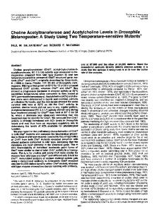
FIG. 1. Acrylamide gel electrophoresis of purified CATase (20
,gg of protein per gel). (Upper) Gradient gel at pH 4.3. (Lower) NaDodSO4 gel migration was from left to right. Two distinct protein bands are visible under both conditions: A, band A; B, band B.
Neurobiology:
Cozzari and Hartman
Proc. Natl. Acad. Sci. USA 77 (1980)
7455
of CATase activity from 0.2% Triton X-100 extracts of caudate, in which essentially 100% of CATase activity is solubilized, although in the presence of detergent approximately 3 times as much antiserum was required per unit of enzyme activity. This experiment indicates that all CATase present in caudate $ ~~~~~~~~~~10 is recognizable by the antisera. Double Diffusion Experiments. Immunoprecipitin lines were first observed when antisera obtained 1 week after the 60 second enzyme injection were run in double-diffusion plates Cd~~~~~~~~~~~~~~~~~~~~~~~~C against enzyme preparations. These lines became more intense -0 P.~~~~~~~ as the antibody titer increased. A single precipitin line of immunological identity formed when either antiserum was run ~~~~~~~~~~~0against the enzyme fractions obtained at various stages of pu0 rification (Fig. 3). It was not possible to demonstrate additional immunoprecipitin lines by altering the relative concentrations of either enzyme or antisera. It thus appears that a single anti4.0 2.0 3.0 0 1.0 system was present in each of the antisera. In order to be body Antiserum, gIl certain that the antibody system visible in double diffusion was FIG. 2. Indirect immunotitration ot CATase with antisera to band indeed the result of CATase-anti-CATase interaction, a douA (om) and to band B (0,0). Variable quantities of antisera were experiment was carried out in which CATase ble-diffusion mixed with normal rabbit serum to maintain a constant quantity (4 in agar segments. CATase activity was measured was activity id) of serum [diluted with 50 mM potassium phosphate buffer (pH concentrated in the agar slices that contained the immu7.4) containing 0.5 mg of bovine serum albumin per ml] to a final noprecipitin lines (Fig. 4). volume of 250 ,id (i.e., the first point on the curve contained 4 id of normal serum and no anti-CATase). To these solutions 50 Al of soThe relationship between the antisera to band A and band lution containing 15 X 10-3 units of partially purified CATase (after B was investigated by running the two antisera against purified Sephadex G-100 chromatography) was added; the mixture was inCATase (containing both A and B forms of the enzyme). The cubated 45 mmn at room temperature and 18 hr at 40C. Sufficient two precipitin lines joined without evidence of crossing or anti-rabbit IgG was then added to precipitate the IgG in 4 isl of serum. spurring (Fig. 5). This test confirms the results of immunotiSamples were centrifuged, and aliquots of the supernatant (em) and tration of CATase activity, both experiments indicating that of the resuspended washed immunoprecipitates (0,0) were assayed the two electrophoretically separable forms of the enzyme have for CATase activity. antigenic sites in common. It should be emphasized that this does not demonstrate antigenic identity of the two forms. By using direct immunoprecipitation titration of CATase The induction of minor contaminating antibody systems can activity (i.e., by incubation with specific antisera only) it was be a serious problem when antigens used for inoculation are also possible to achieve quantitative precipitation of enzyme difficult to obtain in pure form. Immunohistochemical appliactivity. After centrifugation, however, no immunoprecipitates cations are especially affected because all antigens that have were observable and no attempt was made to recover the preinduced antibodies may be localized. Similarly, it is important cipitated activity. This is not surprising because 15 X 10-3 unit to rule out the possibility that significant crossreactivity occurs would only contain 0.12 ,ug of enzyme protein. In direct titrawith other proteins in the system. The presence of a single tions, approximately 4-5 times as much antisera was required precipitin line against the CATase protein pools at different to precipitate the same amount of activity as in the indirect method. This fact is consistent with the increased antibody required to form an insoluble enzyme-antibody complex. The immunotitration experiments indicated that band A and band B contained sufficient CATase to induce the production of antibodies directed against the enzyme. The antiserum to band B exhibited a higher antibody titer than did that to band A. The titer of specific antibodies was estimated, by microimmu6 s 6 2 noprecipitation in capillary tubes, to be approximately 0.5 and 0.8 mg/ml for anti-band A and anti-band B, respectively. A.A Because the overall yield of the enzyme preparation was only .-1 4m.. about 6%, it was important also to demonstrate that the purified CATase was immunologically representative of caudate 5 3 3 5 CATase in general. After the removal of lipids by acetone extraction (the second step in purification), the yield of soluble 4 CATase was 75% of the activity in the initial homogenate. 4. When enzyme at this stage of purification was subjected to indirect immunoprecipitation, the quantity of the anti-band B required to precipitate an equivalent amount of enzyme FIG. 3. Double-diffusion immunoprecipitin patterns in 0.5% agar activity was the same as shown in Fig. 2 (ije., 0.6 ,u1 of anti-band of antibodies against band A (A) and band B (B) run against CATase B for 50% precipitation). This relationship betweeen the at various stages of purification. The outer wells were filled with CATase preparations after the indicated steps of purification: 1, quantity of antiserum and the amount of enzyme activity concentrated supernatant from acetone powder extraction; 2, Sephprecipitated remained constant for all subsequent stages of adex G-100 3, first CM-cellulose; 4, second CM-cellulose; 5, Sephadex purification, indicating that the specific activity of the CATase G-150 (superfine); 6, naphthylvinylpyridine affinity chromatograat the final stage of purification is representative of the enzyme phies. Single immunoprecipitation bands of identity are observed that- produces 75% of the total activity in the initial homogenate. against enzyme at all stages of purity. Gel plates were stained with It was possible to achieve quantitative immunoprecipitation Coomassie blue after 2 days of incubation at 40C.
i'UL
-c
..eIS1 --
pi
I; I
II I
2.1
I I
I
I3.. .,I
It
-
~rlts_,n.7mS ~ I
Neurobiology: Cozzari and Hartman
7456
I
X6
I
I
I
I
It .1 N
a-
..N * as
I .4
__
II
[ '
I IX~ I
%I I1 A
I I
II
I
I I 'l
/~~~~~~~~~~~~~ X~~~~~~~~~~
s-
I I
I
Proc. Natl. Acad. Sci. USA 77 (1980)
AS.~~~~~~~~~~~~
/~~~~~~~
Amd R. w~w ~~~ ~ ~ ~ ~ ~~~~~~~i:AT
w
[-~~~~~~~~~~~~~~~~ § 1 a-'o '' I t' ''''''Y''
II
1.2 Said
''
w
I lI T''
1 4
94
FIG. 4. Demonstration of CATase activity in immunoprecipitin lines cut from 0.5% agar strip shown schematically at the bottom of the figure (dark shading shows the position of the precipitin lines). After incubation for 4 days at 40C, the gel strip was exhaustively washed and cut into 24 1-mm slices that were assayed for CATase activity. CATase activity was concentrated in those slices containing the immunoprecipitin lines. CAT, concentrated enzyme after second CM-cellulose chromatography. Anti A and anti B represent 25 ul of antisera to band A and band B, respectively.
stages of enzyme purification rules out contamination of the enzyme fractions by detectable amounts of impurities or crossreacting proteins. A more sensitive test for the absence of contaminating antibody system was the demonstration that the present antisera did not produce immunoprecipitin lines against any non-CATase-containing protein fractions at any stage of enzyme purification. These fractions usually will contain pos-
O'
ANTIB 'V
FIG. 5. Double diffusion in agar of anti-band A (anti A) and anti-band B (anti B) against purified CATase (after affinity chromatography; contains both A and B forms). The absence of spurs indicates that there are antigenic sites common to both A and B forms of CATase. Gel plates were stained with Coomassie blue.
sible contaminants in higher concentrations than will fractions containing enzyme. In our experience these negative data are a stringent indication of purity and specificity of the antisera. These data coupled with the observation of single precipitin lines against the CATase-containing fractions at all stages of purification and the demonstration of CATase activity in the immunoprecipitin lines are strong evidence for the purity and specificity of the present antisera to CATase. Immunoelectrophoresis of both antisera was performed against purified CATase and a crude preparation of CATase at pH 6.0. In all cases a single immunoprecipitin line was observed with the same electrophoretic mobility. No evidence of additional immunological reactions was observed at any position in the gels, confirming the absence of either contaminating antibody systems or significant crossreaction of the antibody with other proteins in the system (data not shown). Immunohistochemical localization of CATase The antisera have been used for localization of the enzyme in beef brain by the technique of immunofluorescence. The initial results of these studies are given to demonstrate the feasibility' of localization of the cholinergic system by using this technique. The antibodies reacted exclusively with neuronal elements within the brain. When anti-band A and anti-band B were applied to adjacent tissue sections, the same groups of neurons exhibited a positive reaction and the same groups of neurons were negative. The only discernible differences between the localization observed with the two antisera were that the A form of the enzyme was more clearly observed in the dendritic tree of the neurons and the B form of the enzyme exhibited brighter fluorescence in terminals. Thus, although both antisera demonstrated the same neuronal populations, subtle differences were observed in the intensity of fluorescence in different segments of the neuron (Fig. 6). DISCUSSION the Through application of the rather complex purification procedure outlined, it was possible to obtain preparations of CATase from bovine caudate nucleus with a specific activity of 120-160 units/mg. This represents a substantially higher specific activity than has been reported for the enzyme: approximately 5-fold greater than the highest value reported for mammalian brain [20 units/mg in rat brain (12) and 30 units/mg in bovine brain (13)] and 2 to 6 orders of magnitude greater than other reported values [0.012 unit/mg in human brain (14), 0.039 unit/mg in human placenta (15), 0.73 unit/mg in rat brain (16), 1.45 units/mg in bovine brain (17), and 4 units/mg in rat brain (18, 19)]. In fact, this specific activity is twice that reported for CATase purified from squid head ganglion (8). The enzyme assay conditions and the enzyme sources were not identical in each case, however, so that the relative purity of the various preparations is not necessarily accurately reflected from the reported specific activities alone, although one would expect that, at least within mammalian species, the degree of purity would roughly correlate with specific activity. In contrast to early reports, CATase appears to be a relatively good antigen when injected in pure form. Significant titers of anti-CATase always developed after two injections. It is also clear that several potential contaminants are substantially more antigenic than CATase because additional antibody systems could be detected in antisera prepared from purified CATase that had not been further purified by using immunoadsorption with antibodies prepared to these contaminants. This observation emphasizes the need to use pure enzyme for inoculation. Stringent criteria were applied to demonstrate the purity and specificity of the present antisera.
Neurobiology:
Cozzari and Hartman
Proc. Natl. Acad. Sci. USA 77 (1980)
7457
I
FIG. 6. Immunohistochemical localization of CATase in bovine brain. (A) Magnocellular neurons of pontine reticular formation, after incubation with anti-band A. (B) Neurons of the same type after incubation with anti-band B. (C) Neurons in the medullary reticular formation after incubation with a mixture of anti A and anti B. Fine fluorescent dots are visible in the gray matter between bundles of myelinated fibers. This localization presumably represents a cholinergic terminal area. (Scale bar = 20 am.)
In terms of the ability to precipitate enzyme activity, the antisera prepared to these preparations of CATase compare favorably to other antisera available. Rossier (20) compared the relative ability of various antisera to precipitate or inhibit 50% of 0.25 X 10⁻³ unit of CATase activity. Rossier's most potent antisera required 0.1 Al, compared to 2 pl for that of MaltheSorrenssen et al. (13) (our calculation) and 10 Al for that of Singh and McGeer (14) and of Eng et al. (21) who were unable to achieve 50% loss of enzyme activity with any quantity of antisera. The present antisera required 0.06 1ul of anti-band A and 0.05 MI of anti-band B to precipitate this quantity of enzyme activity in the direct precipitation titration and 0.012 and 0.010 AM, respectively, by the indirect method. The immunohistochemical localization observed with the application of the two antisera was interesting because, although positive (or negative) reactions occurred in the same cell populations, the two antisera produced different intensities of fluorescence within different parts of the neurons exhibiting a positive reaction. The immunochemical evidence clearly indicates that both antisera contain antibodies to antigenic sites common to both forms of CATase. However, immunohistochemical localization indicates that the separate antisera also recognize sites that are not common to both A and B forms of the enzyme. These differences can only be tested immunochemically when sufficient A and B forms have been separated to permit double-diffusion tests against the individual antisera.
The fact that anti-band A reacts immunohistochemically most intensely in the cytoplasm of the perikarya and the dendrites whereas anti-band B intensely stains the terminals as well cell bodies suggests that the two forms are not artifacts of purification but may have physiological significance. as
We gratefully acknowledge the excellent technical assistance of Delores J. Blehm and Sandra J. Kalmbach. This work was supported in part by Grants MH-24604, NS-12311, and NS-13672 and Research Scientist Development Award MH-70451 (B.K.H.).
1. Nachmansohn, D. & Machado, A. L. (1943) J. Neurophysiol. 6, 397-403. 2. Schrier, B. K. & Shuster, L. (1967) J. Neurochem. 14, 977985. 3. Stadtman, E. R. (1957) Methods Enzymol. 111, 936. 4. Geiger, P. |. & Bessman, S. P. (1972) Anal. Biochem. 49, 467473. 5. Reisfeld, R. A:, Lewis, U. J. & Williams, D. E. (1962) Nature (London) 4838, 281-283. 6. Weber, K. & Osborn, M. (1969) J. Biol. Chem. 244, 44064412. 7. King, T. P. (1972) Biochemistry 77, 367-371. 8. Husain, S. S. & Mautner, H. G. (1973) Proc. Natl. Acad. Sci. USA
70,3749-3753. 9. Cuatrecasas, P. & Anfinsen, C. B. (1971) Methods Enzymol. 22, 345. 10. Hartman, B. K. & Udenfriend, S. (1969) Anal. Biochem. 30, 391-394. 11. Hartman, B. K. (1973) J. Histochem Cytochem. 21, 312332. 12. Rossier, J. (1976) J. Neurochem. 26, E 13-548. 13. Malthe-Sorenssen, D., Lea, T., Fonnum, F. & Eskeland, T. (1978) J. Neurochem. 30,35-46. 14. Singh, V. K. & McGeer, P. L. (1974) Life Sci. 15,901-913. 15. Roskoski, R., Jr., Tiong Lim, C. & Roskoski, L. M. (1975) J. Biochem. 14,5105-5110. 16. Potter, L. T., Glover, V. A. S., Jeffrey, K. & Saelens, J. K. (1968) J. Biol. Chem. 243,3864-3870. 17. Chao, L. P. & Wolfgram, F. (1973) J. Neurochem. 20, 10751081. 18. Malthe-Sorenssen, D., Eskeland, T. & Fonnum, F. (1973) Brain Res. 62, 517-522. 19. Wenthold, R. J. & Mahler, H. R. (1975) J. Neurochem. 24, 963-967. 20. Rossier, J. (1975) Brain Res. 98, 619-622. 21. Eng, L. F., Uyeda, C. T., Chao, L. P. & Wolfgram, F. (1974) Nature (London) 250,243-245.