ARTIGO/ARTICLE
Revista da Sociedade Brasileira de Medicina Tropical 38(6):496-502,nov-dez, 2005
Prevalence of hepatitis C virus infection and HCV genotypes among hemophiliacs in the State of Bahia, Northeastern Brazil: analysis of serological and virological parameters Prevalência da infecção pelo vírus da hepatite C e genótipos entre hemofílicos no Estado da Bahia, nordeste do Brasil: análise de parâmetros sorológicos e virológicos Luciano Kalabric Silva1, Maria Betânia Souza da Silva1, Gisele Barreto Lopes1, Itatiana Ferreira Rodart1, Fernando Quadros Costa1, Nelma P. Santana2, Raymundo Paraná3, Aurelino Santana4 and Mitermayer Galvão dos Reis1, 3, 4
ABSTRACT The objective of the present study was to analyze HCV serological and virological parameters from hemophiliacs in the State of Bahia. Anti-HCV was investigated by ELISA in a cohort of 268 hemophiliacs A/B who were followed-up in a reference unit for hemotherapy in the State of Bahia. HCV viremia and genotypes were also determined from a subset of 66 anti-HCV seropositive hemophiliacs. Seroprevalence among hemophiliacs was 42.2% (95% CI 36.5-48.1) and was significantly higher (p10 years, presence of factor VIII/IX inhibitory antibodies and other infection markers. None of the hemophiliacs less than 5 years of age were anti-HCV seropositive. Viremia was detectable in 77.3% (51/66). HCV genotype 1 (74%) was the most prevalent followed by genotype 3 (22%) and genotype 2 (4%). Our results indicate that HCV prevalence is still high among hemophiliacs, although HCV transmission was not observed in young hemophiliacs. Key-words: Hepatitis C virus. Hemophilia. Prevalence. Genotype. Bahia. RESUMO O objetivo deste estudo foi analisar parâmetros sorológicos e virológicos em hemofílicos no Estado da Bahia. O anti-VHC foi investigado por ELISA em uma coorte de 268 hemofílicos A/B sob acompanhamento em uma unidade de referência do Estado da Bahia. A viremia do VHC e genótipos foram determinados em um subgrupo de 66 hemofílicos soropositivos para o antiVHC. A soroprevalência do anti-VHC entre os hemofílicos foi de 42,2% (IC 95% 36,5-48,1) e foi associada significativamente (p10 anos, presença de anticorpos antifator VIII/IX e outros marcadores sorológicos de infecção. Nenhum dos hemofílicos com idade inferior a 5 anos foram anti-VHC positivos. A viremia foi detectada em 77,3% (51/66), sendo o genótipo 1 do VHC (74%) o mais prevalente, seguido pelos genótipos 3 (22%) e 2 (4%). Nossos resultados indicam que a prevalência do VHC é ainda alta entre os hemofílicos, muito embora a transmissão não tenha sido observada entre os menores de 5 anos. Palavras-chaves: Vírus da hepatite C. Hemofilia. Prevalência. Genótipos. Bahia.
1. Laboratory of Pathology and Molecular Biology, Gonçalo Moniz Research Center, Fundação Oswaldo Cruz, Salvador, BA, Brazil. 2. Hemotransfusion and Hemotherapy Foundation (HEMOBA), Secretaria de Saúde do Estado da Bahia, Salvador, BA, Brazil. 3. School of Medicine, Universidade Federal da Bahia, Salvador, BA, Brazil. 4. School of Medicine and Public Health, FDC, Salvador, BA, Brazil. Informed consent was obtained from all subjects who participated in this study. Guidelines of the Ethical Committee of the Gonçalo Moniz Research Center, FIOCRUZ, were followed in the conduct of this research. Financial support: This work was partially supported by research grants from CNPq, FIOCRUZ, FAPESB, and CAPES/FIOCRUZ (doctorate scholarship, between March 1999 to March 2003). Address to : Prof. Mitermayer G. Reis. Centro de Pesquisas Gonçalo Moniz/FIOCRUZ. R. Waldemar Falcão 121, Brotas, 40295-001 Salvador-BA, Brasil. Tel: 55 71 3356-4320; ext: 205; Fax: 55 71 3356-4292. e-mail:
[email protected] Recebido para publicação em 28/9/2004 Aceito em 20/8/2005
496
Silva LK et al
Hepatitis C virus (HCV), a positive-stranded RNA virus, has been identified as the major causative agent of non-A non-B post transfusion hepatitis8 17. Prior to the introduction of screening candidate blood donors for antibody to hepatitis C virus (anti-HCV) and inactivation methods for pooled plasma products, nearly all hemophiliacs became infected with HCV soon after their first exposure23. In the early 1990s, data from around the world showed that the seroprevalence of anti-HCV antibodies in hemophiliacs could reach up to 90% 11 21 26 31 47 48 , because virtually all clotting factor concentrates manufactured before the late 1980s were contaminated22. As a consequence, HCV is by far the most common cause of infection among hemophiliacs. Since the mid 1970s, with evidence of the existence of non-A non-B hepatitis viruses and the early 1980s with the epidemic of human immunodeficiency virus (HIV), physicians involved in hemophiliac care and manufacturers of clotting factors have been aware of the risk of transmission of unknown viruses12 13 15. Consequently, methods have been developed to inactivate these viruses, which became accessible in the developed countries in the mid 1980s. It is now known that some of these methods such as pasteurization in liquid state (10 hr at 60°C), viral inactivation by the so-called solvent detergent (SD) and dry heat treatment up to 80°C were effective, while others were inappropriate9 14 15 30 32 41. Other risk factors for HCV infection in hemophiliacs include age, severity of disease, previous blood transfusion, annual quantity of clotting factor and infection with human immunodeficiency virus (HIV)6 34. In the majority of the developing countries, these inactivated products were introduced only in the mid 1990s, substituting locally produced cryoprecipitate and fresh-frozen plasma. In Brazil, seroprevalence of HCV infection in hemophiliacs can range from 44.6% to 60% according to studies conducted in Southeastern and Central areas of Brazil6 24 34. Carmo et al 6 published a follow-up study from the State of Minas Gerais, Southeastern Brazil, revealing a tendency towards a decrease in the general seroprevalence of HCV infection among hemophiliacs. However, the majority of seropositive hemophiliacs identified were viremic, indicating the necessity of specialized health care and treatment. The study of the genetic variability of HCV strains have led to a consensus classification of six major genotypes, many of which include a number of closely related subtypes 7 44. Studies suggest that the clinical features of liver disease depend on HCV genotypes 20 33 42 49. It is also noteworthy that the success of interferon treatment seems to be genotype related 16 . These observations make the identification of infecting HCV genotypes from different geographical regions and groups under risk of great interest. Furthermore, genotypes can be a very useful tool for molecular epidemiology purposes. Thus, the primary objective of the present study was to determine the hepatitis C virus (HCV) seroprevalence, viremia and genotypes isolated from a cohort of hemophiliacs in the State of Bahia, Northeastern Brazil.
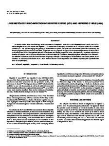
PATIENTS AND METHODS Patients. Between November 1999 and August 2000, the Hemotransfusion and Hemotherapy Foundation of Bahia (HEMOBA) attended a total of 339 patients with hemophilia A or B. However only 268 were tested in HEMOBA for the presence of anti-HCV by ELISA (3rd Generation, Abbott Murex, IL, USA) and became eligible for the study. Laboratory data. Clinical and laboratory data were collected from patients’ records and are summarized in Table 1. Serological tests for anti-HCV (Abbott Murex, IL, USA), HBsAg/ anti-HBc (Biorad, CA, USA), anti-HIV (Biorad, CA, USA), antiHTLV I/II (Abbott Murex, IL, USA), Chagas’ disease (Embrabio, SP, Brazil) and Syphilis (Weiner Lab, Argentine) were based on ELISA according to each manufacturer’s instructions. To test the coagulation factors and the presence of inhibitor antibody activity, the functional method based on the comparative activity assays against a factor deficient plasma was used according to the kit instructions (Biomérieux, NC, USA). Samples for molecular assays. Of the total of anti-HCV positive hemophiliacs, only 66 patients returned to HEMOBA to receive treatment during the period of this study and were interviewed and had their serum collected for molecular assays. The Institution Ethical Committee approved this study and informed consent was obtained from all subjects enrolled in the study. Within 2 hours after venopuncture all samples were aliquoted and stored immediately at -70ºC until use. Aliquots were not thawed more than once prior to analysis. Extraction of HCV RNA and cDNA synthesis. Two hundred microliters of serum were used for HCV RNA extraction using Trizol LS reagent (Invitrogen Life Technologies, CA, USA) following the manufacturer’s instructions and were precipitated with ethanol and then dried36. HCV RNA was immediately transcribed into cDNA using random primers (Amersham Biosciences, NJ, USA). Samples with HCV RNA undetectable by nested-PCR described below were extracted twice in different experiments. Even if they were confirmed negative, all these patients were recalled to repeat the blood collection within six months after the first examination in order to avoid false negative results. HCV RNA detection and genotyping. cDNA was targeted by a nested-PCR directed to the 5’UTR using specific primers 939, 209, 940 and 211 as described previously7. The size of the nestedPCR product was 251 bp. Positivity was confirmed by identification of this fragment after electrophoresis on a 1.5% routine agarose gel and ethidium bromide staining under UV light. Positive samples were genotyped by RFLP10 28. Briefly, restriction digests were carried out on the 251 bp PCR products for 4-16h after adjustment with 10x enzyme reaction buffer as appropriate. Reactions were at 37°C in the presence of 10 units each of (a) RsaI and HaeIII, and (b) HinfI and MvaI. Digestion products were visualized under UV light after electrophoresis through a 4% Metaphor agarose gel (BMA, ME, USA) in 1 x Tris-borate buffer containing 0.5mg/ml ethidium bromide. Figure 1 illustrates the band pattern consistently produced by RFLP in different genotypes. Genotypes were determined according to Simmond’s classification44.
497
Revista da Sociedade Brasileira de Medicina Tropical 38(6):496-502, nov-dez, 2005
1
2 3 4 5 6
7
8
9 (A) 251 (origin) 114/115 hp 44/56 26
(B) 198/251 (origin) 142/143 94 63 53/56 48
Figure 1- Electrophoresis through a 4% Metaphor agarose of restriction digests carried out on the 251 bp PCR fragment. Reactions were at 37°C in the presence of 10 units each of (A) RsaI and HaeIII, (B) HinfI and MvaI as described by McOmish et al28 and Davidson et al10 28. Lane 1 - blank control; lane 2 - genotype 2 control; lane 3 - genotype 1 control; lane 4 - genotype 3 control; lanes 5, 6, 9 genotype 1 samples and lanes 7, 8 - genotype 3 samples. Genotype was deduced from the banding patterns produced by the two restriction enzyme combinations.
RESULTS Baseline characteristics of the 268 hemophiliacs participating in this study are shown in Table 1. The mean age for all hemophiliacs was 19.5 ± 12.1 years old, age range from