DOI: 10.7589/2015-02-048
Journal of Wildlife Diseases, 52(1), 2016, pp. 000–000 # Wildlife Disease Association 2016
PREVALENCE, PATHOLOGY, AND RISK FACTORS ASSOCIATED WITH STREPTOCOCCUS PHOCAE INFECTION IN SOUTHERN SEA OTTERS (ENHYDRA LUTRIS NEREIS), 2004–2010 Georgina Bartlett,1,2 Woutrina Smith,3 Clare Dominik,1 Francesca Batac,1 Erin Dodd,1 Barbara A. Byrne,4 Spencer Jang,5 David Jessup,1 Julian Chantrey,2 and Melissa Miller1,3,6 1
Marine Wildlife Veterinary Care and Research Center, California Department of Fish and Wildlife, 1451 Shaffer Road, Santa Cruz, California 95060, USA National Centre for Zoonosis Research, University of Liverpool, School of Veterinary Science, Leahurst Campus, Chester High Road, Neston CH64 7TE, UK 3 Wildlife Health Center, School of Veterinary Medicine, University of California, Davis, 1089 Veterinary Medicine Drive, Davis, California 95616, USA 4 Department of Pathology, Microbiology and Immunology, School of Veterinary Medicine, University of California, Davis, One Shields Avenue, Davis, California 95616, USA 5 William R. Pritchard Veterinary Medical Teaching Hospital, School of Veterinary Medicine, University of California, Davis, 1 Garrod Drive, Davis, California 95616, USA 6 Corresponding author (email:
[email protected]) 2
ABSTRACT:
Recent studies have implicated beta-hemolytic streptococci as opportunistic pathogens of marine mammals, including southern sea otters (Enhydra lutris nereis), but little is known about their prevalence or pathophysiology. Herein, we focus on risk factors for sea otter infection by a single beta-hemolytic streptococcal species, Streptococcus phocae. Streptococcus phocae was first identified as a marine mammal pathogen in 1994, and the first report in southern sea otters was in 2009. Its broad host range encompasses fish, pinnipeds, cetaceans, and mustelids, with S. phocae now recognized as an important pathogen of marine species worldwide. We assessed risk factors and lesion patterns for S. phocae infection in southern sea otters. Using archival necropsy data, S. phocae prevalence was 30% in fresh dead otters examined 2004–2010. Skin trauma of any type was identified as a significant risk factor for S. phocae infection. The risk of infection was similar regardless of the cause and relative severity of skin trauma, including mating or fight wounds, shark bite, and anthropogenic trauma. Streptococcus phocae–infected sea otters were also more likely to present with abscesses or bacterial septicemia. Our findings highlight the importance of S. phocae as an opportunistic pathogen of sea otters and suggest that the most likely portal of entry is damaged skin. Even tiny skin breaks appear to facilitate bacterial colonization, invasion, abscess formation, and systemic spread. Our data provide important insights for management and care of marine species. Key words: Abscess, beta-hemolytic streptococci, mating trauma, sepsis, skin wound, southern sea otter (Enhydra lutris nereis), Streptococcus phocae.
Bacterial streptococci are common pathogens of terrestrial animals and humans (Henton et al. 1999). Streptococcus spp. are classified by their hemolytic properties on conventional blood agar as alpha-, beta-, or gamma-hemolytic. Beta-hemolytic streptococci are further subdivided via Lancefield serotyping, based on carbohydrate expression on the bacterial cell wall. Twenty serotypes span Lancefield groups A through V (excluding I and J), with some association between Lancefield grouping, host range, and disease expression. In human medicine, key pathogens include alpha- and beta-hemolytic streptococci of Lancefield groups A and B.
INTRODUCTION
The southern sea otter (Enhydra lutris nereis) is a federally listed threatened species that inhabits the coastline of central California, USA. Despite 100 yr of legal protection, this population has been slow to expand, in part, due to high mortality and habitat limitations. Many otters die following exposure to opportunistic pathogens, including land-based parasites and fungi. Although much has been learned about terrestrial pathogens of sea otters, little is known about the pathogenicity of marine and estuarine bacteria that infect this species. 1
2
JOURNAL OF WILDLIFE DISEASES, VOL. 52, NO. 1, JANUARY 2016
Human diseases associated with infection by group A streptococci include streptococcal pharyngitis (strep throat), toxic shock syndrome, necrotizing fasciitis, pneumonia, septicemia, and rheumatic fever. Infection by group B streptococci can trigger pneumonia, meningitis, and septicemia, especially in the elderly and neonates. Group B streptococci also infect terrestrial and aquatic animals, including camels (Camelus dromedaries; Fischer et al. 2013), dogs (Canis lupus familiaris; Lamm et al. 2010), cats (Felis catus; Tillman et al. 1982), crocodiles (Crocodylus porosus; Bishop et al. 2007), and fish (Kusuda and Kawai 1982; Gonzales-Contreras et al. 2011). In exposed animals, group B streptococci can be significant veterinary pathogens, causing mastitis (Keefe 1997), pyometra (Hueffer et al. 2011), abscesses (Imai et al. 2009), and septicemia (Burek et al. 2005). Streptococci are recognized as important marine mammal pathogens. Infection by Streptococcus infantarius subsp. coli and other pathogenic streptococci has been associated with fatal vegetative valvular endocarditis, septicemia, and thromboembolic disease in southern and northern (Enhydra lutris kenyoni) sea otters, especially for otters stranding in Alaska (Burek et al. 2005; Counihan-Edgar et al. 2012). More recently, the facultatively anaerobic, beta-hemolytic, pyogenic bacterium Streptococcus phocae (Skaar et al. 1994; Romalde et al. 2008) has been reported from numerous marine animals. First reported from harbor seals (Phoca vitulina) with pneumonia in northwestern Europe (Skaar et al. 1994), S. phocae is now recognized as an opportunistic pathogen for marine species worldwide, including grey seals (Halichoerus grypus; Vossen et al. 2004), ringed seals (Phoca hispida; Romalde et al. 2008), Cape fur seals (Arctocephalus pusillus pusillus; Henton et al. 1999), Caspian seals (Pusa caspica; Imai et al. 2009), spotted seals (Phoca largha; Hueffer et al. 2011), and California sea lions (Zalophus californianus; Johnson et al.
2006). Streptococcus phocae has also been isolated from a harbor porpoise (Phocoena phocoena; Romalde et al. 2008) and farmed Atlantic salmon (Salmo salar), with mortality rates as high as 25% (Romalde et al. 2008). Although S. phocae can be isolated from the respiratory and gastrointestinal tracts of healthy marine animals (Imai et al. 2009; Miller et al. 2010), infection is commonly associated with pneumonia and septicemia (Henton et al. 1999), neoplasia (Johnson et al. 2006), opportunistic infections (Imai et al. 2009), and pyometra (Hueffer et al. 2011). In southern sea otters, S. phocae infection was first identified during studies of bacterial flora (Imai et al. 2009). Most sea otter–derived, beta-hemolytic streptococci expressed group G, F, and C Lancefield surface antigens. Some additional sea otter isolates were untypeable via the Lancefield system but were assigned to S. phocae by using molecular characterization (Imai et al. 2009). However, despite the wide geographic distribution of S. phocae and its broad host range, potential environmental sources, routes of infection, and common case presentations were unreported (Gonzalez-Contreras et al. 2011). A study of the adherence and invasion properties of S. phocae isolates from salmon revealed nonspecific adherence to eukaryotic cells but limited ability to invade intact tissue (Gonzalez-Contreras et al. 2011), suggesting that skin wounds and other breaks in host defenses could facilitate S. phocae infection in salmonids. Based on these reports, we predicted that S. phocae infection would be common in southern sea otters and would be associated with dermal or mucous membrane lesions. Wounds sustained during fights, copulation, and predation are common in sea otters (Staedler and Riedman 1993; Kreuder et al. 2003), providing a portal for S. phocae infection. Breaks in mucosal barriers lining the respiratory, gastrointestinal, and urogenital tracts might also facilitate infection and systemic spread. We determined the prevalence of S. phocae
BARTLETT ET AL.—STREPTOCOCCUS PHOCAE INFECTION IN SEA OTTERS
infection in southern sea otters by reviewing retrospective necropsy records from 2004 through 2010. Our findings highlight the importance of S. phocae as an opportunistic pathogen of sea otters. We also identify the most likely route of infection and describe S. phocae–associated lesions in sea otters. Our findings will inform care and management of diverse marine species, especially threatened sea otters. MATERIALS AND METHODS Case selection and S. phocae detection
Enrolled southern sea otters included fresh (,72 h postmortem under refrigeration) animals from central California with full necropsies performed at the Marine Wildlife Veterinary Care and Research Center. Cases were defined as S. phocae–positive otters that were not treated with antibiotics prior to euthanasia or death and had at least one aerobic bacterial isolate cultured from lesions, tissue, internal fluids, digesta, or feces following necropsy. Controls satisfied the previously mentioned selection criteria, except that they were culture negative for S. phocae and related beta-hemolytic streptococci. Cases were excluded from the study if detailed necropsy or risk factor data were unavailable and if bacterial isolate identity as S. phocae was undetermined. Fetuses were excluded due to lack of exposure to most trauma types that were being assessed and the absence of a fully formed immune response. Sea otters necropsied prior to 2004 were also excluded due to lower precision of detection and identification of beta-hemolytic streptococci and S. phocae. Swabs for bacterial culture were maintained on Amies transport medium (Becton, Dickinson and Company, Sparks, Maryland, USA), cooled, and mailed overnight to the University of California Davis (UCD) Veterinary Medical Teaching Hospital for bacterial isolation and identification by using standard media and enrichment techniques (Miller et al. 2010). Inoculated sheep blood agar plates (UCD Biological Media Service, Davis, California, USA) were incubated aerobically at 35 C in 5% CO2 for up to 5 d. Small beta-hemolytic colonies staining as grampositive cocci were selected for further biochemical identification. Streptococcus phocae isolates were further characterized by the presence of a soluble hemolysin in sheep blood broth (UCD Biological Media Service), a negative catalase test, lack of utilization of lactose, sorbitol, or trehalose, and agglutination to Lancefield groups C, F, or G antiserum. A subset of
3
isolates was also characterized by using an API Strep Identification Kit (BioMérieux, Hazelwood, Missouri, USA). We performed PCR amplification and partial sequencing of 16s ribosomal DNA on isolates that gave uncertain biochemical testing results (Miller et al. 2010). Characterization of risk factors and data analysis
Potential risk factors for S. phocae infection were defined for each case, including sex (male or female), age (immature: pups, juveniles, and subadults; adult: adults and aged adults), year (sample years 2004 to 2010: year 1 through year 7, respectively), and season (dry season: April through October; wet season: November through March). Breaks in host defenses were defined as grossly apparent, acute to subacute skin trauma (external lesions), or antemortem perforations of the gastrointestinal, respiratory, or urogenital mucosa (internal lesions) that were documented in necropsy reports and photographs. Trauma type was categorized as mating or fighting trauma; great white shark (Carcharodon carcharias) predation; and anthropogenic trauma, such as boat strike, gunshot, or fishing line or hook entanglement. Associations between culture-confirmed S. phocae infection and detection of abscesses or septicemia at necropsy were also evaluated as described in the following. The prevalence of S. phocae infection in sea otters was estimated as the number of S. phocae cases divided by the total number of animals tested in the study population. Univariable and multivariable logistic regression approaches were used in a forward-stepping manner to investigate associations between risk factors and S. phocae detection. First, each risk factor or observed lesion was evaluated individually in relation to S. phocae detection (or absence). Then logistic regression models were used to produce adjusted odds ratios that measured the strength of associations for multiple risk factors on the odds of S. phocae detection in the multivariable model. P,0.05 was considered statistically significant.
RESULTS Case selection and S. phocae detection
Necropsied sea otters consisted of 90 S. phocae–positive cases and 132 culturenegative controls (Table 1). Both groups were broadly distributed across sample years and seasons (wet or dry season), and both contained approximately equal numbers of
4
JOURNAL OF WILDLIFE DISEASES, VOL. 52, NO. 1, JANUARY 2016
TABLE 1. Risk factors and lesion characteristics for necropsied, Streptococcus phocae–infected southern sea otter (Enhydra lutris nereis) cases, compared with S. phocae–negative controls (central California, USA, 2004–2010). Risk factor category
No. of cases
% cases
Break in host external or skin or internal or mucosal defenses Yes 82 90 No 9 10 Location of break in host defenses External 35 38 Internal 14 15 Both 33 36 None 9 10 Trauma type Mating or fight 44 48 Shark 17 19 Anthropogenic 9 10 None 21 23 Abscesses identified at necropsy Yes 42 46 No 49 54 Sepsis identified at necropsy Yes 59 76 No 32 35 Sex Male 51 56 Female 40 44 Age Immature 23 25 Adult 68 75 Sample year 2004 14 15 2005 12 13 2006 19 21 2007 17 19 2008 9 10 2009 7 8 2010 13 14 Season Wet 32 35 Dry 59 65
males and females (Table 1). Both S. phocae and untypeable beta-hemolytic streptococci were isolated from the same sample for two cases, indicative of concurrent infection. Eighty-four S. phocae–positive otters were cultured at greater than one sample site, and 60% (n550) were S. phocae–positive at more than one site. Of 90 S. phocae–positive otters, 75% (n568) had skin lesions at the time of necropsy, 65% (n559) were diagnosed with bacterial sepsis, and 47% (n542) had bacterial abscesses, with 25% (n523) of cases producing S. phocae from abscesses by culture. Of 23 otters with S. phocae–positive
No. of controls
% controls
95 37
72 28
28 49 18 37
21 37 14 28
25 14 10 83
19 11 8 63
19 113
14 86
55 77
42 58
80 52
61 39
56 76
42 58
30 22 38 20 10 8 4
23 16 28 15 13 6 3
51 81
39 61
abscesses, pure growth of S. phocae was noted for 10 animals (mainly lymph nodes, joints, and internal organs), while 13 abscesses produced mixed bacteria, including S. phocae (mainly external wounds and abscesses). Bacterial culture results often correlated with histopathology findings, with mixed bacteria apparent in skin wounds and abscesses, and a predominance of bacterial cocci in internal tissues. Characterization of risk factors and data analysis
Cases contained a higher proportion of adults, while controls were more evenly
BARTLETT ET AL.—STREPTOCOCCUS PHOCAE INFECTION IN SEA OTTERS
5
TABLE 2. Significant risk factors and lesion characteristics (univariable analyses) for Streptococcus phocae infection in southern sea otters (Enhydra lutris nereis) from central California, USA, necropsied from 2004 to 2010.
TABLE 3. Significant risk factors and lesion characteristics (multivariable analyses) for Streptococcus phocae infection in southern sea otters (Enhydra lutris nereis) from central California, USA, necropsied from 2004 to 2010.
Risk factors (reference group)
Significant risk factors (reference group)
External or skin break in host defenses (no external break) Trauma type Intraspecific mating or fight trauma (no trauma) Shark trauma (no trauma) Anthropogenic trauma (no trauma) Bacterial abscesses (no bacterial abscesses) Bacterial septicemia (no septicemia) Immature otters (adults) Otter stranding during 2010 (2004) a
Odds ratio
P
95% CIa
5.1
,0.001
2.8–9.0
6.6
,0.001
3.4–13.0
4.9
,0.001
2.1–11.2
3.2
0.023
1.2–8.9
5.1
,0.001
2.7–9.6
2.6
0.001
1.5–4.5
0.5 6.9
0.009 0.003
0.3–0.8 1.9–25.2
Trauma type Intraspecific mating or fight trauma (no trauma) Shark trauma (no trauma) Anthropogenic trauma (no trauma) Bacterial septicemia (no septicemia) Bacterial abscesses (no bacterial abscesses) Sample year 2010 (2004) a
CI 5 confidence interval.
distributed between immature animals and adults. Cases also appeared to be overrepresented in the categories of external trauma, external breaks in host defenses, abscesses, and sepsis, when compared with controls. Significant associations for univariable analyses are summarized in Table 2. Factors that were not significantly associated with enhanced risk of S. phocae infection included sex (P50.495), season of stranding (P50.54), and internal breaks in host defenses (P50.95). Based on a multivariable logistic regression model that includes multiple risk factors, all forms of external (skin) trauma were significantly associated with S. phocae detection (Table 3). When compared with animals with no trauma, otters with intraspecific mating or fight trauma had 6.7 times greater odds (P,0.001), those with shark bite trauma had 8.4 times greater odds (P,0.001), and animals with anthropogenic trauma had 5.6 times greater odds (P50.005) of S. phocae detection.
Odds ratio
P value
95% CIa
6.7
,0.001
3.1–14.9
8.4
,0.001
3.0–23.2
5.6
0.005
1.7–18.8
2.9
0.003
1.5–5.8
3.9
,0.001
1.8–8.5
6.1
0.014
1.4–26.0
CI 5 confidence interval.
DISCUSSION
Our study has confirmed that skin wounds, but not internal mucosal lesions, are important risk factors for sea otter infection by S. phocae. Skin traumas due to fights, mating wounds, shark bite, boat strike, and fishing line or hook entanglement were all associated with significantly increased, but roughly equivalent, risk of S. phocae infection. This suggests that epithelial breaks of any kind that expose the dermis and subcutis significantly increase the risk of S. phocae invasion and can promote localized or systemic bacterial spread. This finding has important ramifications for managing animal transport, animal exhibits, surgery, or other activities where skin trauma could occur. Skin trauma resulting from mating activity and fighting is common on the nose, paws, flippers, genitals, and tail of sea otters (Fig. 1A, D). These lesions are often mild but can be severe. During copulation, males grasp the female’s nose with their teeth and may cause severe trauma (Staedler and Riedman 1993). Females are commonly in poor nutritional condition at the time of copulation, and animals with nose
6
JOURNAL OF WILDLIFE DISEASES, VOL. 52, NO. 1, JANUARY 2016
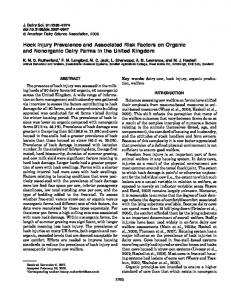
FIGURE 1. Gross lesions associated with Streptococcus phocae infection in necropsied southern sea otters (Enhydra lutris nereis) from central California, USA, 2004–2010. (A) Normal sea otter copulatory behavior results in skin wounds on the rostral face and planum nasale of females, because the male grasps this area with his teeth during mating. Occasionally, these lesions can be severe, and in this example, mating trauma has resulted in partial disruption of the planum nasale. Bar52 cm. (B) Severe suppurative cellulitis, fasciitis, and abscessation of the subcutis of the lateral head and lower jaw of an adult female sea otter. Streptococcus phocae was isolated on bacterial culture, and numerous bacterial cocci were observed in affected tissues on histopathology. This demonstrates regional tissue invasion by S. phocae following colonization of a mating-associated nose wound. Bar54 cm. (C) A deep skin laceration on a sea otter’s neck due to chronic fishing line entanglement. Bar52 cm. (D) Intraspecific fighting, especially between competing males, commonly results in small punctures and skin lacerations on the flippers (seen here), face, paws, and genitalia. These lesions, although comparatively mild, often become infected secondarily by S. phocae and other opportunistic bacterial pathogens. Bar54 cm. (E) The two dark, linearly arranged, “stab-like” lesions on the dorsal abdominal subcutis of a sea otter are typical of a great white shark (Carcharodon carcharias) bite. Although southern sea otters are commonly bitten by great white sharks, these animals are rarely, if ever consumed, and death often results from secondary bacterial infection, especially S. phocae. Bar53 cm. (F) A large, subacute, purulent abscess on the dorsal thoracic subcutis of a sea otter. The location of this abscess is typical of otters that survive a great white shark bite but succumb later to secondary bacterial infection, especially S. phocae. Streptococcus phocae infection was confirmed in this case via culture and histopathology. Bar51 cm.
BARTLETT ET AL.—STREPTOCOCCUS PHOCAE INFECTION IN SEA OTTERS
wounds often develop secondary bacterial infections (Fig. 1B; Kreuder et al. 2003). Anthropogenic trauma, including gunshot, fishing line entanglement, and lacerations from boat propellers can also cause skin trauma (Fig. 1C). Great white sharks often attack southern sea otters, but rarely, if ever, consume them (Ames and Morejohn 1980), so despite significant injuries, animals often survive for days or weeks following shark attack (Fig 1E). As a result, bacterial infection is a common cause of death following shark bite (Fig 1F; Kreuder et al. 2003). Increased S. phocae detection during the final year of study could be due to enhanced case recognition, random chance, or factors that were not assessed during the study. Great white shark–associated mortality appears to be increasing in southern sea otters over time (Tinker et al. 2015) and could also lead to increased S. phocae infection. Studies are in progress to address this question. Detection of abscesses or septicemia at necropsy was also predictive of S. phocae infection: sea otters with abscesses were nearly four times more likely, and septicemic animals three times more likely to be S. phocae–infected than those without either condition. Pure S. phocae septicemia (defined as no other bacteria isolated in culture or detected on histopathology in animals with a diagnosis of septicemia) was the primary or contributing cause of death for 10% of S. phocae–positive otters, suggesting that this bacterium is an important opportunistic pathogen of sea otters. As further evidence, gram-positive bacterial cocci, arranged in small groups or short chains suggestive of streptococci, are the most common bacterial morphology observed on microscopic examination of infected wounds, abscesses, and septicemic southern sea otters (M.M. unpubl. data). Associations between skin or mucosal trauma and streptococcal infection have also been reported in fish (GonzalezContreras et al. 2011) and humans (Libertin et al. 1985). In one prior report
7
(Libertin et al. 1985), human infection by group F streptococci rarely occurred in the absence of preexisting breaks in host defenses, with five of seven cases concurrent with gastrointestinal perforation. Similar to our findings in sea otters, abscessation and bacteremia or sepsis are relatively common sequelae following beta-hemolytic streptococcal colonization of breaks in host defenses for humans (Libertin et al. 1985). Prior studies also provide clues regarding potential mechanisms of S. phocae attachment, invasion, and systemic spread. In fish, S. phocae utilizes hydrophobic interactions to assist with nonspecific adhesion to the epithelium (Doyle 2000). These bacteria cannot invade an intact epidermis and instead survive at the surface, feeding on mucus. However, epidermal breaks and the disruption of mucosal defenses appear to facilitate S. phocae invasion in fish (Gonzalez-Contreras et al. 2011). A recent study of alpha-hemolytic Streptococcus infantarius subspecies coli isolates derived from sea otters with bacterial endocarditis revealed that all strains adhered significantly to exposed extracellular matrix components in vitro, including collagen IV, fibronectin, laminin, and hyaluronic acid (Edgar 2010). Bacterial survival was also documented following phagocytosis by macrophages. These same mechanisms could be used by the related bacterium, S. phocae, to attach to exposed extracellular matrix and spread systemically. Potential environmental sources of S. phocae exposure include the marine environment and seawater (Kusuda and Kawai 1982; Interaminense et al. 2010), endogenous oral-fecal flora of sea otters and other animals (Johnson et al. 2006; Imai et al. 2009; Miller et al. 2010), and traumatic inoculation, with studies of bacterial flora from the teeth and oral cavities of great white (Buck et al. 1984), tiger (Galeocerdo cuvier), and bull sharks (Carcharhinus leucas; Interaminense et al. 2010) confirming that streptococci could be inoculated into wounds during a shark attack. However, our data suggest that the risk of S. phocae
8
JOURNAL OF WILDLIFE DISEASES, VOL. 52, NO. 1, JANUARY 2016
infection in sea otters is not associated with any single source of trauma but instead is associated with skin lesions of any type. This information, combined with culture data suggesting that S. phocae is not a common commensal bacterium of live wild, apparently healthy, southern sea otters (M.M. unpubl. data), and reports of fatal S. phocae infections of marine animals worldwide (Skaar et al. 1994; Henton et al. 1999; Vossen et al. 2004; Imai et al. 2009; Hueffer et al. 2011) suggest that S. phocae is ubiquitous in the marine environment, and infection is associated with opportunistic colonization of damaged skin. Studies of streptococcal infections in farmed fish also support the concept of marine environmental exposure. Streptococci can survive up to 42 d in marine water with salinity from 0 to 7%, and pH ranges from 3.5 to 10. Bacterial proliferation can occur in the water column and in benthic mud at water temperatures from 10 to 45 C (Kusuda and Kawai 1982). Human beta-hemolytic streptococcal infections are also often associated with “wounds received in the marine environment” (Interaminense et al. 2010). It is common for fishermen, divers, marine mammal care workers, and aquarium staff to develop bacterial skin infections after working in marine environments, including handling live or dead animals, especially if there are preexisting skin breaks. The associated infectious cellulitis, often called “seal finger,” is commonly attributed to infection by Mycoplasma phocacerebrale, atypical mycobacteria, Vibrio vulnificus, other Vibrio spp., or Erysipelothrix rhusiopathiae (White and Jewer 2009). Based on their high prevalence of infection in marine animals and fish, their global distribution and propensity for colonizing skin breaks, it is possible that some cases of seal finger in humans are unrecognized infections by S. phocae or other beta-hemolytic streptococci. Our findings confirm that skin wounds and associated abscesses are a common portal of invasion and systemic spread by S. phocae and other beta-hemolytic
streptococci in sea otters. These findings benefit sea otter care and management by highlighting the importance of these in‐ fections, facilitating case recognition and guiding antibiotic selection. Significant associations between skin trauma and fatal S. phocae infection in necropsied sea otters also suggest that efforts to minimize trauma during capture activities, transport, washing of oiled animals, and captive care are advisable. Because exposed dermis and subcutis appear to facilitate streptococcal invasion and spread, careful closure of wounds and surgical sites can also help minimize risk. Finally, studies of the potential for S. phocae and other betahemolytic streptococci to infect humans may reveal that these bacteria are more common human pathogens than is currently recognized. ACKNOWLEDGMENTS
We thank staff at the University of Liverpool, National Centre for Zoonosis Research for their support. We thank Sara Miller for her assistance with preparation of the figures. We also thank the staff at California Department of Fish and Wildlife–Marine Wildlife Veterinary Care and Research Center for their assistance with project completion, along with all organizations and individuals that have submitted stranded southern sea otters for postmortem examination. G.B. thanks Alicia Coupe for her relentless support and the Wellcome Trust for support that facilitated travel abroad to complete this study. LITERATURE CITED Ames JA, Morejohn GV. 1980. Evidence of white shark, Carcharodon carcharias, attacks on sea otters, Enhydra lutris. Calif Fish Game 66:196–209. Bishop EJ, Shilton C, Benedict S, Kong F, Gilbert GL, Gal D, Godoy D, Spratt BG, Currie BJ. 2007. Necrotizing fasciitis in captive juvenile Crocodylus porosus caused by Streptococcus agalactiae: An outbreak and review of the animal and human literature. Epidemiol Infect 135:1248–1255. Buck JD, Spotte S, Gadbaw JJ Jr. 1984. Bacteriology of the teeth from a great white shark: Potential medical implications for shark bite victims. J Clin Microbiol 20:849–851. Burek KA, Gill VA, Doroff AM, Tuomi P, Goldstein T, Miller MA, Jang SS, Shewmaker L, Bodkin
BARTLETT ET AL.—STREPTOCOCCUS PHOCAE INFECTION IN SEA OTTERS
JL. 2005. Valvular endocarditis and septicemia due to Streptococcus infantarius ssp. coli organisms in stranded northern (Enhydra lutris kenyoni) and southern sea otters (Enhydra lutris nereis). In: Proceedings of the international association for aquatic animal medicine 36th annual conference, XXXXX XXXX, Seward, Alaska, 14– 18 May, pp. 215–217. Counihan-Edgar K , Gill V, Doroff A, Burek K, Miller W, Shewmaker P, Jang S, Goertz C, Tuomi P, Miller M, et al. 2012. Genotypic characterization of Streptococcus infantarius subsp. coli isolates from sea otters with infective endocarditis and/ or septicemia and from environmental mussel samples. J Clin Microbiol 50:4131–4133. Doyle RJ. 2000. Contribution of the hydrophobic effect to microbial infection. Microbes Infect 2:391–400. Edgar KL. 2010. Pathogenesis of Streptococcus infantarius subspecies coli valvular endocarditis in sea otters. PhD Dissertation, University of California, Davis, Davis, California, 96 pp. Fischer A, Liljander A, Kasper H, Muriuki C, Fuxelius HH, Bongcam-Rudloff E, de Villiers EP, Huber CA, Frey J, Daubenberger C, et al. 2013. Camel Streptococcus agalactiae populations are associated with specific disease complexes and acquired the tetracycline resistance gene tetM via a Tn916-like element. Vet Res 44:86. Gonzales-Contreras A, Magarinos B, Godoy M, Irgang R, Toranzo AE, Avendano-Herrera R. 2011. Surface properties of Streptococcus phocae strains isolated from diseased Atlantic salmon, Salmo salar L. J Fish Dis 34:203–215. Henton MM, Zapke O, Basson PA. 1999. Streptococcus phocae infections associated with starvation in Cape fur seals. J S Afr Vet Assoc 70:98–99. Hueffer K, Lieske CL, Mcgilvary LM, Hare RF, Miller DL, O’Hara TM. 2011. Streptococcus phocae isolated from a spotted seal (Phoca largha) with pyometra in Alaska. J Zoo Wildl Med 42:108–112. Imai D, Jang SS, Miller MA, Conrad PA. 2009. Characterization of beta-hemolytic streptococci isolated from southern sea otters (Enhydra lutris nereis) stranded along the California coast. Vet Microbiol 136:378–381. Interaminense JA, Nascimento DCO, Ventura RF, Batista JEC, Souza MMC, Hazin FHV, PontesFilho NT, Lima-Filho JV. 2010. Recovery and screening for antibiotic susceptibility of potential bacterial pathogens from the oral cavity of shark species involved in attacks on humans in Recife, Brazil. J Med Microbiol 59:941–947. Johnson S, Lowenstein L, Gulland F, Jang S, Imai D, Almy F, Delong R, Gardner I. 2006. Aerobic bacterial flora of the vagina and prepuce of California sea lions (Zalophus californianus) and
9
investigation of associations with urogenital carcinoma. Vet Microbiol 114:94–103. Keefe GP. 1997. Streptococcus agalactiae mastitis: A review. Can Vet J 38:429–437. Kreuder C, Miller MA, Jessup DA, Lowenstein LJ, Harris MD, Ames JA, Carpenter TE, Conrad PA, Mazet JAK. 2003. Patterns of mortality in southern sea otters (Enhydra lutris nereis) from 1998–2001. J Wildl Dis 39:495–509. Kusuda R, Kawai K. 1982. Characteristics of Streptococcus sp. pathogenic to yellowtail. Fish Pathol 17:11–16. Lamm CG, Ferguson AC, Lehenbauer TW, Love BC. 2010. Streptococcal infection in dogs: A retrospective study of 393 cases. Vet Pathol 47:387. Libertin CR, Hermans PE, Washington JA. 1985. Beta-hemolytic group F streptococcal bacteremia: A study and review of the literature. Rev Infect Dis 7:498–503. Miller MA, Byrne BA, Yang SS, Dodd EM, Dorfmeier E, Harris MD, Ames JA, Paradies D, Worcester K, Jessup DA, et al. 2010. Enteric bacterial pathogen detection in southern sea otters (Enhydra lutris nereis) is associated with coastal urbanization and freshwater runoff. Vet Res 41:1–12. Romalde JL, Ravelo C, Valdes I, Margarinos B, Del La Fuente E, San Martin C, Avendano-Herrera R, Toranzo AE. 2008. Streptococcus phocae, an emerging pathogen for Salmonid culture. Vet Microbiol 130:198–207. Skaar I, Gaustad P, Tonjum T, Holm B, Stenwig H. 1994. Streptococcus phocae sp. nov., a new species isolated from clinical specimens from seals. Int J Syst Bacteriol 44:646–650. Staedler M, Riedman M. 1993. Fatal mating injuries in female sea otters (Enhydra lutris nereis). Mammalia 57:135–139. Tillman PC, Dodson ND, Indiveri M. 1982. Group G streptococcal epizootic in a closed cat colony. J Clin Microbiol 16:1057–1060. Tinker, T, Hatfield, B, Harris M, Ames J. 2015. Dramatic increase in sea otter mortality from white sharks in California. Marine Mammal Science doi: 10.1111/mms.12261. Vossen A, Abdulmawjood A, Lämmler C, Weiß R, Siebert U. 2004. Identification and molecular characterization of beta-hemolytic streptococci isolated from harbor seals (Phoca vitulina) and grey seals (Halichoerus grypus) of the German North and Baltic seas. J Clin Microbiol 42:469–473. White CP, Jewer DD. 2009. Seal finger: A case report and review of the literature. Can J Plast Surg 17:133–135. Submitted for publication 20 February 2015. Accepted 5 June 2015.
5