Cancer Res Treat. 2009;41(1):36-44
DOI 10.4143 / crt.2009.41.1.36
Proapoptotic Ginsenosides Compound K and Rh2 Enhance Fas-induced Cell Death of Human Astrocytoma Cells through Distinct Apoptotic Signaling Pathways Kyungsun Choi, Ph.D.1 Chulhee Choi, M.D., Ph.D.1,2,3
Purpose Malignant astrocytomas are among the commonest primary brain tumors and they have a grave prognosis, and so there is an urgent need to develop effective treatment. In this study, we investigated the molecular mechanisms that are responsible for the anti-tumor effect of ginsenosides on human astrocytoma cells.
1
Materials and Methods We tested 13 different ginsenosides for their anti-tumor effect on human malignant astrocytoma cells in conjunction with Fas stimulation. In addition, the cell signaling pathways were explored by using pharmacological inhibitors and performing immunoblot analysis. DCF-DA staining and antioxidant experiments were performed to investigate the role of reactive oxygen species as one of the apoptosis-inducing mechanisms.
Laboratory of Computational Cell Biology, Department of Bio and Brain Engineering, 2 Graduate School of Medical Science and Engineering, and 3KI for the BioCentury, KAIST, Daejeon, Korea
+Correspondence + + + + + + + + + + Choi, + + + + +Ph.D. + + + + + + + + + + :+Chulhee + + + + + +M.D., + + + + + +Laboratory + + + + of + Computational + + + + + + Cell + +Biology, + + + + + +Department + + + + +of +Bio + and + +Brain + + Engineering, + + + + + + + + + + + + + + + + + + + + + + + + + + +KAIST, + + + 335 + +Gwahangno, + + + + + + + + + + + + + +Yuseong-gu, + + + + +Daejeon + + + 305-701, + + + +Korea + + + + + + + + + + + + + + + + + + + + + + + + + +Tel: + +82-42-350-4321 + + + + + + + + + + + + + + + + +Fax: + +82-42-350-4380 + + + + + + + + + + + + + + + + +E-mail: + +
[email protected] + + + + + + + + + + + + + + + + + + + + + + + + + + + + + + + + + + +Received + + + +February + + + +21,+2009 + + + + + + + + + +Accepted + + + +March + + +2, +2009 + + + + + + + + + + + + + + + + + + + + + + + + + + + + + +This + +work + +is+supported + + + +by+a+grant + +(R01-2008+ + + + + +000-10701-0) + + + + + from + + the + +Korea + + Science + + + +and+ + + + + + + + + + + + + + + + + + + + + + +Engineering + + + + +Foundation + + + + (to + +K.C.) + +and + +the+ + + +Ginseng + + + Grant + + +from + +the+ Korea + + +Food + +Research + + + + +Institute + + + + C.C.), + + + + + + +of+Korea. + + + + + + + + + + (to + + + +Republic + + + + + + + + + + + + + + + + + + + + + + + + + + + + + +
+ + + + + + + + + + + + + + + + + + + + + + +
Results Among the 13 different ginsenoside metabolites, compound K and Rh2 induced apoptotic cell death of the astrocytoma cells in a caspase- and p38 MAPK-dependent manner, yet the same treatment had no cytotoxic effect on the primary cultured human astrocytes. Combined treatment with ginsenosides and Fas ligand showed a synergistic cytotoxic effect, which was mediated by the reduction of intracellular reactive oxygen species. Conclusion These results suggest that ginsenoside metabolites in combination with Fas ligand may provide a new strategy to treat malignant astrocytomas, which are tumors that are quite resistant to conventional anti-cancer treatment.
Introduction Glioblastoma multiforme (GBM) is the most malignant and common brain tumor and it comprises ~23% of all primary brain tumors in adults. These malignancies are refractory to all the current therapeutic approaches, including surgery, radiotherapy and chemotherapy. Fas (CD95 or APO-1) is a member of the TNF/NGF receptor family, and Fas induces caspase-dependent apoptotic death in various transformed cells (1,2). Fas ligation with natural ligand or agonistic anti-Fas antibody is followed by recruitment of proapoptotic adaptor molecules such as Fas-associated death domain (FADD) to transduce the apoptotic signals through the caspase cascades (3). In some cells, Fas efficiently activates caspase-8 and it subsequently activates caspase-3 or 7, while other types of Fasinduced apoptosis are mediated by cytochrome-C release from the mitochondria and this is inhibited by the over-expression of anti-
36
CANCER RESEARCH AND THREATMENT
Key words Apoptosis, Ginsenoside, Fas, Reactive oxygen species, Astrocytoma
apoptotic bcl-2 family members (4). Panax Ginseng is known for its biological and pharmacological activities such as its anti-cancer, anti-aging, anti-inflammatory and anti-oxidant properties in the nervous, immune and circulatory systems (5). These diverse physiological activities of ginseng are mainly mediated by saponin, which is a ginsenoside. Especially, the metabolites of ginsenosides that are formed by enteric bacteria have been focused on for their pharmacological activities. Among them, compound K (C-K) is known to be formed by enteric bacterial fermentation of Rb1, Rb2 and RC, and C-K has been reported to suppress tumor metastasis and inflammatory responses (6,7). Another ginsenoside Rh2, a metabolite of Rg3, is also known for its tumor suppression by inducing apoptosis or retarding growth signals (8). We have previously shown that human malignant astrocytoma cells are quite resistant to Fas-induced apoptosis even though these
Kyungsun Choi, Chulhee Choi_Multiple Modes of Cell Death by Ginsenosides
cells express functional Fas on their surface (2,9). Even though the role of reactive oxygen species (ROS) has been controversial in terms of receptor-induced apoptosis, it has been shown that the inhibition of receptor-induced ROS generation augmented the Fasmediated apoptosis in human astrocytoma cells, and this suggests the anti-apoptotic role of ROS. In this study, we investigated the molecular mechanisms that are responsible for killing of tumor cells by pro-apoptotic ginsenosides and the augmentation of Fas-induced cell death in human astrocytoma cells.
Materials and Methods
3 Measurement of the intracellular ROS levels To detect intracellular ROS, an oxidation-sensitive probe 2, 7dichlorofluorescein-diacetate (DCF-DA) was used as previously described (9). To study the time course of Fas-mediated ROS production, the cells were incubated with 2 μmol/L of DCF-DA for 10 min and then they were treated with CH-11 (500 μg/L) for varying time periods (0~60 min). To investigate the effect of NAC and CAPE on Fas-induced ROS production, the CRT-MG cells were maintained in serum-free media for 16 h, incubated in the absence or presence of these inhibitors for 1 h and then they were treated with CH-11 (500 μg/L) for an additional 15 min. The cellular fluorescence was measured using an inverted epifluorescence microscope (Zeiss, Germany).
1 Cell culture Human astrocytoma CRT-MG cells were grown in RPMI 1640 medium that was supplemented with 10% heat-inactivated fetal bovine serum (FBS), penicillin G (100 U/ml), streptomycin (100 μg/ o ml) and L-glutamine (2 mmol/L) in a 5% CO2 incubator at 37 C, as previously described (10). Other human astrocytoma cell lines, U251-MG and U87-MG cells, were maintained in Dulbecco’s modified Eagle media (JBI, Korea) that was supplemented with 10% FBS and penicillin G (100 U/ml). Primary human fetal astrocytes were obtained from therapeutically aborted fetal brains and they were maintained in Dulbecco’s modified Eagle media that was supplemented with 10% heat-inactivated fetal bovine serum (FBS), penicillin G (100 U/ml) and 1% nonessential amino acids (Gibco-BRL, Grand Island, NY), as previously described (11).
2 Reagents Ginseng saponin ginsenosides (F1, Ro, Rc, Re, Rd, Rf, C-K, Rh2, Rg1, Rg2, Rg3, Rb1 and Rb2) were obtained from KT&G (Daejeon, Korea). N-acetyl cysteine (NAC), 3-(4, 5-dimethylthiazol-2-yl)-2, 5diphenyltetrazolium bromide (MTT) and diphenyl iodonium (DPI) were all purchased from Sigma (St. Louis, MO). Dichlorodihydrofluorescein diacetate (DCF-DA) and tetramethylrho-damine ethyl ester (TMRE) were purchased from Molecular Probe (Eugene, OR). An agonistic IgM type anti-Fas antibody (CH-11) was obtained from Upstate Biotechnology (Lake Placid, NY). Human recombinant TNF-αand Fas ligand were purchased from R&D Systems (Minneapolis, MN). Caffeic acid phenethyl ester (CAPE) and SB202190, SP100625 and U0126, which are pharmacological inhibitors of p38 MAPK, JNK and ERK, respectively, were obtained from Calbiochem (La Jolla, CA).
4 Measurement of cell death Cell death was determined by staining with Annexin V (PharMingen), which is a 35.8-kDa protein that has a strong affinity for phosphatidylserine (12). The cells were washed with PBS, trypsinized, suspended in 200 μL of binding buffer and stained with 0.5 ng of Annexin V-fluorescein isothiocyanate (FITC) and 2.5 ng of propidium iodide (PI). Ten thousand cells were analyzed with using a FACStar (Becton Dickinson, Mountain View, CA) within 30 min after staining. Cell death was defined as those cells that were positive for Annexin V and/or PI. The mitochondrial transmembrane potential (Δψm) was assessed after staining with TMRE. The cell suspension was incubated in 2 μmol/L of TMRE for 30 min at room temperature and then it was analyzed with a FACStar. The MTT (3-(4, 5-dimethylthiazol-2-yl)-2, 5-diphenyltetrazolium bromide) assay was also used to determine cell viability, as described previously (13). Briefly, MTT is a yellow, water-soluble, tetrazolium salt. Metabolically active cells are able to convert this dye into a water-insoluble, dark-blue formazan by reductive cleavage of the tetrazolium ring. The formazan crystals can then be dissolved and quantified by measuring the absorbance of the solution at 595 nm, and the resultant value is related to the number of living cells.
5 Subcellular fractioning The cells were washed with ice-cold PBS and then vortexed in the hypotonic buffer solution that contained 500 mmol/L sucrose, protease inhibitors and Na3VO4 (10 mmol/L Hepes, 2 mmol/L MgCl2, 25 mmol/L KCl, 0.5 mmol/L EDTA, 0.5 mmol/L EGTA). Then NP-40 was added to the final concentration of 0.05%, and the mixture was centrifuged at 15,000g for 15 min. The supernatants were assayed for the cytosolic fractions, and the precipitants were analyzed for the mitochondrial fractions.
VOLUM 41 NUMBER 1 MARCH 2009
37
Cancer Res Treat. 2009;41(1):36-44
6 Immunoblot analysis Immunoblot analysis for caspases was performed as previously described (14). The cell lysates (20 μ g) were electrophoresed in 10% SDS-PAGE gels, the proteins were transferred to nitrocellulose membranes and these were probed with antibodies against human caspase-3, PARP, p38 MAPK, phosphor-specific p38 MAPK and β -actin (Cell Signaling, Danvers, MA). The blots were developed by chemiluminescence (AbFrontier, Korea).
7 Statistical analysis The data is presented as means ±SDs. Comparisons between two different samples were analyzed by the Student t-test, and comparisons between more than three different samples were done by ANOVA (analysis of variance) with applying Tukey’s post-hoc test to the significant main effects or interactions (SPSS 12.0K for Windows, SPSS, Chicago, IL).
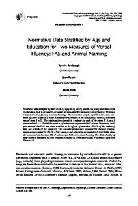
cultured human astrocytes even at higher concentrations of C-K and Rh2 (Fig. 1B). Interestingly, the cells treated with C-K showed vacuolar changes even with using concentrations that had little toxicity. To test what type of cell death was induced by ginsenosides, the cells treated with the ginsenosides were stained for Annexin V and propidium iodide (Fig. 1C). As expected, treatment with C-K and Rh2 induced typical apoptotic cell death, which was confirmed by the positive staining with Annexin V. Pretreatment with a broad-spectrum caspase inhibitor z-VAD-Fmk markedly reduced the ginsenoside induced cell death, confirming that ginsenosides induced caspase-dependent apoptotic cell death in the malignant astrocytoma cells. Since it has been reported that the mitochondrial intermembrane potential (Δψ m) is selectively decreased during the activation of the apoptotic cascades (15), we further investigated whether treatment with ginsenosides affected the Δψm by staining with TMRE (Fig. 1D). In concordance with the Annexin V staining results, treatment with C-K and Rh2 induced a rapid depolarization of the Δψm, and this was nearly completely reversed by pretreatment with z-VAD-fmk. These results clearly indicate that the ginsenosides C-K and Rh2 induced caspasedependent apoptotic cell death in human malignant astrocytoma cells.
Results 2 The signaling pathways involved in ginsenosides1 Cell death of the malignant astrocytoma cells by pro-
apoptotic ginsenosides We screened thirteen ginsenosides for their anti-cancer effect on human malignant astrocytoma cells (CRT-MG, U87-MG and U251-MG cells). Among the 13 ginsenosides, compound K (C-K) and Rh2 induced marked time-dependent cytotoxicity even at a concentration as low as 25 mg/L, while the other ginsenosides showed marginal cytotoxic effects at a concentration as high as 50 mg/L (Fig. 1A). The cytotoxic effect of C-K and Rh2 was only observed for the transformed tumor cells, but not for the primary
38
CANCER RESEARCH AND THREATMENT
induced apoptosis We next investigated the signaling pathways that are responsible for the ginsenoside-induced apoptotic cell death. Since we have observed that inhibition of caspases significantly suppressed the ginsenoside-induced cell death, we first tested the involvement of specific caspases by performing western blot analysis (Fig. 2A, B). Caspase-3 is known as the common executioner of apoptotic cell death, and it was cleaved into active fragments (17/19 kDa) in a time-dependent manner by treatment with C-K or Rh2. We observed the proteolytic cleavage of caspase-3 as early as 12 h after treatment with ginsenosides, while the maximal cleavage was observed 24 h
Kyungsun Choi, Chulhee Choi_Multiple Modes of Cell Death by Ginsenosides
Fig. 1. Caspase-dependent apoptosis by the ginsenosides CK and Rh2 in human astrocytoma cells. (A) The cells were treated with varying doses (0~50 mg/L) of 13 different ginsenosides, and the cell death was determined by MTT assay. *, significantly different from the control sample without any treatment (p