the expression of DDr and the prognostic implication in gBM has yet to be published. .... horse and goat serum (Dako corp., carpinteria, cA, UsA) as the primary ...
oncology reports 26: 423-430, 2011
Prognostic implications of the DNA damage response pathway in glioblastoma Ho Jun Seol1*, Hae Yong Yoo3*, Juyoun Jin1,3,4*, Kyeung Min Joo4,5, Doo-Sik Kong1, SU JIN YOON3, Heekyoung Yang1,3,4, Wonyoung Kang1,3,4, Do-Hoon Lim2, Kwan Park1, Jong Hyun Kim1, Jung-II Lee1 and Do-Hyun Nam1,3,4 Departments of 1Neurosurgery and 2Radiation Oncology, 3Samsung Biomedical Research Institute and 4 Cancer Stem Cell Research Center, Samsung Medical Center, Sungkyunkwan University School of Medicine, 50 Ilwon-Dong, Gangnam-Gu, Seoul 135-710; 5Department of Anatomy, Seoul National University College of Medicine, 28 Yeongeon-dong, Jongno-gu, Seoul 110-799, Republic of Korea Received February 2, 2011; Accepted March 28, 2011 DOI: 10.3892/or.2011.1325 Abstract. Genomic instability and resistance to genotoxic therapies for glioblastoma (GBM) suggest aberrant DNA damage response (DDR), since DDR maintains the genomic integrity against genotoxic insults including anti-tumor therapies. To elucidate the biological and clinical meaning of DDR in GBM, we retrospectively investigated the immunohistochemical expression of DDR proteins (ATM, Chk1, Chk2, TopBP1, Rad17, p53, Nbs1, MDC1, γH2AX and RPA1) in 69 GBM surgical samples and their relation with GBM patient survival. Remarkably, higher expression of ATM revealed significantly longer overall survival (OS) and progression-free survival (PFS) (p70 Location Supratentorial Infratentorial Extent of resection TR PR Biopsy Adjuvant treatments RT only CCRT+TMZ RT+TMZ MGMT statusa Unmethylated Methylated
69 40/29 54.4 (12-79) 562 (51-1558) 410 (357-462) 204 (180-227)
28 41 66 3 51 17 1 16 26 27 23 21
F/U, follow-up; 95% CI, 95% confidence interval; KPS, Karnofsky performance status; TR, total resection; PR, partial resection; RT, radiation therapy; CCRT, concomitant chemoradiotherapy; TMZ, temozolomide chemotherapy. aAvailable tissue samples (N=44) were included.
2006. Patients were managed according to established diagnostic and therapeutic protocols, including surgical resection and subsequent chemoradiotherapy. A macroscopic total resection was performed in 51 of 69 patients (73.8%), a partial resection in 17 of 69 patients (24.6%), and a biopsy only in 1 of 69 patients (1.4%). All patients underwent subsequent radiotherapy (60 Gy in 2 Gy fractions) after surgery. For concurrent chemoradiotherapy or adjuvant chemotherapy, 53 of 69 patients received temozolomide with a median of 4 cycles (range, 1-9 cycles). The remaining 16 patients (23.2%) did not receive chemotherapy because of clinical deterioration during radiotherapy. Tumor samples were re-evaluated by two neuropathologists to confirm the diagnosis according to the World Health Organization criteria. Tumor samples were obtained during surgical treatment and were embedded in paraffin for histological studies. Written informed consent was obtained from all patients, and tissue collection was approved by the institutional review board. Immunohistochemical (IHC) studies were performed in a double-blinded manner, without prior knowledge of clinical outcome.
Immunohistochemical study. Four-micrometer-thick sections sliced from paraffin-embedded specimens were prepared on the slide. Sections were immunostained with antibodies for ATM (1:50, Santa Cruz Biotechnology, CA, USA), Chk1 (1:50, Santa Cruz Biotechnology), Chk2 (1:50, Santa Cruz Biotechnology), TopBP1 (1:50, Bethyl Laboratories, Inc., Montgomery, TX, USA), Rad17 (1:50, Santa Cruz Biotechnology), p53 (1:5000, Santa Cruz Biotechnology), Nbs1 (1:500, GeneTex, Irvine, CA, USA), MDC1 (1:250, Bethyl Laboratories, Inc.), γH2AX (1:500, Abcam, Cambridge, MA, USA), RPA1 (1:100, Calbiochem, San Diego, CA, USA), p21 (1:40, Lab Vision, Fremont, CA, USA), p18 (1:20, Lab Vision), MDM2 (1:100, Lab Vision) and p27 (1:40, Lab Vision). Tumor-containing sections were baked at 56˚C for 30 min, deparaffinized in xylene and rehydrated in graded concentrations of ethanol. Endogenous peroxidase activity was blocked by incubation in 0.3% hydrogen peroxide in methanol and with heat-induced antigen retrieval [for p53, 10 mM citrate buffer (pH 6.0) for 25 min in a vegetable steamer]. Immunostaining involved sequential applications of primary antibody for 16 h at 4˚C, followed by biotinylated secondary antibodies (Vector Laboratories, Orton Southgate, UK) at 1:200 for 1 h and avidin (Elite ABC; Vector Laboratories) for 1 h. Negative control slides received normal horse and goat serum (Dako Corp., Carpinteria, CA, USA) as the primary antibody. Diaminobenzidine tetrahydrochloride was used as the enzyme substrate to observe the specific antibody localization, and Harris hematoxylin was used as a nuclear counterstain. Sections were examined for immunoreactivity for the proteins by an observer who was unaware of the pathological diagnoses, outcomes or clinical features. Tumors were categorized as follows: Grade 0 for those expressing no protein, Grade 1 for those expressing in 75% of cells, based on the expression level in nucleus visualized in a high-power field in areas with maximal staining. The expression of the proteins was analyzed as a dichotomous covariate: no or little immunoreactivity (Grade 0 or 1) vs. overexpression (Grades 2-4). Bisulfite modification and methylation-specific PCR. DNA (1 µg) from the tumor was denatured by sodium hydroxide and modified by sodium bisulfite. Methylation-specific PCR was performed as previously described (14). Cell culture and cell lines. Human glioma cell lines, U138MG, U373MG, U87MG, U251MG and control human osteosarcoma cells (U2OS) (American Type Culture Collection) were maintained in Dulbecco's Modified Eagle's Medium (DMEM) containing 10% fetal bovine serum (HyClone Laboratories, Inc., Logan, UT, USA), 100 U/ml penicillin and 100 µg/ml streptomycin. Cells were cultured in a 37˚C, 5% CO2 humidified chamber. Ionizing radiation and Western blotting. Cells were lysed and prepared for Western blot analysis as previously described (15). Cells were processed 6 h after ionizing radiation (IR) (10 Gy), washed (PBS) and harvested. Anti-ATM, -ATR, -Chk1 anti-
oncology reports 26: 423-430, 2011
425
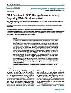
Figure 1. Overall survival (OS) (a) and progression-free survival (PFS) (b) of 69 glioblastoma patients. OS curve according to age (c), number of tumors (d) and extent of resection (e) demonstrated significance (p