Comm. Appl. Biol. Sci, Ghent University, 70/3, 2005
101
PSEUDOMONADS ASSOCIATED WITH MIDRIB ROT AND SOFT ROT OF BUTTERHEAD LETTUCE AND ENDIVE B. COTTYN1, K. VANHOUTEGHEM2,4, J. HEYRMAN3, P. BLEYAERT2, J. VAN VAERENBERGH1, P. DE VOS3, M. HÖFTE4 & M. MAES1 1
Department of Crop Protection, Agricultural Research Centre Burg. Van Gansberghelaan 96, BE-9820 Merelbeke, Belgium, 2 Provincial Research and Advisory Centre for Agriculture and Horticulture Ieperseweg 87, BE-8800 Rumbeke, Belgium 3 Laboratory of Microbiology, Ghent University K.L. Ledeganckstraat 35, BE-9000 Gent, Belgium 4 Laboratory of Phytopathology, Ghent University Coupure Links 653, BE-9000 Gent, Belgium E-mail:
[email protected]
ABSTRACT During the past ten years, bacterial soft rot and midrib rot of glasshouse-grown butterhead lettuce (Lactuca sativa L. var. capitata) and field-grown endive (Cichorium endivia L.) has become increasingly common in the region of Flanders, Belgium. Severe losses and reduced market quality caused by bacterial rot represent an important economical threat for the production sector. Symptoms of midrib rot are a brownish rot along the midrib of one or more inner leaves, often accompanied by soft rot of the leaf blade. Twenty-five symptomatic lettuce and endive samples were collected from commercial growers at different locations in Flanders. Isolations of dominant bacterial colony types on dilution plates from macerated diseased tissue extracts yielded 282 isolates. All isolates were characterized by colony morphology and fluorescence on pseudomonas agar F medium, oxidase reaction, and soft rot ability on detached chicory leaves. Whole-cell fatty acid methyl esters profile analyses identified the majority of isolates (85 %) as belonging to the Gammaproteobacteria, which included members of the family Enterobacteriaceae (14%) and of the genera Pseudomonas (73%), Stenotrophomonas (9%), and Acinetobacter (3%). Predominant bacteria were a diverse group of fluorescent Pseudomonas species. They were further differentiated based on the nonhost hypersensitive reaction on tobacco and the ability to rot potato slices into 4 phenotypic groups: HR-/P- (57 isolates), HR-/P+ (54 isolates), HR+/P- (16 isolates) and HR+/P+ (35 isolates). Artificial inoculation of suspensions of HR-, pectolytic fluorescent pseudomonads in the leaf midrib of lettuce plants produced various symptoms of soft rot, but they did not readily cause symptoms upon spray inoculation. Fluorescent pseudomonads with phenotype HR+ were consistently isolated from typical dark midrib rot symptoms, and selected isolates reproduced the typical midrib rot symptoms when spray-inoculated onto healthy lettuce plants.
INTRODUCTION Over the last ten years, midrib rot of butterhead lettuce (Lactuca sativa L. var. capitata) grown in glasshouses has emerged as a serious threat for the lettuce production sector in Flanders, Belgium. Disease build-up can be rapid and consistently has been associated with nearly mature plants but can also occur postharvest on apparently healthy plants during cold storage or transportation. Symptoms are a brownish rot in and along the midrib of one or more leaves, often accompanied by rotting of the leaf blade. Two types of midrib rot can be distinguished and often are found in combination. The first type, referred to as soft rot in this study, usually affects the
102
outermost leaves of the head and symptoms appear as slimy light brown to reddish brown discolorations of the midrib commonly accompanied by a slimy decay of the leaf blades. The second type, referred to as typical midrib rot, is characterized by dark brown to greenish black discolorations on the midrib of one or more inner wrapper leaves. The symptoms always are inside the head, the outermost leaves of the head usually are not affected. Typically, lesions initiate as water-soaked, greenish brown blotches along the midrib at the point where it bends up. Lesions expand as dark brown streaks onto the midrib and into the lateral veins bordering the midrib, and darken until they are almost black. At the initial stages of symptom development, scattered dark brown spots on the leaf surface are also observed, resembling symptoms of varnish spot (Grogan et al., 1977). However, these brown spots are of minor importance compared to the main rot symptoms developing on the midrib. If symptoms develop on several inner leaves, the crop loses market quality and will not be harvested. Damage can be extensive and usually results in partial or total loss of crops. Although bacterial soft rot on the outer leaves of the lettuce head may resemble bottom rot caused by Rhizoctonia solani, neither R. solani or any other fungal agent was isolated from lettuce midrib rot in a previous study by Bleyaert et al. (1999). However, fluorescent pseudomonads were consistently isolated from lettuce leaves showing these soft rot and midrib rot lesions. These results suggested a possible bacterial etiology for the disease. Our present aim is to make a more detailed study of the disease problem, with special emphasis on greenhouse-produced butterhead lettuce and fieldgrown endive (Cichorium endivia L.). The objectives of this study were to (i) identify and characterize bacteria from lettuce and endive plants displaying midrib rot symptoms, (ii) set up a culture collection of bacteria isolated from symptomatic plants, and (iii) determine which fluorescent pseudomonads are associated with lettuce midrib rot. MATERIALS AND METHODS Isolation of bacteria In autumn and winter 2004, lettuce and endive plants with leaf midrib or soft rot symptoms, or a combination of both, were obtained from local growers in Flanders, Belgium (Table 1). Collected diseased samples were routinely examined under a stereomicroscope for fungal growth to exclude symptoms possibly attributed to Rhizoctonia solani or Botrytis cinerea. Symptomatic leaves were surface disinfested by shortly dipping in 70 % ethanol and drywiped with tissue paper. Then, small pieces of tissue were aseptically excised from the margins of lesions and macerated in 2 ml sterile 0,05 M potassium phosphate buffer (PB, pH 7.0). Tenfold serial dilutions from the macerate were prepared with PB and 50 µl aliquots of each dilution (10-3, 10-4 and 10– 5) were replicate spread on pseudomonas agar F medium (PAF; Difco Laboratories, Detroit, MI) amended with 0.01 % cycloheximide to prevent fungal contamination. The plates were incubated for 3 days at 28°C before bacterial colonies were counted and examined on morphological appearance. Dominant colony types were picked and purified by re-streaking on PAF medium.
Comm. Appl. Biol. Sci, Ghent University, 70/3, 2005
103
The isolates were kept on tryptic soy agar (TSA; Sigma-Aldrich) slant tubes for routine use, or in PB with 20% glycerol at –70°C for long-term storage. Characterization of the bacteria All bacteria isolated from symptomatic tissue were tested for morphological, biochemical, and phytopathological characteristics. Colony morphology was determined visually and under the dissecting microscope by examining 4days-old cultures on PAF medium for colony shape, texture and pigmentation. Fluorescent pigment production on PAF medium (King et al, 1954) was visually evaluated under UV light (254-366 nm). As a routine test, all isolates were checked for the ability to cause rotting on detached cichory leaves. Aqueous suspensions of 12 h old bacterial cultures grown on TSA slant tubes were adjusted to a concentration of 106 cfu/ml by dilution, and 25 ul of each bacterial suspension was inoculated with a Pipetman into the midrib of a detached chicory leaf. Isolates that caused rot to some extent on chicory leaves (i.e. score 3, on a scale from 0-5) were further evaluated for LOPAT (Lelliot & Stead, 1987) characteristics, which included oxidase reaction, ability to rot potato, and induction of the nonhost hypersensitive reaction in tobacco (Nicotiana tabacum cv. Xanthi NN). The oxidase reaction was scored by use of Bactident oxidase strips (Merck, Germany) according to manufacturer’s instructions. For the potato rot test, 300 µl of aqueous bacterial suspensions (approximately 106 cfu/ml) were spread on potato slices placed on moistened filterpaper in petridishes, the plates were incubated at 28°C and examined for soft rot after 2 days. The ability to cause the nonhost hypersensitive reaction (Klement et al., 1964) was tested by infiltration of bacterial suspensions (107–108 cfu/ml) by hypodermic syringe into alternate leaf panels of expanded tobacco leaves. A positive hypersensitive reaction (HR+) was scored if the zone of infiltrated tissue collapsed and became brown and papery 3 days after inoculation. For fatty acid analyses, the bacterial isolates were grown on trypticase soy broth agar at 28°C for 24 h, then extracted for fatty acid methyl esters using a standard method (Sasser, 1990), and analysed by an automated HewlettPackard gas chromatograph. Fatty acid patterns of all isolates were compared with the commercial TSBA fatty acid database provided by MIDI (Microbial ID Inc., Newark, DE). The analyses included assessing the degree of similarity of fatty acid composition. Pathogenicity tests Pathogenicity of the isolates was tested by growing inoculum in tryptic soy broth (TSB; Sigma-Aldrich) shake cultures for 24 h, suspending bacterial cultures in sterile water, diluting to 106 cfu/ml (as determined by dilution plating), and injecting 0.2 ml by a hypodermic syringe into the leaf midrib of 5 week old lettuce plants (cv. Burgia). In addition to the midrib inoculations by injection, nearly mature lettuce plants (2 months old) were sprayinoculated until runoff with a hand-held mister. Negative control plants were inoculated with water. All plants were kept in a greenhouse and examined for disease symptoms after 3 to 10 days.
104
RESULTS AND DISCUSSION Over the past 10 years, damage by bacterial rot has become increasingly common on leafy vegetables grown in greenhouses and the field. Particularly the incidence of midrib rot of greenhouse-produced butterhead lettuce has increased to a serious threat for the production sector in Flanders, Belgium. The aim of this study was to gain a better understanding of the bacteria associated with midrib rot and soft rot symptoms on butterhead lettuce and endive plants. Twenty-one greenhouse-produced lettuce and 4 field-grown endive symptomatic samples were collected from commercial growers in Flanders (Table 1). All collected samples were nearly mature plants that developed rotting symptoms near harvest, except for 3 lettuce samples with postharvest damage by soft rot after cold transportation. Isolations of dominant colony morphotypes on dilution plates from macerated diseased pith, petiole and leaf tissue extracts yielded 282 isolates. Whole-cell fatty acid methyl esters profile analyses using the Microbial Identification System (Microbial ID Inc., Newark, DE) identified the isolates as members of the classes Gammaproteobacteria (85 %), Betaproteobacteria (9 %), Alphaproteobacteria (1 %), Flavobacteria (1 %), Sphingobacteria (1 %), Bacilli (1 %) and Actinobacteria (2 %). Numbers of isolates and their taxonomic classifications appear in Table 2. Table 1. Butterhead lettuce and endive samples with leaf midrib and soft rot symptoms collected during autumn and winter season of 2004 Sample
Location
Origin
Crop
Symptoms
No. of isolates nonfluor fluora 0001 – 0005 St-Kathelijne Waver field endive midrib and soft rot 6 16 0006 Putte glasshouse lettuce soft rot 4 1 0007 – 0008 St-Kathelijne Waver glasshouse lettuce soft rot 5 5 0009 -0014 St-Kathelijne Waver glasshouse lettuce midrib and soft rot 11 5 0015 – 0019 Roeselare cold transport lettuce soft rot 5 4 0020 – 0024 Fijnaard (N) field endive early soft rot 5 6 0027 Westrozebeke glasshouse lettuce early midrib rot 1 0029 – 0033 Torhout field endive midrib and soft rot 3 11 0034 – 0039 Houthulst glasshouse lettuce midrib and soft rot 5 14 0040 – 0042 Koolskamp glasshouse lettuce early midrib rot 6 5 0044 – 0046 Hooglede glasshouse lettuce minor brown blotch 4 2 0053 – 0057 Reninge glasshouse lettuce midrib rot 4 11 0059 Berlaar glasshouse lettuce midrib rot 2 0063 – 0065 Mechelen glasshouse lettuce midrib rot 2 3 0066 – 0067 Mechelen glasshouse lettuce midrib rot 2 4 0068 – 0069 Mechelen glasshouse lettuce midrib rot 1 6 1001 – 1007 Westrozebeke cold transport lettuce soft rot 10 8 1008 – 1011 Wingene cold transport lettuce soft rot 10 7 1012 – 1017 Haasdonk glasshouse lettuce midrib rot 9 2 1018 – 1025 Nevele field endive soft rot 8 13 1026 – 1028 Putte glasshouse lettuce midrib rot 2 5 1029 – 1030 Rummen glasshouse lettuce soft rot 5 2 1031 – 1034 Moerzeke glasshouse lettuce midrib and soft rot 2 7 1040 – 1047 Merelbeke glasshouse lettuce midrib rot 9 17 1048 – 1049 Merelbeke glasshouse lettuce soft rot 2 5
Fluorescence was determined under UV-light (254-366 nm) on Pseudomonas agar F medium
a
Comm. Appl. Biol. Sci, Ghent University, 70/3, 2005
105
Table 2. Bacteria isolated from leaf midrib and soft rot lesions on butterhead lettuce and endive samples collected from 25 local growers in Flanders, Belgium Taxonomic groupsa Alphaproteobacteria (1%) Methylobacteriaceae Betaproteobacteria (9%) Comamonadaceae
Oxalobacteraceae Gammaproteobacteria (85%) Alteromonadaceae Enterobacteriaceae
Moraxellaceae Pseudomonadaceae Xanthomonadaceae Flavobacteria (1%) Flavobacteriaceae Sphingobacteria (1%) Sphingobacteriaceae Bacilli (1%) Bacillaceae Actinobacteria (2%) Brevibacteriaceae Microbacteriaceae
Identificationb
No. of isolates
Roseomonas fauriae Acidovorax avenae Comamonas testosteroni Delftia acidovorans Variovorax paradoxus Duganella zoogloeoides Janthinobacterium lividum Pseudoalteromonas haloplanktis Enterobacter intermedius Pantoea agglomerans Pantoea ananatis Pectobacterium carotovorum Serratia odorifera Acinetobacter calcoaceticus Pseudomonas spp. Stenotrophomonas maltophilia
7 6 3 8 2 3 4 9 2 13 5 7 173 20
Chryseobacterium balistinum Flavobacterium johnsoniae Myroides odoratus
1 2 1
Pedobacter heparinus Sphingobacterium multivorum
1 2
Bacillus pumilus, megaterium
3
Brevibacterium liquefaciens 1 Clavibacter michiganensis 1 Microbacterium esteraromaticum 1 Micrococcaceae Arthrobacter agilis 2 Kocuria kristinae 1 a Taxonomic classification according to Bergey’s Manual (Garrity et al., 2004); percentage of the group on total bacteria isolated given in parenthesis b Best matching identifications obtained by fatty acid methyl esters profile analyses using the Microbial Identification System (Microbial ID Inc., Newark, DE).
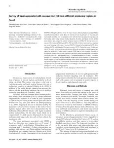
The majority of bacteria isolated from symptomatic tissue were identified as Pseudomonas species, other bacteria regularly present were Stenotrophomonas maltophilia and members of the family Enterobacteriaceae (Fig. 1A). A total of 173 isolates were identified as Pseudomonas species. Of these, 162 isolates produced colonies on PAF medium that were green to blue fluorescent under UV light. They were represented by more than 20 morphologically different colony types. All produced a positive reaction in the oxidase test, except for 9 isolates. The fluorescent isolates were further differentiated based on the nonhost hypersensitive reaction (HR) on tobacco and the ability to rot potato slices (P) into 4 phenotypic groups: HR-/P-, HR-/P+, HR+/P- and HR+/P+ (Fig. 1B). Of the 162 fluorescent isolates, 111 isolates did not induce a hypersensitive reaction upon infiltration into tobacco leaves but 54 of these
106
isolates did rot potato slices. The remaining 51 fluorescent isolates induced a hypersensitive reaction on tobacco leaves, and 35 of these isolates were also pectolytic on potato slices.
(A) Gamma-proteobacteria Acinetobacter 3% Pseudoalteromonas
Stenotrophomonas 9%
1%
Enterobacteriaceae
14%
Pseudomonas 73%
(B) Fluorescent pseudomonads
+
HR /P 22%
+
HR /P 10%
+ -
HR /P 35%
-
-
-
HR /P 33%
+
Figure 1.A. Majority of bacteria (85 %) isolated from symptomatic lettuce and endive plants belonged to the class of Gamma-proteobacteria. Predominantly isolated bacteria were identified as members of the genus Pseudomonas, which included 162 fluorescent and 11 non-fluorescent isolates. Figure 1.B. The group of fluorescent pseudomonads was differentiated into four phenotypic groups based on their hypersensitive reaction (HR) on tobacco leaves and their pectolytic (P) ability on potato slices. In lettuce inoculation tests by leaf midrib injections of bacterial suspensions (106 CFU/ml), isolates of phenotype HR-/P+ caused rotting symptoms with variable severity, isolates of phenotypes HR+/P- and HR+/P+ consistently caused midrib rot symptoms and also produced midrib rot upon sprayinoculation of mature lettuce plants.
Comm. Appl. Biol. Sci, Ghent University, 70/3, 2005
107
In the lettuce inoculation tests, the 57 isolates of phenotype HR-/P- did not produce symptoms upon injection of the leaf midrib and were considered saprophytic pseudomonads. The 54 isolates of phenotype HR-/P+ caused rotting symptoms with various severities upon injection of the leaf midrib. However, they did not cause symptoms when spray-inoculated onto healthy lettuce plants, and were considered pectolytic pseudomonads with low disease potential. All isolates that induced a hypersensitive reaction on tobacco leaves (HR+ phenotypes) consistently caused symptoms upon injection of the leaf midrib, and selected isolates produced midrib rot symptoms identical to those observed on collected samples when spray-inoculated onto mature lettuce plants (Fig. 2). In compliance with Koch’s postulate, fluorescent pseudomonads were recovered from midrib rot symptoms of spray-inoculated plants and were characterized and confirmed to be identical to the inoculated strains. Control plants inoculated with water did not develop symptoms. The 35 isolates with phenotype HR+/P+ were clearly the most aggressive isolates in the collection, and were considered pectolytic plantpathogenic pseudomonads.
Figure 2. Typical dark brown midrib rot symptoms produced by a phenotype HR+/P+ fluorescent isolate 10 days after spray-inoculation on lettuce.
It is clear that fluorescent pseudomonads figure prominently in the collection of isolates from symptomatic lettuce and endive plants. Bacterial soft rot, the leading biological cause of postharvest losses of fruits and vegetables, is caused by a diverse group of pectolytic plant pathogens, which includes Erwinia carotovora and pectolytic Pseudomonas species (Hansen, 1985; Liao & Wells, 1987). Pectolytic activity has been reported for various fluorescent pseudomonads, such as P. fluorescens, P. marginalis, P. putida, P. aureofaciens, P. tolaassii, P. viridiflava, and some pathovars of P. syringae (Janse et
108
al., 1992; Wells et al., 1993). Isolates identified as Pectobacterium carotovorum were found in association with two of the postharvest lettuce samples with soft rot damage after cold transportation, the third postharvest sample yielded only HR-, pectolytic pseudomonads. Our results indicate that Erwinia spp. are not directly involved in the development of midrib rot symptoms on nearly mature lettuce and endive plants. Also other studies have reported on bacterial rot caused solely by pseudomonads (Charron et al., 2002). Pseudomonas populations are known to form part of the lettuce native bacterial flora, including types considered in this study as pectolytic pseudomonads with low disease potential (Magnuson et al., 1990). The isolated fluorescent pseudomonads with phenotype HR-/P+ represented a diverse group comprised of various colony morphotypes and displayed a wide variation in pectolytic activity. They were commonly found throughout this study, and were the only bacteria with disease potential isolated from samples exhibiting exclusively soft rot symptoms. Samples with dark brown to green-black midrib rot symptoms, whether or not in combination with soft rot, additionally yielded fluorescent pseudomonads with phenotypes HR+/P- and HR+/P+. These aggressive pathogenic pseudomonads were represented by 3 main colony morphotypes and formed a homogeneous group based on fatty acids analyses. It has been reported that midrib rot causing pathogens are possibly introduced by overhead spray irrigation (Van Vaerenbergh & Seynnaeve, 2003). The fact that these aggressive pathogens were only found in association with typical midrib rot symptoms in our study, could further support the suggestion that they originate from an external inoculum source. In conclusion, evidence is provided that midrib and soft rot symptoms are caused by fluorescent Pseudomonas species. This is based on the consistent association of fluorescent pseudomonads with symptomatic plants, the ability of selected isolates to induce these symptoms, and characterization of the isolates based on morphological, fatty acid, and phytopathological properties. Further efforts to identify these Pseudomonas isolates at the species level by ribosomal DNA sequence analyses and DNA-DNA hybridisations are currently ongoing. A better understanding of the epidemiology of these pectolytic Pseudomonas in relation to the induction of symptoms only in the late plant growth stage needs further research. ACKNOWLEDGEMENT This work was supported by the Flemish Institute for the Stimulation of Scientific and Technological Research in Industry (IWT, Brussels, Belgium).
REFERENCES BLEYAERT P., VAN VAERENBERGH J. & KINT S. (1999) Identificatie van Pseudomonas spp. als oorzaak en sulfaat als promotor van nerfrot in groene botersla onder glas in België. Parasitica, 55(2-3):73-83. CHARRON C.S., SAMS C.E. & CANADAY C.H. (2002) Impact of glucosinolate content in broccoli (Brassica oleracea (Italica group)) on growth of Pseudomonas marginalis, a causal agent of bacterial soft rot. Plant Disease, 86:629-632. GARRITY G.M., BELL J.A. & LILBURN T.G. (2004) Taxonomic outline of the Prokaryotes. Bergey’s Manual of Systematic Bacteriology, Second Edition, Release 5.0, SpringerVerlag, New York. DOI: 10.1007/bergeysoutline200405
Comm. Appl. Biol. Sci, Ghent University, 70/3, 2005
109
GROGAN R.G., MISAGHI I.J., KIMBLE K.A., GREATHEAD A.S., RIRIE D. & BARDIN R. (1977) Varnish spot, destructive disease of lettuce in California caused by Pseudomonas cichorii. Phytopathology, 67:957-960. HANSEN J.D. (1985) Common names for plant diseases. Plant Disease, 69:649-676. JANSE J.D., DERKS J.H.J., SPIT B.E. & VAN DER TUIN W.R. (1992) Classification of fluorescent soft rot Pseudomonas bacteria, including P. marginalis strains, using whole cell fatty acid analysis. Systematic and Applied Microbiology, 15:538-553. KING E.O., WARD M.K. & RANEY D.E. (1954) Two simple media for the demonstration of pyocyanin and fluorescein. J. Lab. Clin. Med., 44:301-307. KLEMENT Z., FARKAS G.L. & LOVREKOVICH L. (1964) Hypersensitive reaction induced by phytopathogenic bacteria in the tobacco leaf. Phytopathology, 54:474-477. LIAO C.H. & WELLS J.M. (1987) Diversity of pectolytic fluorescent pseudomonads causing soft rots of fresh vegetables at produce markets. Phytopathology, 77:673-677. LELLIOT R.A. & STEAD D.E. (1987) Methods for the diagnosis of bacterial diseases of plants. Blackwell Scientific Publications, Ltd., Oxford. MAGNUSON J.A., KING JR. A.D. & TÖRÖK T. (1990) Microflora of partially processed lettuce. Appl. Environ. Microbiol., 56:3851-3854. SASSER M. (1990) Identification of bacteria through fatty acid analysis. Pages 199-204 in: Methods in Phytobacteriology. Z. Klement, K. Rudolph & D. Sands, eds. Akademiai Kiado, Budapest. VAN VAERENBERGH J. & SEYNNAEVE M. (2003) Opsporing van nerfrotbacteriën van groene botersla in gietwater. Proeftuinnieuws, 13(5):22-23. WELLS J.M., BUTTERFIELD J.E. & REVEAR L.G. (1993) Identification of bacteria associated with postharvest diseases of fruits and vegetables by cellular fatty acid composition: an expert system for personal computers. Phytopathology, 83:445-455.
110