PMC Canada Author Manuscript
PubMed Central CANADA Author Manuscript / Manuscrit d'auteur Biochem Cell Biol. Author manuscript; available in PMC 2011 January 20. Published in final edited form as: Biochem Cell Biol. 2010 April ; 88(2): 203–210. doi:10.1139/o09-129.
Membrane catalysis of peptide-receptor binding David N. Langelaan1 and Jan K. Rainey1,2,* 1 Department of Biochemistry & Molecular Biology, Dalhousie University, Halifax, Nova Scotia B3H 1X5 CANADA 2
Department of Chemistry, Dalhousie University, Halifax, Nova Scotia B3H 1X5 CANADA
Abstract
PMC Canada Author Manuscript
The membrane catalysis hypothesis states that a peptide ligand activates its target receptor after an initial interaction with the surrounding membrane. Upon membrane binding and interaction, the ligand is structured such that receptor binding and activation is encouraged. As evidence for this hypothesis, there are numerous studies concerning the conformation that peptides adopt in membrane mimetic environments. This mini-review analyzes the features of ligand peptides with available high-resolution membrane-induced structure and a characterized membrane-binding region. At the peptide-membrane interface, both amphipathic helices and turn structures are commonly formed in peptide ligands and both hydrophobic and electrostatic interactions can be responsible for membrane binding. Apelin is the ligand to the G-protein coupled receptor (GPCR) named APJ, with various important physiological effects, which we have recently characterized both in solution and bound to anionic micelles. The structural changes that apelin undergoes when binding to micelles provide strong evidence for membrane catalysis of apelin-APJ interactions.
Keywords Membrane catalysis; peptide ligand; membrane proteins; peptide-lipid interactions
Studying peptide-receptor interactions
PMC Canada Author Manuscript
Signaling to cellular receptors has many essential physiological roles that can be influenced to treat disease or pathological conditions. Currently, approximately 48% of known drugs target ligand activated membrane proteins. For example, G-protein coupled receptors (GPCRs) are the therapeutic target of ~1/4 of all top-selling prescription drugs, even though only ~10% of GPCRs are drug targets (Overington et al. 2006). One reason that so few receptors are therapeutic targets, despite their pharmacological importance, is that little is known about the details of interactions between receptors and their ligands. Major contributors to this lack of knowledge are the facts that membrane proteins are difficult to express, purify and study using standard biophysical methods, making drug design difficult. In fact, although membrane proteins constitute 20–30% of the proteins encoded in various genomes (Bernsel and Von Heijne 2005, Kahsay et al. 2005, Krogh et al. 2001, Wallin and Von Heijne 1998), < 1% of the protein structures in the Protein Data Bank (Berman et al. 2000) are of membrane proteins (White 2009). Given these difficulties in studying membrane proteins, it follows that specific interactions between a peptide ligand and a receptor are often difficult to characterize since accurate structural or dynamic detail of the
*
Corresponding author: Dr. Jan Rainey, Department of Biochemistry & Molecular Biology and Department of Chemistry, Dalhousie University, Tupper Medical Building, Room 10-N1, 5850 College St., Halifax, NS B3H 1X5 Canada. Phone: (902) 494-4632; Fax: (902) 494-1355;
[email protected].
Langelaan and Rainey
Page 2
PMC Canada Author Manuscript
bound state is simply not available. Despite these difficulties, there have been successful structural studies of full length membrane proteins or of a ligand bound to its receptor (Cherezov et al. 2007, Grace et al. 2007, Inooka et al. 2001, Kallen et al. 2002, Li et al. 2004, Lopez et al. 2008, Mowbray and Cole 1992, Palczewski et al. 2000, Rasmussen et al. 2007, Somers et al. 1994, Warne et al. 2008). However, notwithstanding these successes, there is still limited structural information for the vast majority of membrane proteins.
PMC Canada Author Manuscript
Due to the difficulties inherent in studying a peptide ligand in the presence of its receptor, simplifications are often made. For example, there have been numerous studies where the structure of the ligand is determined in the presence of a membrane or in membrane mimetic conditions (Carotenuto et al. 2004, Carpenter et al. 1998, Chandrashekar and Cowsik 2003, Deber and Behnam 1984, Dike and Cowsik 2005, 2006b, Gao and Wong 1998, Grace et al. 2003, Grace et al. 2001, Gremlich et al. 1983, 1984, Gysin and Schwyzer 1984, Hashimoto et al. 1999, Kohda and Inagaki 1992, Lee et al. 1990, Lind et al. 2006, Lopes et al. 2005, Malikayil et al. 1992, Mantha et al. 2004, Marcotte et al. 2004, Motta et al. 1998, Nanga et al. 2009, Neidigh et al. 2001, Pellegrini and Mierke 1997, Piserchio et al. 2000, Scrima et al. 2007, Tessmer and Kallick 1997, Vignal et al. 1998, Wilson et al. 1994 and references in Table 1), most commonly using dodecylphosphocholine (DPC) micelles. The justification for this approach is that when a ligand is bound to a receptor, it may also be interacting with the surrounding lipid bilayer. Therefore, when a ligand interacts with a membrane-like environment, it will likely adopt a conformation similar to the receptor-bound state. Inooka and coworkers (2001) demonstrated this concept in their study of pituitary adenylate cyclase. When bound to its GPCR, pituitary adenylate cyclase has an α-helix in the Cterminal region that is also present when the peptide is bound to micelles. However, receptor binding also induces the formation of a β-coil in the N-terminal region that is not induced by micelles (Inooka et al. 2001). Functional correlations to membrane-induced structures of peptides can also be made. In the case of calcitonin, a peptide partly responsible for calcium homeostasis, an a-helix is induced in the presence of DPC micelles. However, when F16 is deleted, both the function and the induced helical structure are disrupted (Findlay et al. 1983, Motta et al. 1998). Overall, there are numerous studies demonstrating the usefulness of membrane mimetic conditions for structural and functional studies of ligands and their receptors.
The membrane catalysis hypothesis
PMC Canada Author Manuscript
Moving beyond simply allowing induction of a structure similar to that of the receptorbound state, the membrane catalysis hypothesis advocates that a peptidic ligand must interact with the membrane before receptor binding. This hypothesis, which was developed by Sargent and Schwyzer (Sargent and Schwyzer 1986), has several key elements. First, under physiological conditions, it suggests that a peptide is much more likely to interact with the lipid bilayer in which a receptor is embedded, rather than the receptor directly, since the receptor occupies a comparatively small surface area. In addition to physical evidence such as correlations between membrane interactions and activity (Gremlich et al. 1983, 1984), there are also kinetic arguments for membrane binding by ligands (Sargent and Schwyzer 1986). Membrane interactions by the peptide ligand will act to increase its local concentration at the membrane surface (Gremlich et al. 1981), thereby acting to increase the rate of receptor activation. Also, once a peptide binds to a membrane, diffusion to its receptor becomes two-dimensional instead of three-dimensional, making it more likely for a peptide to find and bind its target (Adam and Delbrück 1968). It should be noted that the idea of two-dimensional diffusion increasing ligand-receptor binding rates has come under some criticism (Mccloskey and Poo 1986). Although membrane catalysis may not be a general mechanism for all peptide-receptor binding, it does help to explain why many
Biochem Cell Biol. Author manuscript; available in PMC 2011 January 20.
Langelaan and Rainey
Page 3
PMC Canada Author Manuscript
peptide hormones can have very rapid and pronounced effects despite extremely low concentrations (Muhlradt et al. 1997, Szokodi et al. 2002, Wang et al. 2000). Beyond improving the odds of the ligand-receptor encounter, Sargent and Schwyzer also suggest that the membrane induces conformation changes in the peptide upon binding. This lipid-bound conformation is proposed to “prime” the peptide for recognition by the receptor. In this way, the membrane can act as a true catalyst by breaking down the energetic penalty for the peptide to adopt the proper conformation for receptor activation into several smaller steps (Sargent and Schwyzer 1986). By taking into account the various influences that membranes may have on ligand binding, Sargent and Schwyzer demonstrate that the kinetics of receptor binding can be increased by a factor of 105.
PMC Canada Author Manuscript
There has been considerable experimental work to validate or refute the membrane catalysis hypothesis. The theory that peptide ligands change conformation upon membrane binding is now well established through many studies using techniques such as nuclear magnetic resonance (NMR), fluorescence and circular dichroism (CD) spectroscopy. However, proving that these membrane-induced conformations increase peptide recognition by and activation of the receptor is more difficult. Although unambiguous experimental proof for this has remained elusive, membrane catalysis is a tantalizing idea that appears very likely to occur.
Peptide-membrane interactions
PMC Canada Author Manuscript
Despite the many studies now performed which detail conformational changes in a peptide ligand upon membrane binding, relatively few provide both accurate structural information and an estimate of the actual location of the peptide-lipid interface. The studies where both structure and binding interface are defined are summarized in Table 1. The structures of each of these peptides, superposed over the structurally converged regions (where coordinate files are available), are shown in Figure 1, with residues identified or suggested to be membrane bound indicated with blue side chains. Note that all of the peptides in Table 1 interact with the surface of the membrane and do not insert deeply into it, in contrast to many antimicrobial peptides (Melo et al. 2009). The majority of peptides shown in Figure 1 and listed in Table 1 interact with the micelle surface via the formation of an amphipathic helix. In fact, out of the peptides listed in Table 1, only the amylin nucleation site (Mascioni et al. 2003), apelin (Langelaan and Rainey 2009) and neurotensin (Coutant et al. 2007) do not form a helix at the peptide-micelle interface. Although, at first glance, it appears that the majority of studies demonstrate the formation of an α-helix at the micelle-peptide interface, this structure may be overrepresented. In particular, this binding motif is extremely common in peptides with neurological effects such as bovine pancreatic polypeptide (Lerch et al. 2002), neuromedin B (Lee and Kim 1999), neuropeptide K (Dike and Cowsik 2006a), porcine neuropeptide Y (Bader et al. 2001,Lerch et al. 2004), porcine peptide YY (Lerch et al. 2004) and pituitary adenylate cyclase activating polypeptide (Inooka et al. 2001). Since many of the conformational studies of peptide-membrane interactions to date have focused on neuropeptides, the frequency of amphipathic helices may actually be overrepresented in literature. Furthermore, there is evidence that harsh detergents such as SDS can induce nonnative helical structuring of proteins (Michaux et al. 2008,Otzen 2002). There also appears to be no single trend for the type of interaction, electrostatic or hydrophobic, that is responsible for peptide binding to micelles. Although hydrophobic interactions are commonly observed between amphipathic helices and the membrane, they are often initiated by charged residues (Lerch et al. 2002, Lerch et al. 2004, Lucyk et al. 2006). Likewise, turn structures which interact with membranes can be both charged (Langelaan and Rainey 2009) or non-polar (Coutant et al. 2007) in character. Overall, both
Biochem Cell Biol. Author manuscript; available in PMC 2011 January 20.
Langelaan and Rainey
Page 4
PMC Canada Author Manuscript
hydrophobic and electrostatic interactions can be responsible for a peptide binding to a membrane. Through the many structural studies demonstrating peptide-membrane binding, significant evidence has accumulated supporting the membrane catalysis hypothesis. Our recent characterization of the interactions between apelin, the ligand for the GPCR named APJ, and various micelles (Langelaan and Rainey 2009), adds to this arsenal. A comparison of the solution (Langelaan et al. 2009) and micelle-bound (Langelaan and Rainey 2009) structures of apelin provides insight into the membrane catalysis hypothesis.
Membrane catalysis of apelin-APJ interactions
PMC Canada Author Manuscript
As we recently demonstrated using a combination of CD and NMR spectroscopy, apelin-17 (a 17 residue bioactive apelin isoform) interacts favorably with anionic micelles of 1palmitoyl-2-hydroxy-sn-glycero-3-[phospho-rac-(1-glycerol)] (LPPG) and sodium dodecylsulphate (SDS) but not with zwitterionic DPC micelles (Langelaan and Rainey 2009). A combination of structural determination and paramagnetic relaxation enhancement by NMR spectroscopy allowed full characterization of the SDS micelle bound state of apelin-17 (Fig. 2). This membrane bound structure of apelin-17 is completely different from its structure in solution at 35°C, which only shows moderate structural convergence from R6-L9, with the rest of the peptide being unstructured (Langelaan et al. 2009). The peptidemicelle interface was determined to span from residues R6-L9, a region typically observed to be in a type I β-turn in the NMR ensemble. A second β-turn is initiated at S10. Both of these turns are micelle-induced structures which are in close contact with the surface of the membrane. Since the peptide-micelle interface from R6-L9 is moderately structured in solution, there is a lowered entropic penalty for structuring of this region upon binding to a membrane. An interesting feature of apelin-17 is that, when binding to SDS micelles, there is a second area of induced structure from M15-F17, which is unlikely to be caused by the presence of the micelle surface. This is strong evidence for a membrane-induced conformation of apelin-17 that is likely to increase receptor affinity and provides the basis for the formulation of a two-step binding mechanism of apelin to APJ. In the first step, apelin may interact with the surface of the membrane inducing the conformations observed in the presence of micelles (Langelaan and Rainey 2009). This induced structure may then allow apelin-17 to bind to its receptor APJ where it would be expected to undergo further conformational changes. Instead of the apparently common amphipathic helix motif, apelin-17 interacts with membranes through electrostatic interactions and shows no evidence of becoming membrane-embedded, even though the 4 C-terminal residues of apelin-17 are relatively hydrophobic. Consequently, with bioactive peptides, it can be difficult to predict which type of interaction will dominate membrane binding.
PMC Canada Author Manuscript
Conclusions Although not necessarily intuitive at first glance, the membrane catalysis hypothesis contributes a lot to the understanding of the mechanisms involved in receptor activation by peptides. Two common structural motifs that allow binding to membranes are amphipathic helices and turn structures that may bind through electrostatic or hydrophobic interactions. As an example, apelin-17 is a small hydrophilic peptide that binds to anionic micelles through electrostatic interactions. Upon micelle binding, structure is induced in apelin-17 at both the peptide-micelle interface and in the C-terminal region of the peptide, which suggests that apelin-17 undergoes functionally essential conformational changes in the presence of membranes. Structural studies of peptides bound to membranes or in membrane mimetic conditions are a powerful method to determine the mechanisms by which peptide ligands locate and bind to their receptors.
Biochem Cell Biol. Author manuscript; available in PMC 2011 January 20.
Langelaan and Rainey
Page 5
PMC Canada Author Manuscript
Acknowledgments The research described herein was supported by startup funding from Dalhousie University as well as a Canadian Institutes of Health Research Regional Partnership Program Operating Grant (ROP-91807) matched by the Nova Scotia Health Research Foundation, the Dalhousie Cancer Research Program and Dalhousie University’s Department of Biochemistry. D.N.L. is supported by a Canada Graduate Scholarship through the Natural Sciences and Engineering Research Council of Canada.
References
PMC Canada Author Manuscript PMC Canada Author Manuscript
Adam, G.; Delbrück, M. Structural Chemistry & Molecular Biology. San Francisco, U.S.A: 1968. Reduction of dimensionality in biological diffusion processes. Bader R, Bettio A, Beck-Sickinger AG, Zerbe O. Structure and dynamics of micelle-bound neuropeptide Y: comparison with unligated NPY and implications for receptor selection. J Mol Biol 2001;305(2):307–329. [PubMed: 11124908] Berman HM, Westbrook J, Feng Z, Gilliland G, Bhat TN, Weissig H, Shindyalov IN, Bourne PE. The Protein Data Bank. Nucleic Acids Res 2000;28(1):235–242. [PubMed: 10592235] Bernsel A, Von Heijne G. Improved membrane protein topology prediction by domain assignments. Protein Sci 2005;14(7):1723–1728. [PubMed: 15987901] Braun W, Wider G, Lee KH, Wuthrich K. Conformation of glucagon in a lipid-water interphase by 1H nuclear magnetic resonance. J Mol Biol 1983;169(4):921–948. [PubMed: 6631957] Braun W, Bosch C, Brown LR, Go N, Wuthrich K. Combined use of proton-proton Overhauser enhancements and a distance geometry algorithm for determination of polypeptide conformations. Application to micelle-bound glucagon. Biochim Biophys Acta 1981;667(2):377–396. [PubMed: 6260218] Carotenuto A, Grieco P, Campiglia P, Novellino E, Rovero P. Unraveling the active conformation of urotensin II. J Med Chem 2004;47(7):1652–1661. [PubMed: 15027856] Carpenter KA, Wilkes BC, Schiller PW. The octapeptide angiotensin II adopts a well-defined structure in a phospholipid environment. Eur J Biochem 1998;251(1–2):448–453. [PubMed: 9492317] Chandrashekar IR, Cowsik SM. Three-dimensional structure of the mammalian tachykinin peptide neurokinin A bound to lipid micelles. Biophys J 2003;85(6):4002–4011. [PubMed: 14645089] Cherezov V, Rosenbaum DM, Hanson MA, Rasmussen SG, Thian FS, Kobilka TS, Choi HJ, Kuhn P, Weis WI, Kobilka BK, Stevens RC. High-resolution crystal structure of an engineered human beta2-adrenergic G protein-coupled receptor. Science 2007;318(5854):1258–1265. [PubMed: 17962520] Collaborative Computational Project, N. The CCP4 suite: programs for protein crystallography. Acta Crystallogr D Biol Crystallogr 1994;50(Pt 5):760–763. [PubMed: 15299374] Coutant J, Curmi PA, Toma F, Monti JP. NMR solution structure of neurotensin in membrane-mimetic environments: molecular basis for neurotensin receptor recognition. Biochemistry 2007;46(19): 5656–5663. [PubMed: 17441729] Deber CM, Behnam BA. Role of Membrane-Lipids in Peptide-Hormone Function - Binding of Enkephalins to Micelles. Proc Natl Acad Sci U S A 1984;81(1):61–65. ISI:A1984RZ93700014. [PubMed: 6320173] Dike A, Cowsik SM. Membrane-Induced Structure of Scyliorhinin I: A Dual NK1/NK2 Agonist. Biophys J 2005;88(5):3592–3600. [PubMed: 15731392] Dike A, Cowsik SM. Three-dimensional structure of neuropeptide k bound to dodecylphosphocholine micelles. Biochemistry 2006a;45(9):2994–3004. [PubMed: 16503654] Dike A, Cowsik SM. Solution structure of amphibian tachykinin Uperolein bound to DPC micelles. J Struct Biol 2006b;156(3):442–452. [PubMed: 16979908] Findlay DM, Michelangeli VP, Orlowski RC, Martin TJ. Biological activities and receptor interactions of des-Leu16 salmon and des-Phe16 human calcitonin. Endocrinology 1983;112(4):1288–1291. [PubMed: 6299689] Gao X, Wong TC. Studies of the binding and structure of adrenocorticotropin peptides in membrane mimics by NMR spectroscopy and pulsed-field gradient diffusion. Biophys J 1998;74(4):1871– 1888. [PubMed: 9545049] Biochem Cell Biol. Author manuscript; available in PMC 2011 January 20.
Langelaan and Rainey
Page 6
PMC Canada Author Manuscript PMC Canada Author Manuscript PMC Canada Author Manuscript
Grace CR, Perrin MH, Gulyas J, Digruccio MR, Cantle JP, Rivier JE, Vale WW, Riek R. Structure of the N-terminal domain of a type B1 G protein-coupled receptor in complex with a peptide ligand. Proc Natl Acad Sci U S A 2007;104(12):4858–4863. [PubMed: 17360332] Grace RC, Lynn AM, Cowsik SM. Lipid induced conformation of the tachykinin peptide Kassinin. J Biomol Struct Dyn 2001;18(4):611–621. 623–615. [PubMed: 11245256] Grace RC, Chandrashekar IR, Cowsik SM. Solution structure of the tachykinin peptide eledoisin. Biophys J 2003;84(1):655–664. [PubMed: 12524318] Gremlich HU, Fringeli UP, Schwyzer R. Conformational changes of adrenocorticotropin peptides upon interaction with lipid membranes revealed by infrared attenuated total reflection spectroscopy. Biochemistry 1983;22(18):4257–4264. [PubMed: 6313037] Gremlich HU, Fringeli UP, Schwyzer R. Interaction of adrenocorticotropin-(11-24)-tetradecapeptide with neutral lipid membranes revealed by infrared attenuated total reflection spectroscopy. Biochemistry 1984;23(8):1808–1810. [PubMed: 6326811] Gremlich HU, Ha TK, Zumofen G, Buhler RE. Stability of a Transient Ion-Pair (Ccl3+. Cl−) in Irradiated Non-Polar Liquids - a Quantum Chemical and Electrostatic Model Calculation. J Phys Chem 1981;85(10):1336–1340. ISI:A1981LQ49400013. Gysin B, Schwyzer R. Hydrophobic and electrostatic interactions between adrenocorticotropin-(1-24) tetracosapeptide and lipid vesicles. Amphiphilic primary structures. Biochemistry 1984;23(8): 1811–1818. [PubMed: 6326812] Hashimoto Y, Toma K, Nishikido J, Yamamoto K, Haneda K, Inazu T, Valentine KG, Opella SJ. Effects of glycosylation on the structure and dynamics of eel calcitonin in micelles and lipid bilayers determined by nuclear magnetic resonance spectroscopy. Biochemistry 1999;38(26): 8377–8384. [PubMed: 10387083] Inooka H, Ohtaki T, Kitahara O, Ikegami T, Endo S, Kitada C, Ogi K, Onda H, Fujino M, Shirakawa M. Conformation of a peptide ligand bound to its G-protein coupled receptor. Nat Struct Biol 2001;8(2):161–165. [PubMed: 11175907] Kahsay RY, Gao G, Liao L. An improved hidden Markov model for transmembrane protein detection and topology prediction and its applications to complete genomes. Bioinformatics 2005;21(9): 1853–1858. [PubMed: 15691854] Kallen JA, Schlaeppi JM, Bitsch F, Geisse S, Geiser M, Delhon I, Fournier B. X-ray structure of the hRORalpha LBD at 1.63 A: structural and functional data that cholesterol or a cholesterol derivative is the natural ligand of RORalpha. Structure 2002;10(12):1697–1707. [PubMed: 12467577] Kohda D, Inagaki F. Structure of epidermal growth factor bound to perdeuterated dodecylphosphocholine micelles determined by two-dimensional NMR and simulated annealing calculations. Biochemistry 1992;31(3):677–685. [PubMed: 1731923] Krogh A, Larsson B, von Heijne G, Sonnhammer EL. Predicting transmembrane protein topology with a hidden Markov model: application to complete genomes. J Mol Biol 2001;305(3):567–580. [PubMed: 11152613] Langelaan DN, Rainey JK. Headgroup-Dependent Membrane Catalysis of Apelin-Receptor Interactions Is Likely. J Phys Chem B. 2009 In press. Langelaan DN, Bebbington EM, Reddy T, Rainey JK. Structural Insight into G-Protein Coupled Receptor Binding by Apelin. Biochemistry 2009;48(3):537–548. [PubMed: 19123778] Lee S, Kim Y. Solution structure of neuromedin B by (1)H nuclear magnetic resonance spectroscopy. FEBS Lett 1999;460(2):263–269. [PubMed: 10544247] Lee SC, Russell AF, Laidig WD. Three-dimensional structure of bradykinin in SDS micelles. Study using nuclear magnetic resonance, distance geometry, and restrained molecular mechanics and dynamics. Int J Pept Protein Res 1990;35(5):367–377. [PubMed: 2165467] Lerch M, Mayrhofer M, Zerbe O. Structural similarities of micelle-bound peptide YY (PYY) and neuropeptide Y (NPY) are related to their affinity profiles at the Y receptors. J Mol Biol 2004;339(5):1153–1168. [PubMed: 15178255] Lerch M, Gafner V, Bader R, Christen B, Folkers G, Zerbe O. Bovine pancreatic polypeptide (bPP) undergoes significant changes in conformation and dynamics upon binding to DPC micelles. J Mol Biol 2002;322(5):1117–1133. [PubMed: 12367532]
Biochem Cell Biol. Author manuscript; available in PMC 2011 January 20.
Langelaan and Rainey
Page 7
PMC Canada Author Manuscript PMC Canada Author Manuscript PMC Canada Author Manuscript
Li J, Edwards PC, Burghammer M, Villa C, Schertler GF. Structure of bovine rhodopsin in a trigonal crystal form. J Mol Biol 2004;343(5):1409–1438. [PubMed: 15491621] Lind J, Graslund A, Maler L. Membrane interactions of dynorphins. Biochemistry 2006;45(51): 15931–15940. [PubMed: 17176116] Lopes SC, Fedorov A, Castanho MA. Lipidic membranes are potential “catalysts” in the ligand activity of the multifunctional pentapeptide neokyotorphin. Chem Bio Chem 2005;6(4):697–702. Lopez JJ, Shukla AK, Reinhart C, Schwalbe H, Michel H, Glaubitz C. The Structure of the Neuropeptide Bradykinin Bound to the Human G-Protein Coupled Receptor Bradykinin B2 as Determined by Solid-State NMR Spectroscopy. Angew Chem Int Ed Engl 2008;47:1668–1671. [PubMed: 18236494] Lucyk S, Taha H, Yamamoto H, Miskolzie M, Kotovych G. NMR conformational analysis of proadrenomedullin N-terminal 20 peptide, a proangiogenic factor involved in tumor growth. Biopolymers 2006;81(4):295–308. [PubMed: 16315141] Malikayil JA, Edwards JV, McLean LR. Micelle-bound conformations of a bombesin/gastrin releasing peptide receptor agonist and an antagonist by two-dimensional NMR and restrained molecular dynamics. Biochemistry 1992;31(31):7043–7049. [PubMed: 1322692] Mantha AK, Chandrashekar IR, Baquer NZ, Cowsik SM. Three dimensional structure of mammalian tachykinin peptide neurokinin B bound to lipid micelles. J Biomol Struct Dyn 2004;22(2):137– 148. [PubMed: 15317475] Marcotte I, Separovic F, Auger M, Gagne SM. A multidimensional 1H NMR investigation of the conformation of methionine-enkephalin in fast-tumbling bicelles. Biophys J 2004;86(3):1587– 1600. [PubMed: 14990485] Mascioni A, Porcelli F, Ilangovan U, Ramamoorthy A, Veglia G. Conformational preferences of the amylin nucleation site in SDS micelles: an NMR study. Biopolymers 2003;69(1):29–41. [PubMed: 12717720] McCloskey MA, Poo MM. Rates of membrane-associated reactions: reduction of dimensionality revisited. J Cell Biol 1986;102(1):88–96. [PubMed: 3001105] Melo MN, Ferre R, Castanho MA. Antimicrobial peptides: linking partition, activity and high membrane-bound concentrations. Nature reviews 2009;7(3):245–250. Michaux C, Pomroy N, Privé G. Refolding SDS-denatured proteins by the addition of amphipathic cosolvents. J Mol Biol 2008;375(5):1477–1488. [PubMed: 18083190] Motta A, Andreotti G, Amodeo P, Strazzullo G, Castiglione Morelli MA. Solution structure of human calcitonin in membrane-mimetic environment: the role of the amphipathic helix. Proteins 1998;32(3):314–323. [PubMed: 9715908] Mowbray SL, Cole LB. 1.7 A X-ray structure of the periplasmic ribose receptor from Escherichia coli. J Mol Biol 1992;225(1):155–175. [PubMed: 1583688] Muhlradt PF, Kiess M, Meyer H, Sussmuth R, Jung G. Isolation, structure elucidation, and synthesis of a macrophage stimulatory lipopeptide from Mycoplasma fermentans acting at picomolar concentration. J Exp Med 1997;185(11):1951–1958. [PubMed: 9166424] Nanga RP, Brender JR, Xu J, Hartman K, Subramanian V, Ramamoorthy A. Three-dimensional structure and orientation of rat islet amyloid polypeptide protein in a membrane environment by solution NMR spectroscopy. J Am Chem Soc 2009;131(23):8252–8261. [PubMed: 19456151] Neidigh JW, Fesinmeyer RM, Prickett KS, Andersen NH. Exendin-4 and glucagon-like-peptide-1: NMR structural comparisons in the solution and micelle-associated states. Biochemistry 2001;40(44):13188–13200. [PubMed: 11683627] Otzen DE. Protein unfolding in detergents: effect of micelle structure, ionic strength, pH, and temperature. Biophys J 2002;83(4):2219–2230. [PubMed: 12324439] Overington JP, Al-Lazikani B, Hopkins AL. How many drug targets are there? Nat Rev Drug Discov 2006;5(12):993–996. [PubMed: 17139284] Palczewski K, Kumasaka T, Hori T, Behnke CA, Motoshima H, Fox BA, Le Trong I, Teller DC, Okada T, Stenkamp RE, Yamamoto M, Miyano M. Crystal structure of rhodopsin: A G proteincoupled receptor. Science 2000;289(5480):739–745. [PubMed: 10926528] Pellegrini M, Mierke DF. Threonine6-bradykinin: molecular dynamics simulations in a biphasic membrane mimetic. J Med Chem 1997;40(1):99–104. [PubMed: 9016333]
Biochem Cell Biol. Author manuscript; available in PMC 2011 January 20.
Langelaan and Rainey
Page 8
PMC Canada Author Manuscript PMC Canada Author Manuscript
Piserchio A, Usdin T, Mierke DF. Structure of tuberoinfundibular peptide of 39 residues. J Biol Chem 2000;275(35):27284–27290. [PubMed: 10856302] Rasmussen SG, Choi HJ, Rosenbaum DM, Kobilka TS, Thian FS, Edwards PC, Burghammer M, Ratnala VR, Sanishvili R, Fischetti RF, Schertler GF, Weis WI, Kobilka BK. Crystal structure of the human beta2 adrenergic G-protein-coupled receptor. Nature 2007;450(7168):383–387. [PubMed: 17952055] Sargent DF, Schwyzer R. Membrane lipid phase as catalyst for peptide-receptor interactions. Proc Natl Acad Sci U S A 1986;83(16):5774–5778. [PubMed: 2874556] Scrima M, Campiglia P, Esposito C, Gomez-Monterrey I, Novellino E, D’Ursi AM. Obestatin conformational features: a strategy to unveil obestatin’s biological role? Biochem Biophys Res Commun 2007;363(3):500–505. [PubMed: 17904104] Somers W, Ultsch M, De Vos AM, Kossiakoff AA. The X-ray structure of a growth hormoneprolactin receptor complex. Nature 1994;372(6505):478–481. [PubMed: 7984244] Szokodi I, Tavi P, Foldes G, Voutilainen-Myllyla S, Ilves M, Tokola H, Pikkarainen S, Piuhola J, Rysa J, Toth M, Ruskoaho H. Apelin, the novel endogenous ligand of the orphan receptor APJ, regulates cardiac contractility. Circ Res 2002;91(5):434–440. [PubMed: 12215493] Tessmer MR, Kallick DA. NMR and structural model of dynorphin A (1-17) bound to dodecylphosphocholine micelles. Biochemistry 1997;36(8):1971–1981. [PubMed: 9047294] Vignal E, Chavanieu A, Roch P, Chiche L, Grassy G, Calas B, Aumelas A. Solution structure of the antimicrobial peptide ranalexin and a study of its interaction with perdeuterated dodecylphosphocholine micelles. Eur J Biochem 1998;253(1):221–228. [PubMed: 9578480] Wallin E, von Heijne G. Genome-wide analysis of integral membrane proteins from eubacterial, archaean, and eukaryotic organisms. Protein Sci 1998;7(4):1029–1038. [PubMed: 9568909] Wang HY, Lee DH, Davis CB, Shank RP. Amyloid peptide Abeta(1-42) binds selectively and with picomolar affinity to alpha7 nicotinic acetylcholine receptors. J Neurochem 2000;75(3):1155– 1161. [PubMed: 10936198] Warne T, Serrano-Vega MJ, Baker JG, Moukhametzianov R, Edwards PC, Henderson R, Leslie AG, Tate CG, Schertler GF. Structure of a beta1-adrenergic G-protein-coupled receptor. Nature 2008;454(7203):486–491. [PubMed: 18594507] White, S. Membrane proteins of known 3D structure [online]. 2009. Available from http://blanco.biomol.uci.edu/Membrane_Proteins_xtal.html Wilson JC, Nielsen KJ, McLeish MJ, Craik DJ. A determination of the solution conformation of the nonmammalian tachykinin eledoisin by NMR and CD spectroscopy. Biochemistry 1994;33(22): 6802–6811. [PubMed: 8204614] Ying J, Ahn JM, Jacobsen NE, Brown MF, Hruby VJ. NMR solution structure of the glucagon antagonist [desHis1, desPhe6, Glu9]glucagon amide in the presence of perdeuterated dodecylphosphocholine micelles. Biochemistry 2003;42(10):2825–2835. [PubMed: 12627948]
PMC Canada Author Manuscript Biochem Cell Biol. Author manuscript; available in PMC 2011 January 20.
Langelaan and Rainey
Page 9
PMC Canada Author Manuscript
Fig. 1.
Superposed structures of the peptides listed in Table 1. Where possible, all members of an ensemble have been superposed over the converged regions (Table 1) using LSQKAB (Collaborative Computational Project 1994). Backbone atoms are shown in black and the side chains of residues which are implicated in membrane-binding are blue, whenever the NMR ensemble was well superposed. (Produced using PyMOL (Delano Scientific, San Carlos, CA).)
PMC Canada Author Manuscript PMC Canada Author Manuscript Biochem Cell Biol. Author manuscript; available in PMC 2011 January 20.
Langelaan and Rainey
Page 10
PMC Canada Author Manuscript PMC Canada Author Manuscript
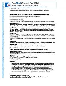
Fig. 2.
PMC Canada Author Manuscript
Superposed structures of apelin-17 in solution at 35°C (Langelaan et al. 2009) and in the SDS micelle-bound state (Langelaan and Rainey 2009). All 80 members of the apelin-17 structural ensemble in solution (BMRB entry 20029) are superposed over the reasonably structurally converged regions of K1-R4 and R6-L9. The 80 members of the apelin-17 structural ensemble bound to SDS micelles (BMRB entry 20082) are superimposed over backbone atoms R6-K12 and all heavy atoms from M15-F17 using LSQKAB (Collaborative Computational Project 1994). Backbone atoms are shown in black, side chains of residues involved with membrane binding in blue and the converged region at the C-terminal region of apelin-17 in red. (Produced using PyMOL (Delano Scientific, San Carlos, CA).)
Biochem Cell Biol. Author manuscript; available in PMC 2011 January 20.
Langelaan and Rainey
Page 11
Table 1
PMC Canada Author Manuscript
Overview of studies on peptide ligands which bind to micelles where both a high-resolution structure is available and where the residues involved in membrane binding were determined.
PMC Canada Author Manuscript Manuscript
Peptide
PDB entry
Micelle type
Residues structured
Residues bound to micelle
Amylin nucleation site (Mascioni et al. 2003)
1KUW
SDS
F23-I26: β–turn
N21, N22, F23, I26, L27
Apelin-17 (Langelaan and Rainey 2009)
20082 a
SDS, DPC and LPPG
R6-K12: β-turns M15-F17: extended
R6, P7, R8 and L9
Bovine pancreatic polypeptide (Lerch et al. 2002)
1LJV
DPC
M17-L31: α-helix
L3, E4, E6, G9, A12, A18, A21, L24, R25, R33, P34, R35
Glucagon (Braun et al. 1981, Braun et al. 1983)b
1KX8
DPC
T5-Y10, L14-R17: extended Y10-L14: helix-like turn R17-T29: irregular α-helix
Hydrophobic residues
Glucagon antagonist (Ying et al. 2003)
1NAU
DPC
T5-L12: Irregular helix A17-T27: α-helix
Y8, Y11, L12, F20, V21, W23, L24, M25
Neuromedin B (Lee and Kim 1999)
1C98
SDS
W4-M10: α-helix
W4, H8 and F9
Neuropeptide K (Dike and Cowsik 2006a)
2B19
DPC
A2-K8: β-turns or 3–10 helix Q9-G18 and H27-V33: α-helix H19-R26: non-canonical β– turn
F32, V33, G34, L35 and M36 (hydrophobic residues)
Neurotensin (Coutant et al. 2007)
1OYV
DPC
L2-N5: extended P10-L13: close to β-turn
P10, Y11, I12 and L13
Pituitary adenylate cyclase activating polypeptide (Inooka et al. 2001)
1D2P
DPC
I5-L27: α-helix
I5, F6, Y10, Y13, M17, Y22, L23, V26 and L27
Porcine Neuropeptide Y (Bader et al. 2001, Lerch et al. 2004)
1F8P
DPC
Y21-T32: α-helix
L17, Y20, Y21, L24, R25, Y27, I28, N29, I31, T32 and Y36
Porcine peptide YY (Lerch et al. 2004)
1RUU
DPC
L17-V31: α-helix
L17, Y20, Y21, L24, Y27, L28, L30, V31, T32, R33, Y36
Proadrenomedullin N terminal peptide (Lucyk et al. 2006)c
2FLY
SDS
R2-A17: α-helix
R2, L3, V5, F9, K12, W13 W16 and R20
Rat islet amyloid polypeptide (Nanga et al. 2009)
2KJ7
DPC
A5-L23: α-helix
N3, T6, A8, T9, L12, A13, L16
a
Apelin-17 structure deposited in BioMagResBank (BMRB) due to current minimum polypeptide size limitations for PDB.
b
Glucagon (PDB entry 1KX8) did not superpose well over the identified converged regions.
c Structure published in PDB (entry 2FLY) in trifluoroethanol, with direct comparison to SDS micelle bound state in paper.
PMC Canada Author Biochem Cell Biol. Author manuscript; available in PMC 2011 January 20.