PMC Canada Author Manuscript
PubMed Central CANADA Author Manuscript / Manuscrit d'auteur J Clin Exp Ophthalmol. Author manuscript; available in PMC 2014 June 26. Published in final edited form as: J Clin Exp Ophthalmol. 2012 January 27; 3: 208–. doi:10.4172/2155-9570.1000208.
Drusen and Pro-inflammatory Mediators in the Post-Mortem Human Eye Kailun Jiang1, Eleanor To2, Jing Z. Cui2, Sijia Cao2, Jiangyuan Gao2, and Joanne A. Matsubara2,* 1Faculty
of Medicine, University of Manitoba, Winnipeg, Canada
2Department
of Ophthalmology and Visual Sciences, University of British Columbia, Vancouver
BC, Canada
PMC Canada Author Manuscript
Introduction Despite significant advances in treatment, age-related macular degeneration (AMD) remains the leading cause of blindness in the elderly of developed nations [1]. Retinal cells experience functional decline with age as a result of metabolic changes and cumulative oxidative stress [2]. Early AMD is characterized by retinal pigment epithelium (RPE) dysfunction [3]. In the early stages of disease, immunomodulatory proteins and inflammatory cells accumulate in the outer retina, with an associated increase in membrane attack complex (MAC) at the RPE and choroidal interface at Bruch’s Membrane [4,5]. In some cases, accumulation of lipo-glyco-proteinacous deposits in the basal lamina, called drusen, become progressively larger and more abundant and may further cause RPE atrophy [6,7].
PMC Canada Author Manuscript
Growing evidence suggests a key role of inflammatory pathways in pathogenesis of AMD, triggered either by an aberrant innate immunity or autoimmunity mechanism. Variants of genes in the complement system, such as complement factor B (CFB), factor I (CFI), factor H (CFH), complement component 2 (C2), component 3 (C3) and component 7 (C7), correlate with either increased or decreased susceptibility to AMD [8,9]. In the AMD retina, immune cells such as macrophages, microglia, and giant cells localize to areas surrounding drusen deposits [10–12]. Activated microglia is capable of inducing direct damage to photoreceptors as shown by in vitro studies [12,13]. Proteomic studies revealed that drusen contain a number of inflammatory molecules, including C3, C5, C5b-9 and MAC [9,14]. Additionally, amyloid-beta (Aβ) and advanced glycation end products (AGE) are present [15]. In an earlier microarray study, we found that human RPE cells treated with Aβ produced strong upregulation of interleukin-1-beta (IL-1β) [16]. IL-1β is an important upstream mediator of inflammatory response and is involved in signaling macrophage activation and production of cytokines. IL-1β is capable of inducing reactive oxygen
Copyright: © 2012 Jiang K, et al. This is an open-access article distributed under the terms of the Creative Commons Attribution License, which permits unrestricted use, distribution, and reproduction in any medium, provided the original author and source are credited. * Corresponding author: Joanne A. Matsubara, Ph.D, Department of Ophthalmology and Visual Sciences, University of British Columbia, 2550 Willow Street, Vancouver, BC V5Z 3N9, Canada, Tel: 604-875-4383; Fax: 604-875-4663;
[email protected].
Jiang et al.
Page 2
PMC Canada Author Manuscript PMC Canada Author Manuscript
species(ROS) in RPE cells [17]. ROS trigger the release of IL-8, which recruits proinflammatory cells such as macrophages [16–18]. Macrophages are present in drusen deposits of AMD eyes and play a key role in promoting neovascular proliferation [19,20]. Recent studies demonstrated the presence of anti-retinal auto-antibodies in early AMD [14]. In these patients, components of drusen such as AGE may be involved in triggering autoimmune responses. In vitro, AGE strongly stimulates chemokine CXC motif ligand 10 and 11 (CXCL-10 and CXCL-11) production in RPE cells (unpublished observation). CXCL-11 recruits activated-Th-lymphocytes and B-cells and may serve as a beacon for Bcell accumulation, activation, and proliferation [21,22]. It is likely that this pathway, along with other similar ones, results in increasing tissue exposure to auto-antibodies. Altogether, our in vitro studies indicate that the components of drusen may be involved in the induction of inflammatory pathways facilitating AMD pathology (unpublished observation) [16]. In particular, IL-1β, CFI, radical S-adenosyl methionine domain containing 2 (RSAD2), and CXCL-10/11 were significantly upregulated. Our data also suggested activation of the JAK/ STAT pathway, involved in the interferon response. STAT3, a member of the JAK/STAT pathway, was found to accumulate in the RPE cells of choroidalneovascular membranes of AMD patients [23]. With age, accumulations of drusen may increase the susceptibility of the retina to damage inflicted by these pathways. Here, we sought to verify the upregulation of the aforementioned inflammation-related gene products in the normal human retina, and to test whether changes in expression of these genes are related to the two major risk factors of AMD, age and presence of drusen.
Methods Eye samples
PMC Canada Author Manuscript
Human donor eyes were obtained from the Eye Bank of British Columbia, Canada. Methods for securing human tissue were in compliance with the Declaration of Helsinki. The protocol was approved by the Clinical Research Ethics Board (CREB) at the University of British Columbia. All tissue samples included in this study were considered normal and excluded tissues from donors with any of the following: evidence of systemic or local infection; progressive central nervous system disease or systemic disease of unknown etiology; lymphoproliferative or myeloproliferative disorders; intrinsic eye disease or previous ocular surgery. All tissues were fixed within 20 hours of death (median time 14 hours). Eyes were divided into two age groups, a “younger” group (n=17), less or equal to 57 years and an “older” group (n=17), greater or equal to 70 years (Table 1). Tissue preparation and immunohistochemistry Eye tissues were prepared as previously described by Seth et al. [4] and embedded in paraffin to obtain 6 μm sections through the pupil and optic nerve axis. Sections that included the macular region were selected for this study. Antibodies against IL-1β, CFI, RSAD2, phosphorylated signal transducer and activator of transcription 3 (p-STAT3), CXCL-10, and CXCL-11 were used to stain sections from both age groups. Antibody dilution and source are documented in Table 2. Paraffin sections were deparaffinized and rehydrated by standard procedures. Sections underwent antigen retrieval
J Clin Exp Ophthalmol. Author manuscript; available in PMC 2014 June 26.
Jiang et al.
Page 3
PMC Canada Author Manuscript
in protease K solution (20 μg/ml, pH 8.0) for 10 minutes at room temperature. Sections were then washed twice in phosphate buffered saline (PBS, pH 7.4), treated with 0.3% H2O2 for 15 minutes, and blocked for 20 minutes with 3% horse or goat serum diluted in 0.3% Triton X (TX)-100-PBS solution to minimize non-specific staining. Tissue sections were allowed to incubate in specific primary antibodies diluted in serum and PBS with 0.3% TX-100 for 24 to 48 hours at 4°C, based on different target antigens. Two methods were employed to obtain negative control sections. In one approach, sections were incubated in the absence of primary antibody. In the second method, sections were treated with an irrelevant IgG isotype at the same concentration and in place of the primary antibody incubation (Table 2). All subsequent steps were identical. Both controls resulted in very low background staining confirming the specificity of the immunohistochemical staining patterns shown here.
PMC Canada Author Manuscript
After incubation in the primary antibodies, sections were thoroughly washed and incubated in secondary antibodies for 30 to 60 minutes at room temperature and rinsed twice before proceeding to alkaline phosphatase (Vector Blue, Vector Laboratories) or 3-Amino-9ethylcarbazole (AEC, Sigma Aldrich) development. For Vector Blue chromogenic reaction, sections were first treated with Avidin-Biotinylated enzyme complex-alkaline phosphatase (ABC-AP) solution for 30 minutes and then developed for 1 hour at room temperature (counterstained with Nuclear Fast Red: Crystal Mount; Biomeda). Sections developed with AEC were pre-incubated for 30 minutes with ABC before incubation with AEC for 5 minutes (counterstained with hematoxylin). Tissue sections from both age groups were developed simultaneously to ensure comparable processing. Statistical analysis
PMC Canada Author Manuscript
Statistical analysis was conducted by examination of four sections per eye per each antibody in a masked manner. For each molecule immunostaining intensities were scored semiquantitatively using 40x objective lens (Eclipse 80i; Nikon, Tokyo, Japan), graded from 0 to 3. A score of 0 indicates no detectable staining above background as determined by comparison with the negative control sections. By systematic scanning through the slides, we identified ones with the strongest staining and classified them as having a score of 3. For staining levels that fall between 0 and 3, a score of 1 was given to samples with the weakest immunolabeling, while a score of 2 represented intermediate immunolabeling. We have also provided a sample range of the semi-quantitative analysis: an example of the strongest immunolabeling is shown in Figure 1, with 1A representing +1, 1B and 1C representing +3. Data were scored in the retina (ganglion cell layer (GCL), inner plexiform layer (IPL), inner nuclear layer (INL), outer plexiform layer (OPL), outer nuclear layer (ONL), inner segment (IS), and outer segment (OS)), retinal pigment epithelium (RPE), Bruch’s membrane, choroid, and drusen sites. Sub-retinal layers were scored individually; scores of all retinal layers were summed and averaged to evaluate expression level in the overall retina. Drusen size was determined using RPE cell size as reference (RPE diameter measured parallel to Bruch’s membrane was estimated at 12–14 μm) [24]. Statistical analysis was done using the Mann-Whitney test (GraphPad Software, Inc., La Jolla, CA). A one-tailed test was selected based on the directional hypothesis that accumulation of pro-inflammatory modulators increased with age and presence of drusen. Significance level was set at P ≤ 0.05.
J Clin Exp Ophthalmol. Author manuscript; available in PMC 2014 June 26.
Jiang et al.
Page 4
PMC Canada Author Manuscript
Categorization of drusen by size Drusen were grouped into three categories based on their size. Tissues were initially classified to contain “observable drusen” if there are discrete lipo-proteinaceous deposits between the basal lamina of the RPE and Bruch’s membrane with diameters ≥ 4 μm. Next, we classified “clinically significant drusen” to be those with a diameter of ≥ 25 μm [24]. This is based on the fact that, during clinical exams, the limit of resolution through fundoscopy is between 25 to 30 μm. Lastly, due to the physiological significance associated with size we further sub-categorized samples into those with drusen ≥ 63 μm in diameter [24,25]. We randomly sampled 20 sections at 20x magnification from each eye to determine the presence and size of drusen in individual eyes. These categories are shown for each eye on Table 1.
Result Younger eyes (≥ 57 years) versus older eyes (≥ 70 years)
PMC Canada Author Manuscript
In order to determine the age-related difference in expression for components of the inflammatory pathways, we compared immunoreactivity in younger eyes (≤ 57 years) with older eyes (≥ 70 years) (Table 3A). Four of the six antigens studied here demonstrated agerelated changes. IL-1β immunoreactivity was significantly stronger in the retina, Bruch’s membrane, choroid and drusen deposits in tissue obtained from older group (Figure 1C, D). Immunoreactivity for p-STAT3, the activated form of STAT3, was significantly stronger in the older group for the combined retinal layers (Figure 2A, C). Specifically, this observation was found to be statistically significant in the GCL, INL, and ONL. For CXCL-11, the overall retinal layers immunostained significantly stronger in the combined retinal layers, particularly OPL of younger eyes. In addition, younger eyes also showed significantly stronger CXCL-11 expression in Bruch’s membrane. Similar to CXCL-11, CXCL-10 immunoreactivity was significantly stronger in the combined retinal layers of younger eyes, specifically in the GCL. There were no discernable differences in immunoreactivity between the two age groups for CFI and RSAD2. Eyes with drusen: ≤25μm versus ≥25 μm (independent of age)
PMC Canada Author Manuscript
Next, we investigated whether the presence of clinically significant drusen (≥ 25 μm) was correlated with changes in expression levels of components of the inflammatory pathway (Table 3B). Antibodies against IL-1β localized strongly to Bruch’s membrane and choroid of eyes with drusen ≥ 25 μm. We did not observe differences in accumulation of IL-1β in the retinal layers or the RPE between the two analyzed groups (Figure 1B, C, E). p-STAT3 stained more robustly in the combined retinal layers of drusen-containing eyes in particular, significantly higher activation of STAT3 was found in the ganglion cell layer, inner and outer nuclear layers of eyes with clinically significant drusen (Figure 2B, D). CXCL-10 demonstrated similar staining patterns to p-STAT3 in retina. RSAD2, similar to IL-1β was most immunoreactive in Bruch’s membrane and through the choroid (Figure 3B, C). In contrast, CFI and CXCL-11 levels did not differ with the presence of clinically significant drusen, in this age independent analysis. J Clin Exp Ophthalmol. Author manuscript; available in PMC 2014 June 26.
Jiang et al.
Page 5
PMC Canada Author Manuscript
Younger eyes (≤ 57 years): ≤ 4 μm drusen versus ≥ 4 μm drusen (controlled for age) Individual components of drusen (Aβ, AGE) have been found to upregulate proinflammatory factors in cultured RPE cells (unpublished observation) [16]. Thus, it is important to evaluate for any correlation between presence of drusen and changes in the expression and/or accumulation of inflammatory factors in the eye. Although drusen ≤ 25 μm are not considered clinically significant, the pro-inflammatory Aβ is present in these deposits [26]. Thus, we compared candidate gene expression levels in younger eyes with (n=10) or without (n=7) drusen (≥ 4 μm). Expression of CXCL-11was observed to be stronger in the drusen group for the combined retinal layers, RPE, Bruch’s membrane, and choroid (Table 3C, Figure 4). Specifically, in the retina, this observation was found to be statistically significant for the ganglion cell layer and outer segments. p-STAT3 was also strong in the retinal layers in young eyes with drusen, while the immunoreactivity against CXCL-10 was strong in the choroid layer of young eyes with drusen (Figure 2, 5). We found no significant difference in IL-1β and CFI immunostaining between the retinas of younger eyes with or without drusen.
PMC Canada Author Manuscript
Eyes with drusen: ≤ 63 μm versus ≥ 63 μm (independent of age) Next, we examined whether any sub-populations exist within eyes carrying drusen. This analysis was motivated by the observation that 95.5% to 98.8% of the human population has one or more drusen [27–30]. While the most frequently observed drusen are ≤ 63μm and considered relatively benign, the prevalence of drusen ≥63 μm increases with age and is considered a risk factor for age related maculopathy [24]. Thus, it is important to discern between these two sizes of drusen. The drusen diameters found in this study range from 4 μm to 126 μm. We observed that drusen ≥ 63 μm are found mostly outside of the macular area at peripheral location, while drusen ≤ 63 μm are distributed more evenly amongst foveal, parafoveal and peripheral regions (data not shown). Using the morphological classification provided by Rudulf et al. [31], drusen ≥ 63 μm identified in our study were predominantly hard drusen.
PMC Canada Author Manuscript
Of the 25 eyes found with drusen deposits, 12 eyes had drusen deposits that were ≥ 63 μm. In this group of eyes (≥ 63 μm), expression of IL-1β was found to be significantly stronger in the combined retinal layers, Bruch’s membrane, choroid, and drusen. Immunoreactivity to CXCL-11 was significantly stronger in the combined retinal layers of eyes with drusen ≥ 63 μm, although significant differences were not observed in individual neuronal layers of the retina (Table 3D). Interestingly, p-STAT3 immunoreactivity was stronger in Bruch’s membrane in eyes with drusen ≤ 63 μm. However, expression of p-STAT3 in combined retinal layers was stronger in eyes with drusen ≥ 63μm. In particular, the ganglion cell and the inner nuclear layers show greater p-STAT3 expression in eyes with drusen ≥ 63 μm (Table 3D). No differences in CFI, RSAD2, and CXCL-10 expression levels were observed between the two groups (Table 3D).
J Clin Exp Ophthalmol. Author manuscript; available in PMC 2014 June 26.
Jiang et al.
Page 6
PMC Canada Author Manuscript
Discussion Here, we examined the expression levels of multiple candidate biomarkers of AMD by immunohistochemistry in human postmortem eye tissues and correlated the results with age and presence of drusen, two known risk factors for AMD development and progression. Most of the targets we tested were positively correlated with age and/or presence of drusen. This would be expected if drusen indeed induces inflammation via Aβ and/or AGEs [16]. IL-1β
PMC Canada Author Manuscript
IL-1β is strongly implicated in the pathogenesis of chronic inflammatory diseases [32]. First, aberrant auto-upregulation of IL-1β leads to excessive inflammation. Second, it promotes angiogenesis through upregulation of VEGF, the target of choice for slowing the exudative or wet form of AMD [33]. In this study, eyes from the older group demonstrated significantly stronger immunoreactivity for IL-1β than those from the younger group. Since the tissue samples were from individuals who had no known or diagnosed eye diseases, this result suggests that older eyes are more prone to an inflamed state. This is also consistent with studies identifying age as one of the biggest risk factors of AMD [1].
PMC Canada Author Manuscript
Additional examination of IL-1β accumulation showed that regardless of age, eyes with clinically significant drusen have significantly stronger immunereactivity localized to Bruch’s membrane and choroid layers (Figure 1). IL-1β is secreted by a number of cell types into local environment and systemic circulation. Serum levels of IL-1β do not change significantly with aging [34]. Also, in vitro stimulation of RPE cells with Aβ resulted in increased IL-1β expression [16,35]. While we cannot rule out an abnormal retinal accumulation of IL-1β derived from systemic circulation, at least part of the IL-1β accumulation with aging and drusen may be explained by increased IL-1β secretion by RPE cells. Furthermore, a sub-analysis of all eyes with drusen indicated that staining for IL-1β was significantly stronger in eyes with large drusen (≥ 63 μm), which we found to be enriched in the peripheral. Peripheral drusen, while producing little structural disruption, may have indirect effects on the cells in the macular region [36]. Interestingly, peripheral drusen ≥ 63 μm is strongly associated with CFHY402H polymorphism [37]. Taken together, these results suggest that, while expression and accumulation of IL-1β in eye tissue is correlated with the presence of drusen deposits, it is drusen ≥ 63 μm that are associated with increased levels of IL-1β. It is important to note that advanced age can potentially confound our conclusions in this analysis because the prevalence of drusen ≥ 63 μm is correlated with increasing age. Thus, further studies designed to control for this confounding factor are warranted. p-STAT3 STAT3 is an oncogene and can be activated by IFNs; it induces transcriptional responses that block apoptosis and promote cell survival during inflammation [38,39]. In tumour cell lines, STAT3 can induce VEGF production either directly via activation of VEGF transcription or indirectly viahypoxia-inducible factor 1α [40]. We observed increased immunoreactivity for p-STAT3 with increasing age and with presence of drusen. The increase in pSTAT3 level is particularly prominent in retina with drusen ≥ 63 μm in
J Clin Exp Ophthalmol. Author manuscript; available in PMC 2014 June 26.
Jiang et al.
Page 7
PMC Canada Author Manuscript
diameter (Figure 2). In an age-controlled analysis, we continued to see STAT3 activation to be associated with presence of drusen ≥ 4 μm. These associations raise the possibility that components of drusen promote the activation of STAT3, which acts to protect the retina from apoptotic damages, and to promote exudative AMD through VEGF upregulation. RSAD2
PMC Canada Author Manuscript
Increased RSAD2 expression has been reported in age-related diseases such as atherosclerosis, the pathology of which is associated with deposition of drusen components such as Aβ and AGE [26,41]. In atherosclerosis, local inflammation could cause increased RSAD2 expression, leading to abnormal lipid accumulation [42]. Lipid accumulation in Bruch’s membrane is associated with upregulation of VEGF in the choriocapillaris of mice [43]. Previously, we found that stimulation of RPE cells with either Aβ or AGE increased RSAD2 expression [16] (unpublished observation). Here, we reported increased RSAD2 immunoreactivity in eyes with drusen, but not in aged eyes. These data indicate that drusen component such as Aβ/AGE might upregulate RSAD2 expression through induction of local inflammation. The consequent lipid accumulation may further lead to VEGF upregulation in the choriocapillaris, a result that is implicated in neovascular AMD. CXCL-10 and CXCL-11 RSAD2 is an antiviral protein, activated by IFN α, β, or γ [44]. Upregulation of RSAD2 suggests a role for interferon in AMD pathogenesis. We further examined this system by looking at the downstream targets of IFN-γ: CXCL-10 and CXCL-11 [21]. Both ligands are agonist of CXC receptor 3 (CXCR3). They promote chemotaxis of T-cells, B-cells, and natural killer cells [21]. Through CXCR3-mediated mechanism, CXCL-10 and 11 inhibit angiogenesis and inhibit endothelial cell proliferation (49, 50 of serum paper). Serum concentration of CXCL-10 increased dramatically with age and with the development of AMD [45,46]. Mo et al. [46] confirmed the accumulation of CXCL-10 in human eye tissue with AMD and proposed that CXCL-10 may function as a biomarker for AMD. Although we found the opposite in terms of age related changes in CXCL-10 concentration, this difference may be accounted for by the different methods used: serum versus histology. However, CXCL-10 accumulation was determined to be greater in eyes with clinically significant drusen. This trend continues in the younger age group.
PMC Canada Author Manuscript
Not much is known about the effects of aging on CXCL-11 levels. CXCL-11 immunohistochemical level is higher in younger eyes and no difference in staining intensity exists between those with and without clinically significant drusen (Table 3). However, CXCL-11 levels were higher in those eyes with drusen ≥ 63 μm. Moreover, CXCL-11 level was higher in tissue of the younger group with drusen (≥ 4 μm). These findings suggest that the relationship between drusen and CXCL-11 is complex. With normal aging, the macrophage population shifts from the pro-inflammatory M1 to the anti-inflammatory M2, which are associated with diminishing CXCL-11 expression [47]. Cytokines such as CXCL-11 and IL-1β are markers for M1 macrophage phenotype. CXCL-11 is also upregulated in AMD [47]. Our data suggest that CXCL-11 is an early precursor of disease progression, upregulated in younger eyes with drusen. This may warrant further investigation to determine its future role as an early indicator of AMD disease progression.
J Clin Exp Ophthalmol. Author manuscript; available in PMC 2014 June 26.
Jiang et al.
Page 8
PMC Canada Author Manuscript
Summary
PMC Canada Author Manuscript
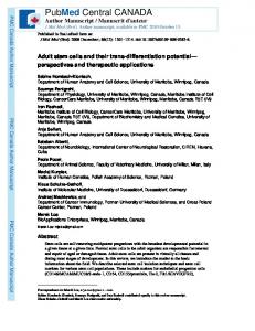
With age and drusen accumulation, the environment of the eye tends to shift more towards a pro-inflammatory state (Figure 6) through either IL-1β and/or possibly IFN pathways. Little change is induced in the complement inhibitor, CFI, in association with drusen, suggesting a failure to activate protective mechanisms against complement activation. Alternatively, the lack of change in CFI accumulation level could indicate that that the pro-inflammatory actions of drusen may not yet be enough to trigger inhibition of the complement cascade in healthy eye tissue. Here, we identified three inflammatory molecules in the postmortem human eye, p-STAT3, CXCL-10, and CXCL-11, to be strongly correlated with the presence of drusen within a population controlled for age (≤57 years old, Figure 6). This pattern of accumulation suggests that these molecules are closely involved with the presence of drusen rather than with aging, and may represent a predisposition toward AMD pathogenesis. A recent microarray study on mouse RPE cells reported expression changes in over 315 genes associated with advanced age [48]. Many pathways they identified were also found in our prior microarray study (unpublished observation) [16]. Similarly, Chen et al. [49] found that the mouse neuroretina displayed age-related upregulation of the complement cascade and activation of retinal microglia [49]. Curiously, neither study found expression changes in any of the gene products examined in this study. It is important to note that drusen-like deposits have not been documented in wild-type-mouse eyes [50]. Thus, one interpretation for this discrepancy may be that the genes identified in our microarray studies, and specifically the gene products studied here reflect a drusen-specific response rather than a general response to aging. Our studies now set the stage for future experiments testing the function of each of these potential mediators in the pathogenesis of AMD. One approach might involve assessing the response of each mediator following Aβ and/or AGE injection into young wild-type mice to determine degree of mimicry to AMD pathology.
Acknowledgments We thank Jonathan Tang, Jonathan Coleman, and Dr. Ian Clark for discussion and editing of the manuscript. This work was funded by Canadian Institutes of Health Research (CIHR-MOP-97806) to J.A.M. Grant Support Supported by Canadian Institutes of Health Research (CIHR) Grant# MOP-97806 (to JAM).
PMC Canada Author Manuscript
References 1. Friedman DS, O’Colmain BJ, Munoz B, Tomany SC, McCarty C, et al. Prevalence of age-related macular degeneration in the United States. Arch Ophthalmol. 2004; 122:564–572. [PubMed: 15078675] 2. Xu H, Chen M, Forrester JV. Para-inflammation in the aging retina. Prog Retin Eye Res. 2009; 28:348–368. [PubMed: 19560552] 3. Binder S, Stanzel BV, Krebs I, Glittenberg C. Transplantation of the RPE in AMD. Prog Retin Eye Res. 2007; 26:516–554. [PubMed: 17532250] 4. Seth A, Cui J, To E, Kwee M, Matsubara J. Complement-Associated Deposits in the Human Retina. Invest Ophthalmol Vis Sci. 2008; 49:743–750. [PubMed: 18235023] 5. Donoso LA, Kim D, Frost A, Callahan A, Hageman G. The Role of Inflammation in the Pathogenesis of Age-related Macular Degeneration. Surv Ophthalmol. 2006; 51:137–152. [PubMed: 16500214]
J Clin Exp Ophthalmol. Author manuscript; available in PMC 2014 June 26.
Jiang et al.
Page 9
PMC Canada Author Manuscript PMC Canada Author Manuscript PMC Canada Author Manuscript
6. Pauleikhoff D, Barondes MJ, Minassian D, Chisholm IH, Bird AC. Drusen as risk factors in agerelated macular disease. Am J Ophthalmol. 1990; 109:38–43. [PubMed: 1688685] 7. Coleman HR, Chan C-C, Ferris FL III, Chew EY. Age-related macular degeneration. Lancet. 2008; 372:1835–1845. [PubMed: 19027484] 8. Klein RJ, Zeiss C, Chew EY, Tsai J-Y, Sackler RS, et al. Complement Factor H Polymorphism in Age-Related Macular Degeneration. Science. 2005; 308:385–389. [PubMed: 15761122] 9. Patel M, Chan C-C. Immunopathological aspects of age-related macular degeneration. Semin Immunopathol. 2008; 30:97–110. [PubMed: 18299834] 10. Dastgheib K, Green WR. Granulomatous Reaction to Bruch’s Membrane in Age-Related Macular Degeneration. Arch Ophthalmol. 1994; 112:813–818. [PubMed: 7516148] 11. Penfold PL, Wong JG, Gyory J, Billson FA. Effects of triamcinolone acetonide on microglial morphology and quantitative expression of MHC-II in exudative age-related macular degeneration. Clin Experiment Ophthalmol. 2001; 29:188–192. [PubMed: 11446466] 12. Ding X, Patel M, Chan CC. Molecular pathology of age-related macular degeneration. Prog Retin Eye Res. 2009; 28:1–18. [PubMed: 19026761] 13. Roque RS, Rosales AA, Jingjing L, Agarwal N, Al-Ubaidi MR. Retina-derived microglial cells induce photoreceptor cell death in vitro. Brain Res. 1999; 836:110–119. [PubMed: 10415410] 14. Wang Y, Wang VM, Chan CC. The role of anti-inflammatory agents in age-related macular degeneration (AMD) treatment. Eye. 2011; 25:127–139. [PubMed: 21183941] 15. Crabb JW, Miyagi M, Gu X, Shadrach K, West KA, et al. Drusen proteome analysis: An approach to the etiology of age-related macular degeneration. Proc Natl Acad Sci U S A. 2002; 99:14682– 14687. [PubMed: 12391305] 16. Kurji K, Cui J, Lin T, Harriman D, Prasad S, et al. Microarray Analysis Identifies Changes in Inflammatory Gene Expression in Response to Amyloid-Beta Stimulation of Cultured Human Retinal Pigment Epithelial Cells. Invest Ophthalmol Vis Sci. 2009; 51:1151–1163. [PubMed: 19797223] 17. Yang D, Elner SG, Bian Z-M, Till GO, Petty HR, et al. Pro-inflammatory cytokines increase reactive oxygen species through mitochondria and NADPH oxidase in cultured RPE cells. Exp Eye Res. 2007; 85:462–472. [PubMed: 17765224] 18. Hwang YS, Jeong M, Park JS, Kim MH, Lee DB, et al. Interleukin-1[beta] stimulates IL-8 expression through MAP kinase and ROS signaling in human gastric carcinoma cells. Oncogene. 2004; 23:6603–6611. [PubMed: 15208668] 19. Espinosa-Heidmann DG, Suner IJ, Hernandez EP, Monroy D, Csaky KG, et al. Macrophage Depletion Diminishes Lesion Size and Severity in Experimental Choroidal Neovascularization. Invest Ophthalmol Vis Sci. 2003; 44:3586–3592. [PubMed: 12882811] 20. Jager MJ, Klaver CCW. Macrophages feel their age in macular degeneration. J Clin Invest. 2007; 117:3182–3184. [PubMed: 17975664] 21. Petkovic V, Moghini C, Paoletti S, Uguccioni M, Gerber B. I-TAC/CXCL11 is a natural antagonist for CCR5. J Leukoc Biol. 2004; 76:701–708. [PubMed: 15178708] 22. Silverman GJ, Carson DA. Roles of B cells in rheumatoid arthritis. Arthritis Res Ther. 2003; 5(Suppl 4):S1–6. [PubMed: 15180890] 23. Fasler-Kan E, Wunderlich K, Hildebrand P, Flammer J, Meyer P. Activated STAT 3 in choroidal neovascular membranes of patients with age-related macular degeneration. Ophthalmologica. 2005; 219:214–221. [PubMed: 16088240] 24. Sarks SH, Arnold JJ, Killingsworth MC, Sarks JP. Early drusen formation in the normal and aging eye and their relation to age related maculopathy: a clinicopathological study. Br J Ophthalmol. 1999; 83:358–368. [PubMed: 10365048] 25. Hageman GS, Luthert PJ, Victor Chong NH, Johnson LV, Anderson DH, et al. An integrated hypothesis that considers drusen as biomarkers of immune-mediated processes at the RPE-Bruch’s membrane interface in aging and age-related macular degeneration. Prog Retin Eye Res. 2001; 20:705–732. [PubMed: 11587915] 26. Mullins RF, Russell SR, Anderson DH, Hageman GS. Drusen associated with aging and agerelated macular degeneration contain proteins common to extracellular deposits associated with
J Clin Exp Ophthalmol. Author manuscript; available in PMC 2014 June 26.
Jiang et al.
Page 10
PMC Canada Author Manuscript PMC Canada Author Manuscript PMC Canada Author Manuscript
atherosclerosis, elastosis, amyloidosis, and dense deposit disease. FASEB J. 2000; 14:835–846. [PubMed: 10783137] 27. Bressler NM, Bressler SB, West SK, Fine SL, Taylor HR. The Grading and Prevalence of Macular Degeneration in Chesapeake Bay Watermen. Arch Ophthalmol. 1989; 107:847–852. [PubMed: 2786408] 28. Klein R, Klein BE, Linton KL. Prevalence of age-related maculopathy. The Beaver Dam Eye Study. Ophthalmology. 1992; 99:933–943. [PubMed: 1630784] 29. Vingerling JR, Dielemans I, Hofman A, Grobbee DE, Hijmering M, et al. The prevalence of agerelated maculopathy in the Rotterdam Study. Ophthalmology. 1995; 102:205–210. [PubMed: 7862408] 30. Mitchell P, Smith W, Attebo K, Wang JJ. Prevalence of age-related maculopathy in Australia. The Blue Mountains Eye Study. Ophthalmology. 1995; 102:1450–1460. [PubMed: 9097791] 31. Rudolf M, Clark ME, Chimento MF, Li CM, Medeiros NE, et al. Prevalence and morphology of druse types in the macula and periphery of eyes with age-related maculopathy. Invest Ophthalmol Vis Sci. 2008; 49:1200–1209. [PubMed: 18326750] 32. Dinarello CA. IL-1: Discoveries, controversies and future directions. Eur J Immunol. 2010; 40:599–606. [PubMed: 20201008] 33. Jung Y, Liu W, Reinmuth N, Ahmad S, Fan F, et al. Vascular endothelial growth factor is upregulated by interleukin-1β in human vascular smooth muscle cells via the P38 mitogenactivated protein kinase pathway. Angiogenesis. 2001; 4:155–162. [PubMed: 11806247] 34. Di Iorio A, Ferrucci L, Sparvieri E, Cherubini A, Volpato S, et al. Serum IL-1beta levels in health and disease: a population-based study. ‘The InCHIANTI study’. Cytokine. 2003; 22:198–205. [PubMed: 12890453] 35. Wang J, Ohno-Matsui K, Yoshida T, Shimada N, Ichinose S, et al. Amyloid-beta up-regulates complement factor B in retinal pigment epithelial cells through cytokines released from recruited macrophages/microglia: Another mechanism of complement activation in age-related macular degeneration. J Cell Physiol. 2009; 220:119–128. [PubMed: 19277984] 36. Al-Hussaini H, Schneiders M, Lundh P, Jeffery G. Drusen are associated with local and distant disruptions to human retinal pigment epithelium cells. Exp Eye Res. 2009; 88:610–612. [PubMed: 18992244] 37. Munch IC, Ek J, Kessel L, Sander B, Almind GJ, et al. Small, Hard Macular Drusen and Peripheral Drusen: Associations with AMD Genotypes in the Inter99 Eye Study. Invest Ophthalmol Vis Sci. 2010; 51:2317–2321. [PubMed: 20007824] 38. Levy DE, Lee CK. What does Stat3 do? J Clin Invest. 2002; 109:1143–1148. [PubMed: 11994402] 39. Kortylewski M, Jove R, Yu H. Targeting STAT3 affects melanoma on multiple fronts. Cancer Metastasis Rev. 2005; 24:315–327. [PubMed: 15986140] 40. Xu Q, Briggs J, Park S, Niu G, Kortylewski M, et al. Targeting Stat3 blocks both HIF-1 and VEGF expression induced by multiple oncogenic growth signaling pathways. Oncogene. 2005; 24:5552– 5560. [PubMed: 16007214] 41. Peppa M, Uribarri J, Vlassara H. The role of advanced glycation end products in the development of atherosclerosis. Curr Diab Rep. 2004; 4:31–36. [PubMed: 14764277] 42. Olofsson PS, Jatta K, Wagsater D, Gredmark S, Hedin U, et al. The antiviral cytomegalovirus inducible gene 5/viperin is expressed in atherosclerosis and regulated by proinflammatory agents. Arterioscler Thromb Vasc Biol. 2005; 25:e113–116. [PubMed: 15890971] 43. Rudolf M, Schloetzer-Schrehardt U, Michels S, Aherrahou Z, Doehring LC, et al. Lipid Accumulation in Bruch’s Membrane Is Associated With an Increased Expression of Vascular Endothelial Growth Factor (VEGF) in the Choriocapillaries. Invest Ophthalmol Vis Sci. 2005; 46:1213. 44. Chin K-C, Cresswell P. Viperin (cig5), an IFN-inducible antiviral protein directly induced by human cytomegalovirus. Proc Natl Acad Sci U S A. 2001; 98:15125–15130. [PubMed: 11752458] 45. Miles EA, Rees D, Banerjee T, Cazzola R, Lewis S, et al. Age-related increases in circulating inflammatory markers in men are independent of BMI, blood pressure and blood lipid concentrations. Atherosclerosis. 2008; 196:298–305. [PubMed: 17118371]
J Clin Exp Ophthalmol. Author manuscript; available in PMC 2014 June 26.
Jiang et al.
Page 11
PMC Canada Author Manuscript
46. Mo FM, Proia AD, Johnson WH, Cyr D, Lashkari K. Interferon gamma-inducible protein-10 (IP-10) and eotaxin as biomarkers in age-related macular degeneration. Invest Ophthalmol Vis Sci. 2010; 51:4226–4236. [PubMed: 20220052] 47. Cao X, Shen D, Patel MM, Tuo J, Johnson TM, et al. Macrophage polarization in the maculae of age-related macular degeneration: a pilot study. Pathol Int. 2011; 61:528–535. [PubMed: 21884302] 48. Chen H, Liu B, Lukas TJ, Neufeld AH. The aged retinal pigment epithelium/choroid: a potential substratum for the pathogenesis of age-related macular degeneration. PLoS One. 2008; 3:e2339. [PubMed: 18523633] 49. Chen M, Muckersie E, Forrester JV, Xu H. Immune activation in Retinal Aging: A Gene Expression Study. Invest Ophthalmol Vis Sci. 2010; 61:528–535. 50. Luhmann UF, Robbie S, Munro PM, Barker SE, Duran Y, et al. The drusenlike phenotype in aging Ccl2-knockout mice is caused by an accelerated accumulation of swollen autofluorescent subretinal macrophages. Invest Ophthalmol Vis Sci. 2009; 50:5934–5943. [PubMed: 19578022]
PMC Canada Author Manuscript PMC Canada Author Manuscript J Clin Exp Ophthalmol. Author manuscript; available in PMC 2014 June 26.
Jiang et al.
Page 12
PMC Canada Author Manuscript PMC Canada Author Manuscript
Figure 1.
PMC Canada Author Manuscript
Immunostaining for IL-1β with AEC development system (red) and counterstained with Mayer’s hematoxylin (blue). IL-1β expression levels in human eye tissue, analyzed by age groups and size of drusen. (A) Younger group (≤ 57 yr) with drusen ≤ 25 μm; note faint immunolabeling pattern (semi-quantitative grading of 1). (B) Eye tissue from the younger group (≤ 57 yr) with drusen ≥ 25 μm (arrows). Note immunoreactivity for IL-1β in drusen and through choroid (semi-quantitative grading of 3). RPE cells are mildly immunostained. (C) Strong immunoreactivity in drusen, Bruch’s membrane, and choroid in eyes from older group (≥ 70 yr, semi-quantitative grading of 3) with drusen ≥ 63 μm (arrows). Scale bar 20 μm. (D) Aside from the retinal pigmented epithelial (RPE) cells, tissue from older group (n = 12) are more immunoreactive for IL-1β (retinal layers through to the choroid) than younger group (n = 15) (black asterisk, p ≤ 0.05). (E) Eyes with clinically significant drusen (≥ 25 μm, n = 15) were more immunoreactive for IL-1β in Bruch’s membrane and choroid compared to eyes without clinically significant drusen (n = 12). (F) Eyes with drusen ≥ 63 μm (n = 12) were more immunoreactive for IL-1β in the retinal layers, Bruch’s membrane, choroid, and drusen than eyes with drusen ≤ 63 μm (n = 8).
J Clin Exp Ophthalmol. Author manuscript; available in PMC 2014 June 26.
Jiang et al.
Page 13
PMC Canada Author Manuscript PMC Canada Author Manuscript PMC Canada Author Manuscript
Figure 2.
p-STAT3 levels in human eye tissue visualized with Vector Blue (blue) and counterstained with neutral red (pink). (A) Cells (arrows) in the ganglion cell layer (GCL), inner nuclear layer (INL), and outer nuclear layer are found to be immunoreactive (vector blue) for phosphorylated (activated) STAT3 in the older group (≥ 70 yr). (B) Similarly, immunolabeling profile is seen in eyes with drusen ≥ 25 μm regardless of age. Scale bar 30 μm. p-STAT3 accumulation significantly differs (p ≤ 0.05, black asterisk) in the retinal layers between the analyzed groups. STAT3 activation is greater in older eyes (C, ≥ 70 yr, n = 14), in eyes with clinically significant drusen (D, ≥ 25 μm, n = 14) and particularly in eyes with large drusen (F, ≥ 63 μm, n = 10). In the younger group (E, ≤ 57 yr, n = 14), the J Clin Exp Ophthalmol. Author manuscript; available in PMC 2014 June 26.
Jiang et al.
Page 14
PMC Canada Author Manuscript
presence of drusen ≥ 4 μm (n = 7) is associated with increased STAT3 activation compared to eyes without drusen (n = 7).
PMC Canada Author Manuscript PMC Canada Author Manuscript J Clin Exp Ophthalmol. Author manuscript; available in PMC 2014 June 26.
Jiang et al.
Page 15
PMC Canada Author Manuscript PMC Canada Author Manuscript
Figure 3.
Immunostaining for RSAD2 with Vector Blue (blue) and counterstained with neutral red (pink). (B) Representative sections from eyes with drusen ≥ 25 μm shown that areas around drusen (≥ 25 μm), in RPE and Bruch’s membrane are more immunoreactive for RSAD2 than (A) the background reactivity seen in tissues with small (≤ 25 μm) or no drusen. Scale bar 25 μm. (C) Eyes with clinically significant drusen (≥ 25 μm, n = 15) are found to have stronger immunostaining for RSAD2 particularly in the Bruch’s membrane and choroid (p ≤ 0.05, black asterisk) than those without clinically significant drusen (n=13).
PMC Canada Author Manuscript J Clin Exp Ophthalmol. Author manuscript; available in PMC 2014 June 26.
Jiang et al.
Page 16
PMC Canada Author Manuscript PMC Canada Author Manuscript Figure 4.
PMC Canada Author Manuscript
Immunolabeling for CXCL-11 with Vector Blue (blue) and counterstained with neutral red (pink). Representative sections from younger eyes (≥ 57 yr, n = 16) are shown. Samples were analyzed for the presence of drusen (≥ 4 μm, n = 9). Within the younger age-group, eyes with drusen (B) have greater accumulation of CXCL-11 throughout the retina and choroid. Scale bar 20μm. (C) Within tissue of younger eyes (≤ 57 yr, n = 16), those with any observable drusen (≥ 4 μm, n = 9) were found to stain stronger (p ≤ 0.05, black asterisk) for chemokine, CXCL-11 from the retina through to the choroid.
J Clin Exp Ophthalmol. Author manuscript; available in PMC 2014 June 26.
Jiang et al.
Page 17
PMC Canada Author Manuscript Figure 5.
In the younger age group (≤ 57 yr, n = 15), immunoreactivity against CXCL-10 is significantly greater in the choroid of eyes with drusen (≥ 4 μm, p ≤ 0.05, n = 9).
PMC Canada Author Manuscript PMC Canada Author Manuscript J Clin Exp Ophthalmol. Author manuscript; available in PMC 2014 June 26.
Jiang et al.
Page 18
PMC Canada Author Manuscript PMC Canada Author Manuscript
Figure 6.
Summary diagram depicting the distribution of IL-1β, RSAD2, p-STAT3, and CXCL-10/11 immunoreactivity in post-mortem eye tissue with respect to aging and present of drusen. Icons denote the relative immunostaining of each molecule in a given retinallocation.
PMC Canada Author Manuscript J Clin Exp Ophthalmol. Author manuscript; available in PMC 2014 June 26.
80
72
73
47
46
46
48
56
16
17
18
19
20
21
22
80
12
15
72
11
70
72
72
74
9
10
13
M
71
8
14
M
72
7
M
J Clin Exp Ophthalmol. Author manuscript; available in PMC 2014 June 26.
F
F
F
F
F
M
M
F
F
M
M
M
F
M
M
74
75
5
M
F
F
M
6
70
4
70
2
71
74
1
3
Age
Donor
Sex
PMC Canada Author
Donor List.
breast cancer with brain metastasis
liver cancer, cirrhosis
brain tumor
lung cancer
lung cancer with mets
lung and liver cancer
renal cancer
not available
large left cerebral vascular accident
cardiogenic arrest
renal failure
respiratory arrest
gastroenteritis and chronic renal failure
bone cancer with metastasis
colon cancer with metastasis
pancreatic cancer
heart disease (cardiac arrest)
bowel cancer
hepatitis B and colon cancer with metastasis
lung cancer
glioblastoma multiform
sub-arachnoid hemorrhage
Diagnosis
X
X
X
X
X
X
X
X
X
X
X
X
X
X
X
IL-1β
X
X
X
X
X
X
X
X
X
X
X
X
X
X
X
X
X
X
X
X
CFI
X
X
X
X
X
X
X
X
X
X
X
X
X
X
X
X
X
X
X
STAT3
X
X
X
X
X
X
≤ 57 yr Old
X
X
X
X
X
X
X
X
X
X
X
X
X
X
X
X
X
X
X
X
X
X
X
X
X
X
X
X
X
X
X
X
X
X
CXCL-10
≥ 70 yr Old
CXCL-11
X
X
X
X
X
X
X
X
X
X
X
X
X
X
X
X
X
X
RSAD2
X
X
X
No Drusen
X
X
X
X
Drusen