Jun 4, 1990 - Cell-associated alkaline phosphatase (ALPase) of Bacteroides gingivalis 381 was found in the outer part ofthe periplasmic space by using an ...
INFECTION AND IMMUNITY, Sept. 1990, p. 2882-2887 0019-9567/90/092882-06$02.00/0 Copyright C 1990, American Society for Microbiology
Vol. 58, No. 9
Purification and Characterization of Alkaline Phosphatase of Bacteroides gingivalis 381 YOSHIHISA YAMASHITA,1 KUNIAKI TOYOSHIMA,2 MIKIKO YAMAZAKI,1 NOBUHIRO HANADA,1 AND TADAMICHI TAKEHARAl* Department of Preventive Dentistry' and Department of Oral Anatomy,2 Kyushu Dental College, Kokurakita-ku,
Kitakyushu 803, Japan Received 23 October 1989/Accepted 4 June 1990
Cell-associated alkaline phosphatase (ALPase) of Bacteroides gingivalis 381 was found in the outer part of the periplasmic space by using an ultracytochemical procedure. Cell-associated ALPase was solubilized by extraction with 1% Triton X-100, and the solubilized enzyme was purified 904-fold with 5.6% recovery by using affinity column chromatography for mammalian intestinal-form ALPase. The purified enzyme gave a single protein band that corresponded to the enzyme activity band on polyacrylamide gel electrophoresis preparations. A single protein band at a molecular weight of 61,000 was observed on sodium dodecyl sulfate-polyacrylamide gel electrophoresis preparations. The molecular weight of the native enzyme was estimated to be 130,000 by gel ifitration with TSK-gel G3000SW. These findings indicate that B. gingivalis ALPase is a homodimer. The optimal pH of the enzyme was between 9.1 and 9.3 in the absence of divalent metal ions and was between 10.1 and 10.3 in the presence of manganese or zinc ions. The apparent Km for p-nitrophenylphosphate was 0.037 ± 0.003 mM (mean ± standard deviation) at pH 9.2 in the absence of divalent metal ions and 0.22 ± 0.02 mM at pH 10.2 in the presence of 1 mM manganese ions. Under both of the conditions described above, the purified enzyme was able to hydrolyze casein and O-phosphoserine, suggesting that B. gingivalis ALPase can act as a phosphoprotein phosphatase. ALPase that immunologically cross-reacted with the purified enzyme was found in the extracellular soluble fraction. This means that ALPase is released from the periplasmic space into the culture supernatant as a soluble form.
Bacteroides species are frequently detected in the microflora of patients with several types of periodontitis (21, 23, 31, 36), and Bacteroides gingivalis in particular is thought to be one of the most important etiological agents of adult periodontitis in humans (24, 28, 32). Some animal experiments have confirmed the pathogenicity of this organism (10, 11, 17, 34), and identification of many virulence factors, such as some kinds of proteases (7, 27, 33) and hemagglutinin (26), from the bacterial cells or their supernatants and characterization of these factors have been achieved. B. gingivalis is known to have high alkaline phosphatase (ALPase) activities (14, 29) in addition to the virulence factors described above. A previous report clarified that most ALPase activity takes place in a cell-associated form and that some ALPase activity is released into the culture medium associated with extracellular vesicles and as a soluble form (20). However, many enzymological properties of these ALPases are still unknown. In this study, we purified cell-associated ALPase from B. gingivalis 381 and characterized this enzyme. In addition, we prepared an antiserum against the purified enzyme and compared the immunological properties of the purified enzyme and the soluble ALPase in the extracellular fraction.
,ug/ml), and hemin (0.5 or 5 ,ug/ml); this medium was designated medium A. Enzyme assay. ALPase activity was assayed at 37°C by using 5 mM p-nitrophenylphosphate (p-NPP) as a substrate in 0.1 M glycine-NaOH buffer (pH 9.2); 1 U of enzyme activity corresponded to 1 p,mol of substrate hydrolyzed per min at 37°C, as described previously (25). All values for enzyme activity given below are the means of three replicate determinations. The effects of various divalent metal ions on enzyme activity were examined by using 0.1 M glycineNaOH buffer (pH 8.5 to 11.7) containing 5 mM p-NPP and 1 mM MnCl2, 1 mM Zn(COOH)2, 1 mM MgCl2, 1 mM CuCl2, or 1 mM CaCl2. The optimal pH was determined by using 0.1 M acetate buffer (pH 5.0 to 6.5), 0.1 M Tris hydrochloride buffer (pH 6.5 to 8.5), and 0.1 M glycine-NaOH buffer (pH 8.5 to 11.7) with or without 1 mM MnCl2. The phosphatase activities of the enzyme were examined by using the following esters: p-NPP, 0-glycerophosphate, D-glucose 6-phosphate, a-naphthylphosphate, ATP, pyridoxal 5'-phosphate, O-phosphoserine, PPi, and casein. The assay solutions were 0.1 M glycine-NaOH buffer (pH 9.2) and 0.1 M glycine-NaOH buffer (pH 10.2) containing 1 mM MnCl2. The amount of free phosphate released was estimated by using the method of Fiske and Subbarow (5). Culture fraction preparation. Bacterial cell, extracellular vesicle, and extracellular soluble fractions were obtained by using the method of Minhas and Greenman (20). Ultracytochemical procedure. After cultivation in medium A containing 0.5 or 5 ,ug of hemin per ml, bacterial cells and extracellular vesicles were obtained as described previously (20) and were washed twice with 0.1 M Tris hydrochloride buffer (pH 7.5) containing 0.15 M NaCl (TBS buffer). The washed cells were incubated for 5 min in a modification of the reaction mixture of Mayahara et al. (16) (50 mM Tris
MATERIALS AND METHODS Bacterial strain and culture conditions. B. gingivalis 381 was kindly provided by T. Koga, The National Institute of Health, Tokyo, Japan. The organism was grown anaerobically in a tryptic soy broth medium (Difco Laboratories, Detroit, Mich.) containing yeast extract (5 g/liter; BBL Microbiology Systems, Cockeysville, Md.), menadione (1 *
Corresponding author. 2882
VOL. 58, 1990
hydrochloride buffer [pH 9.0] containing 5 mM p-NPP as a substrate, 8% sucrose, and 5 mM lead citrate). Cells incubated in the medium without a substrate were used as a control. After incubation, the cells were washed twice with TBS buffer, fixed with a mixture containing 2% para-formaldehyde and 2.5% glutaraldehyde in 0.1 M cacodylate buffer (pH 7.2) for 2 h, and postfixed with 2% osmium tetroxide for 2 h. The postfixed cells were washed with 0.1 M cacodylate buffer (pH 7.4), and 1.5% agar was added to the washed cells. The agar cores were dehydrated in a graded series of ethanol, passed through propylene oxide, and embedded in epoxy resin. Ultrathin sections obtained from the embedded block were examined, with or without additional staining with uranyl acetate and lead citrate, by using a model JEOL-1OOC electron microscope. Extracellular vesicles were examined by using the ultracytochemical procedure described above. Solubilization of ALPase from bacterial cells. Bacterial cells grown in medium A containing 0.5 ,ug of hemin per ml were obtained by centrifugation. The cells were washed twice with TBS buffer and suspended in 50 mM Tris hydrochloride buffer (pH 8.4) containing 2 M KCI; 0.01, 0.1, or 1% Triton X-100; or both 2 M KCI and 1% Triton X-100. After each suspension was stirred at room temperature for 30 min, the supematant was collected by centrifugation at 15,000 x g for 20 min. The EDTA-osmotic shock procedure and lysozyme treatment with 0.2 M MgCl2 or 1 mM EDTA were carried out as described previously (4, 15, 22). Supernatants and periplasmic fractions obtained by using the procedures described above were used as extract preparations for procedures. Purification of ALPase. All purification procedures were carried out at 4°C. Bacterial cells (20 g) grown in medium A containing 0.5 ,g of hemin per ml were harvested from an 8-liter culture of B. gingivalis 381 by centrifugation at 10,000 x g for 20 min. The cells were washed twice with TBS buffer and suspended in 50 mM Tris hydrochloride buffer (pH 8.4) containing 1% Triton X-100 and the following three protease inhibitors: leupeptine (5 ,ug/ml), antipain (5 ,ug/ml), and phenylmethylsulfonyl fluoride (1 mM). All of the purification procedures described below were carried out in the presence of these three protease inhibitors. The suspension was centrifuged at 100,000 x g for 40 min, and the supernatant was collected as the crude enzyme extract. The crude enzyme extract was applied to a DEAE-cellulose column (6 by 20 cm) that was equilibrated with 50 mM Tris hydrochloride buffer (pH 8.4). The column was washed with the equilibration buffer until no A280 was detected in the effluent, and then the column was eluted with a linear 0 to 0.2 M NaCl gradient in 2 liters of the same buffer. The effluent was collected in 20-ml fractions. The ALPase-active fractions (fractions 40 to 50) were pooled and concentrated to a volume of 20 ml by ultrafiltration. The concentrated enzyme solution was applied to a hydroxylapatite column (1.6 by 16 cm) that was equilibrated with 5 mM potassium phosphate buffer (pH 8.4). The column was washed with the equilibration buffer until no A280 was detected in the effluent, and then the column was eluted with 400 ml of a linear 5 to 200 mM potassium phosphate buffer (pH 8.4) gradient. The effluent was collected in 10-ml fractions, and the enzyme-active fractions (fractions 14 to 22) were pooled and concentrated to a volume of 12 ml by ultrafiltration. Ammonium sulfate was added to the concentrated enzyme solution to 30%o saturation, and then the enzyme solution was applied to a Butyl-Toyopearl (TOSOH, Tokyo, Japan) column (1.0 by 8.0 cm) that was equilibrated with 50 mM Tris hydrochloride
B. GINGIVALIS ALPase
2883
buffer (pH 8.4) containing ammonium sulfate at 30% saturation. The column was washed with the equilibration buffer until no A2, was detected in the effluent, and then the column was eluted with a linear gradient of ammonium sulfate at 30 to 0o saturation in the same buffer. The effluent was collected in 10-ml fractions, and the enzyme-active fractions (fractions 15 to 18) were pooled. The enzyme solution was concentrated to a volume of 2 ml and dialyzed against 50 mM Tris hydrochloride buffer (pH 8.4) by using a centrifugal concentrator (model Centricon 30; Amicon Corp., Lexington, Mass.). The enzyme was further purified by using affinity chromatography for mammalian intestinalform ALPase. The affinity column, which contained a tyraminyl-Sepharose derivative coupled to the diazonium salt derived from a 4-(p-aminophenylazo)phenylarsonic acid column, was prepared by using the method of Brenna et al. (3). The enzyme solution was applied to the affinity column (1.6 by 8 cm), which was equilibrated with 50 mM Tris hydrochloride buffer (pH 8.4) containing 0.25 M NaCl, and the unbound protein was washed from the column with the equilibration buffer. Finally, the enzyme was eluted with 300 ml of a linear 0 to 100 mM phosphate gradient in the same buffer. The effluent was collected in 10-ml fractions, and the enzyme-active fractions (fractions 5 to 8) were pooled. The purified enzyme solution was concentrated to a volume of 1 ml and dialyzed against 50 mM Tris hydrochloride buffer (pH 8.4). Preparation of antiserum against the purified ALPase. ALPase that was purified as described above was subjected to sodium dodecyl sulfate (SDS)-polyacrylamide gel electrophoresis (PAGE), and a protein that corresponded to the major band was extracted electrically. An antiserum against the extracted protein was prepared as described previously (35), and a control serum was obtained by bleeding the rabbit prior to immunization. The ALPase in the extracellular soluble fraction was partially purified by salting it out with ammonium sulfate at 80o saturation and DEAE-cellulose column chromnatography. The enzyme-active fractions from DEAE-cellulose column chromatography were concentrated, and the resulting concentrated solution was used as the extracellular soluble ALPase. Determination of the molecular weight of the purified enzyme. The molecular weight of the purified enzyme was estimated by high-performance liquid chromatography in which we used a TSK-gel G3000SW column (TOSOH) and 50 mM Tris hydrochloride buffer (pH 8.4) containing 0.2 M NaCl. The molecular weight standards used were cytochrome c (molecular weight, 12,400), adenylate kinase (32,000), enolase (67,000), lactate dehydrogenase (142,000), and glutamate dehydrogenase (290,000). Amino acid analysis. The purified enzyme was dialyzed against distilled water and then hydrolyzed in 6 M HCI at 110°C for 22 h. The amino acid analysis was performed with a model 835 amino acid analyzer (Hitachi Ltd., Tokyo, Japan). Other procedures. PAGE and isoelectric focusing were performed by using the PhastSystem (Pharmacia LKB Biotechnology, Uppsala, Sweden) with PhastGel Homogeneous 12.5 and PhastGel IEF3-9 (Pharmacia LKB Biotechnology). ALPase activity was detected after PAGE, isoelectric focusing, and immunodiffusion analysis by incubating gels in 0.1 M Tris hydrochloride buffer (pH 9.0) containing 0.2 mg of a-naphthylphosphate per ml and 1 mg of fast violet B salt per ml, as described previously (9). SDS-PAGE was performed by using the method of Laemmli (13). Protein determinations
2884
YAMASHITA ET AL.
INFECT. IMMUN.
A
V..
., ".i
1f K.
'.
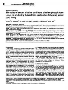
FIG. 1. Localization of ALPase in cells and extracellular vesicles of B. gingivalis 381 grown in medium A containing 0.5 ,ug of hemin per ml by using ultracytochemistry. (A) Bacterial cell. Magnification, x97,000. (B) Extracellular vesicles. Magnification, x 100,000. Details of the procedures which we used are described in the text.
in the presence of Triton X-100 were carried out by using the method of Smith et al. (30), and protein determinations in the absence of Triton X-100 were carried out by using the method of Bradford (2), with bovine serum albumin as a standard. Double immunodiffusion and Western blot analyses and preparation of water-insoluble glucan synthase (GTF-Id) and antiserum against GTF-Id were carried out as described previously (34). RESULTS Most of the ALPase activity of B. gingivalis 381 in log-phase growth was found in the cell-associated form, but 6 and 5% of the enzyme activity were found in the extracellular vesicle and extracellular soluble fractions, respectively. No significant differences in enzyme distribution were observed with the two nutrition conditions which we used (0.5 and 5 ,ug of hemin per ml). Figure 1 shows the localization of ALPase in bacterial cells and extracellular vesicles of the bacterium grown in the presence of 0.5 jig of hemin per ml. Deposition of lead phosphate was clearly confined to the outer part of the periplasmic space between the cytoplasmic membrane and the outer membrane; lead phosphate was also found inside extracellular vesicles. B. gingivalis 381 grown in the presence of 5 ,ug of hemin per ml showed the same ultrastructural localization of ALPase. Of all of the procedures described above, treatment with 1% Triton X-100 was the most effective method for solubilization of ALPase from bacterial cells. The purification of ALPase from Triton X-100 extracts is summarized in Table 1. The enzyme was purified 904-fold from Triton X-100
extracts with 5.6% recovery, and the purified enzyme had a specific activity of 160 U/mg. The purified enzyme produced a single protein band that corresponded to ALPase activity on PAGE gels, and SDS-PAGE yielded a single protein band at a molecular weight of 61,000 (Fig. 2). The enzyme gave a broad protein band that corresponded to the ALPase activity in isoelectric focusing, and the pl of the enzyme ranged from pH 5.1 to 6.1 (data not shown). The molecular weight of the enzyme was estimated to be 130,000 by high-performance liquid chromatography with TSK-gel G3000SW. Although the enzyme was completely inhibited by the addition of 0.1 mM EDTA, enzyme activity was restored when 0.2 mM Zn2+ was added, and a higher level of activity was restored when 0.2 mM Mn2+ was added. The other divalent metal ions had slight effects on the restoration of the enzyme activity. In the absence of divalent metal ions, the optimal TABLE 1. Purification of ALPase from B. gingivalis 381 Step
Total activity
Triton X-100 extract DEAE-cellulose
(U) 205 130
chromatography Hydroxylapatite chromatography Butyl-Toyoperal chromatography Affinity chromatography
Total protein
Sp act
YieldPuication
(mg) 1,160 55.0
(Um)
()(fold)
0.177 100 63.4 2.36
1 13.3
67.1
4.86
13.8
32.7
41.2
0.777
53.0
20.1 299
11.5
0.072 160
78.0
5.6 904
B. GINGIVALlS ALPase
VOL. 58, 1990
2885
B
A 1
2
3
4
5 6
7
8
1 2
3
.0
FIG. 2. (A) PAGE of the enzyme preparation at each purification step. A 32-mU portion of enzyme was applied to each lane of a 12.5% polyacrylamide gel. Lanes 1 to 4 were stained with Coomassie brilliant blue R-250, and the ALPase activities in lanes S to 8 were demonstrated as described in the text. Lanes 1 and 5, Enzyme preparation eluted from the DEAE-cellulose column; lanes 2 and 6, enzyme preparation eluted from the hydroxylapatite column; lanes 3 and 7, enzyme preparation from the Butyl-Toyopearl column; lanes 4 and 8, enzyme preparation eluted from the affinity column. (B) SDS-PAGE of the purified enzyme. Lane 2 contained the purified enzyme (1.5 ,ug), and lanes 1 and 3 contained molecular weight markers. The markers which we used were myosin (molecular weight, 200,000), P-galactosidase (116,250), phosphorylase B (92,500), bovine serum albumin (66,200), and ovalbumin (45,000).
pH of the enzyme was between pH 9.1 and 9.3. The specific activity and apparent Km for p-NPP were estimated to be 160 U/mg and 0.037 + 0.003 mM (mean + standard deviation), respectively, in 0.1 M glycine-NaOH buffer (pH 9.2). Enzyme activity was extremely accelerated when Mn2+ was added, and in the presence of 1 mM Mn2+ the optimal pH of the enzyme was between 10.1 and 10.3. The specific activity of the enzyme and Km for p-NPP were estimated to be 1,190 U/mg and 0.22 + 0.02 mM (mean standard deviation) in 0.1 M glycine-NaOH buffer (pH 10.2) containing 1 mM Mn2~ M2+. The substrate specificity of the enzyme was examined by using nine kinds of phosphate esters under two experimental conditions; the assay buffer used in one set of experiments was 0.1 M glycine-NaOH buffer (pH 9.2) with no divalent metal ions, and the buffer used in the other set of experiments was 0.1 M glycine-NaOH buffer (pH 10.2) containing 1 mM Mn2+ (Table 2). Under both conditions, p-NPP was the best substrate for the enzyme. While pyrophosphatase ±
TABLE 3. Amino acid composition of ALPase from B. gingivalis 381 Amino acid
TABLE 2. Substrate specificity of purified ALPase Assay Al Substrate
p-NPP a-Naphthylphosphate PP1 f3-Glycerophosphate Glucose 6-phosphate Pyridoxal 5'-phosphate ATP
O-Phosphoserine Casein
Assay B
Relative
Sp
act
(U/Mg)
activity
160 153 1.8 44.4 30.2 19.6 19.5 138 59.5
100 95.6 1.1 27.8 18.9 12.3
(%)b
12.2 86.2 37.2
Relative
Sp act (U/Mg)
1,190 235 0 390 148 478 191 89.3 74.4
activity
100 19.7 0 32.8 12.4 40.2 16.1 7.5 6.3
In assay A the assay mixture contained 0.1 M glycine-NaOH (pH 9.2) and no divalent metal ions; in assay B, the assay mixture contained 0.1 M glycine-NaOH (pH 10.2) and 1 mM Mn2+. Relative activity; percentages. b Relative activity compared with the activity for p-NPP. a
activity was not observed in either set of experiments, O-phosphoserine and casein were comparatively effective substrates, especially at lower pH values in the absence of divalent metal ions. The amino acid composition of the purified enzyme is shown in Table 3. The extracellular soluble ALPase produced a broad enzyme-active band having migration faster than that of the purified enzyme on PAGE gels (Fig. 3A). The ALPase in the extracellular soluble fraction was examined by Western blot analysis, using antiserum against ALPase purified from bacterial cells, and a few bands corresponding to lower molecular weights than the molecular weight of the cellassociated ALPase cross-reacted with the antiserum (Fig. 3B). An excess load of the purified ALPase (Fig. 3B, lane 1) revealed that a minor band (indicated by an arrow) with slightly faster migration than the major band cross-reacted with the antiserum.
Asp . Thr . Ser . Glu .
Mol% 10.2
Gly . Ala . Val .
6.2 8.9 8.1 9.5 8.3 6.4
Cys/2 .
0.0
2.8 Met . Ile . 4.1 10.2 Leu .
Tyr .
2.6
Lys .
4.3 5.5 3.1
Arg .
5.2 4.7
Phe . His . Pro .
2886
YAMASHITA ET AL.
INFECT. IMMUN.
A
B
A 1 2
1 2
3 4
5
B
0
N;
Ai
2
-'-
t-
3
3 4
FIG. 3. (A) PAGE of ALPase purified from bacterial cels and the soluble enzyme in the culture supernatant. A 32-mU 'portion of the enzyme was applied to each lane of a 12.5% polyacrylamide gel. Lane 1 c'ontained the purified enzyme, and lane 2 contained the extracellular soluble enzyme. After electrophoresis, the ALPase activities in both lanes were demonstrated as described in the text. (B) Western blot analysis of the ALPase purified from bacterial cells and the extracellular soluble enzyme. Lanes 1 to 3 contained purified enzyme, and lanes 4 and 5 contained the extracellular soluble enzy'me. Lane 1 contained 500 mU of enzyme, and the other lanes contained 50 mU of enzyme. The enzyme in lanes 1, 2, and 4 was detected with antiserum against ALPase purified from bacterial cells., and the enzyme in lanes 3 and 5 was detected with control semum.
Figure 4 shows the results of immunological detection of ALPase in the extracellular soluble fraction. The antiserum against the purified ALPase produced single immunoprecipitin lines with both the purified ALPase and the extracellular soluble ALPase, and the lines fused (Fig. 4A). Both of the precipitin lin'es were demonstrated by enzyme activity staining, while the precip'itin line of GTF-Id was not (Fig. 4B). DISCUSSION
High levels of ALPase activity have been observed in various periodontopathic bacteria, such as Bacteroides and Capnocytophaga species and Actinobacillus actinomycetemcomitans (14, 29). It has been reported that, in advanced cases of human periodontitis, some kinds of bacteria come into close contact with the alveolar bone surfaces and typical bone resorption occurs along the bacterial front (6). Relationships between bacterial ALPase activity and pathological changes to alveolar bone caused by bacteria are very interesting. Recently, ALPase activity in gingival fluid has been shown to be positively associated with periodontal disease activity (1). In this study, we found, by using ultracytochemistry, that
cell-associated ALPase is localized in the outer part of the
periplasmic space (Fig. 1). Because this enzyme was effectively solubilized by treatment with Triton X-100 but not by sonication or some kind of spheroplast formation procedure, the enzyme is thought to be firmly bound to a structural component within the outer part of the periplasmic area. Furthermore, in this study, we succeeded in purifying ALPase by using a modification of the affinity chromatography procedure used for mammalian intestinal-form ALPase.
5
FIG. 4. Double immunodiffusion analysis of purified ALPase and extracellular soluble enzyme. (A) Gel prior to ALPase activity detection. (B) Gel after enzyme activity was detected. ALPase activity was detected as described in the text. The center well contained antiserum against ALPase purified from bacterial cells. Outer wells 1, 2, 3, 4, and 5 contained purified ALPase, extracellular soluble enzyme, control serum, GTF-Id, and antiserum against GTF-Id, respectively.
It is interesting that bacterial ALPase possesses the same affinity as mammalian ALPase, and this fact indicates that the affinity chromatography used in this study should be useful for purification of many kinds of ALPase. The results of SDS-PAGE (Fig. 2B) and high-performance liquid chromatography analysis of the purified enzyme showed that the enzyme consists of two identical subunits having a molecular weight of about 61,000. The purified enzyme was contaminated by trace amounts of ALPase having a slightly lower molecular weight (Fig. 2A, lanes 4 and 8, and Fig. 2B). Western blot analysis revealed that the contaminant was due to the degradation product of the enzyme rather than to contamination of ALPase isozyme (Fig. 3, lane 1). Although the enzyme activity was completely inhibited by 0.1 mM EDTA, ALPase activity was restored when we added some kinds of divalent metal ions, especially Mn2" and Zn2+, and Mn2+ strikingly accelerated the enzyme activity. These findings suggest that the ALPase of B. gingivalis is a metalloenzyme containing Mn2+ or Zn2+. The Km of the enzyme for p-NPP at pH 10.2 in the presence of Mn2+ was fivefold higher than the Km at pH 9.2, and Mn2+ is unstable at alkaline pH values. Thus, the effect of Mn2+ on the enzyme activity does not necessarily reflect the physiological kinetics of the enzyme. The amino acid composition of the enzyme (see above) was similar to the amino acid compositions of other ALPases (18). While many mammalian ALPases are known to hydrolyze PPi (19), B. gingivalis ALPase did not hydrolyze PPi (Table 2). On the other hand, this enzyme hydrolyzes casein and effectively hydrolyzes O-phosphoserine, which is a common component of phosphoprotein. These findings are very important, because phosphoprotein phosphatase can destroy mineralized tissues (12). Furthermore, ALPase activity was found in the extracellular soluble fraction of log-phase cultures. Even though the activity of the extracellular enzyme is much lower than that of the cell-associated form, it is interesting to compare this result with that obtained by Cheng and Costerton (4). Western blot analysis of extracellular soluble ALPase revealed that some proteins smaller than the purified ALPase cross-reacted with the antiserum (Fig. 3). Purified ALPase was not inhibited by the presence of the antiserum against the purified enzyme in the reaction mixture (data not shown) in the same way that mammalian ALPase was not inhibited (8). ALPase activity staining of the precipitin lines (Fig. 4B)
VOL. 58, 1990
confirmed the stability of the enzyme activity against the antiserum and the immunological identity between cellassociated ALPase and extracellular soluble ALPase. Thus, B. gingivalis ALPase was released from the periplasmic space into the extracellular soluble fraction. These findings support the pathogenic role of ALPase in periodontitis.
B. GINGIVALIS ALPase
18. 19.
ACKNOWLEDGMENTS We thank Toshihiko Koga and Tatsuji Nishihara for advice concerning this study.
20.
LITERATURE CITED Binder, T. A., J. M. Goodson, and S. S. Socransky. 1987. Gingival fluid levels of acid and alkaline phosphatase. J. Periodontal Res. 22:14-19. Bradford, M. M. 1976. A rapid and sensitive method for the quantitation of microgram quantities of protein utilizing the principle of protein-dye binding. Anal. Biochem. 72:248-254. Brenna, O., M. Perreila, M. Pace, and P. G. Pietta. 1975. Affinity-chromatography purification of alkaline phosphatase from calf intestine. Biochem. J. 151:291-296. Cheng, K. J., and J. W. Costerton. 1973. Localization of alkaline phosphatase in three gram-negative rumen bacteria. J. Bacteriol. 116:424 440. Fiske, C. H., and Y. Subbarow. 1925. The colorimetric determination of phosphorus. J. Biol. Chem. 66:375-400. Frank, R. M., and J. C. Voegel. 1978. Bacterial bone resorption in advanced cases of human periodontitis. J. Periodontal Res. 13:251-261. Grenier, D., and B. C. McBride. 1987. Isolation of a membrane-
21.
1.
2.
3. 4.
5. 6.
7.
8. 9.
10. 11.
12.
13. 14.
15. 16.
17.
associated Bacteroides gingivalis glycylprolyl protease. Infect. Immun. 55:3131-3136. Harada, M., K. Fukasawa, B. Y. Hiraoka, and K. M. Fukasawa. 1984. Similarity between alkaline phosphatases from bovine dental pulp and liver. J. Dent. Res. 63:28-31. Harkness, D. R. 1968. Studies on human placental alkaline phosphatase. I. Purification and crystallization. Arch. Biochem. Biophys. 126:503-512. HeUl, L., J. Wennstrom, J. Lindhe, and S. S. Socransky. 1980. Periodontal disease in gnotobiotic rats. J. Periodontal Res. 15:405-419. Holt, S. C., J. Ebersole, J. Felton, M. Brunsvold, and K. S. Kornman. 1988. Implantation of Bacteroides gingivalis in nonhuman primates initiates progression of periodontitis. Science 239:55-57. Kreitzman, S. N., M. E. Fritz, and A. J. Saffir. 1970. Enzymatic destruction of bone in vitro. Nature (London) 228:575-576. Laemmli, U. K. 1970. Cleavage of structural proteins during the assembly of the head of bacteriophage T4. Nature (London) 227:680-685. Laughon, B. E., S. A. Syed, and W. J. Loesche. 1982. API ZYM system for identification of Bacteroides spp., Capnocytophaga spp., and spirochetes of oral origin. J. Clin. Microbiol. 15:97102. Malamy, M. H., and B. L. Horecker. 1964. Release of alkaline phosphatase from cells of Escherichia coli upon lysozyme spheroplast formation. Biochemistry 3:1889-1893. Mayahara, H., H. Hirano, T. Saito, and K. Ogawa. 1967. The new lead citrate method for the ultracytochemical demonstration of activity of non-specific alkaline phosphatase (orthophosphoric monoester phosphohydrolase). Histochemie 11:88-96. Mayrand, D., and B. C. McBride. 1980. Ecological relationships
22. 23. 24.
25. 26.
27. 28. 29. 30.
31.
32. 33. 34.
35. 36.
2887
of bacteria involved in a simple, mixed anaerobic infection. Infect. Immun. 27:44-50. McComb, R. B., G. N. Bowers, Jr., and S. Posen. 1979. Structural features, p. 189-228. In Alkaline phosphatase. Plenum Publishing Corp., New York. McComb, R. B., G. N. Bowers, Jr., and S. Posen. 1979. Isoenzymes, p. 373-524. In Alkaline phosphatase. Plenum Publishing Corp., New York. Minhas, T., and J. Greenman. 1989. Production of cell-bound and vesicle-associated trypsin-like protease, alkaline phosphatase and N-acetyl-3-glucosaminidase by Bacteroides gingivalis strain W50. J. Gen. Microbiol. 135:557-564. Moore, W. E. C., L. V. Holdeman, R. M. Smibert, D. E. Hash, J. A. Burmeister, and R. R. Ranney. 1982. Bacteriology of severe periodontitis in young adult humans. Infect. Immun. 38:1137-1148. Neu, H. C., and L. A. Heppel. 1965. The release of enzymes from Escherichia coli by osmotic shock and during the formation of spheroplasts. J. Biol. Chem. 240:3685-3692. Newman, M. G., and S. S. Socransky. 1977. Predominant cultivable microbiota in periodontosis. J. Periodontal Res. 12: 120-128. Nilsson, T., J. Carlsson, and G. Sundqvist. 1985. Inactivation of key factors of the plasma proteinase cascade systems by Bacteroides gingivalis. Infect. Immun. 50:467-471. Noguchi, T., and Y. Yamashita. 1987. The rabbit differs from other mammals in the tissue distribution of alkaline phosphatase isoenzymes. Biochem. Biophys. Res. Commun. 143:15-19. Okuda, K., A. Yamamoto, Y. Naito, I. Takazoe, J. Slots, and R. J. Genco. 1986. Purification and properties of hemagglutinin from culture supernatant of Bacteroides gingivalis. Infect. Immun. 54:659-665. Ono, M., K. Okuda, and I. Takazoe. 1987. Purification and characterization of a thiol-protease from Bacteroides gingivalis strain 381. Oral Microbiol. Immunol. 2:77-81. Slots, J. 1979. Subgingival microflora and periodontal disease. J. Clin. Periodontol. 6:351-382. Slots, J. 1981. Enzymatic characterization of some oral and nonoral gram-negative bacteria with the API ZYM system. J. Clin. Microbiol. 14:288-294. Smith, P. K., R. I. Krohn, G. T. Hermanson, A. K. Mallia, F. H. Gartner, M. D. Provenzano, E. K. Fujimoto, N. M. Goeke, B. J. Olson, and D. C. Klenk. 1985. Measurement of protein using bicinchoninic acid. Anal. Biochem. 150:76-85. Spiegel, C. A., S. E. Hayduk, G. E. Minah, and G. N. Krywolap. 1979. Black-pigmented Bacteroides from clinically characterized periodontal sites. J. Periodontal Res. 14:376-382. Tanner, A. C. R., C. Haffer, G. T. Bratthall, R. A. Visconti, and S. S. Socransky. 1979. A study of the bacteria associated with advancing periodontitis in man. J. Clin. Periodontol. 6:278-307. Tsutsui, H., T. Kinouchi, Y. Wakano, and Y. Ohnishi. 1987. Purification and characterization of a protease from Bacteroides gingivalis 381. Infect. Immun. 55:420427. Van Steenbergen, T. J. M., P. Kastelein, J. J. A. Touw, and J. De Graaif. 1982. Virulence of black-pigmented Bacteroides strains from periodontal pockets and other sites in experimentally induced skin lesions in mice. J. Periodontal Res. 17:41-49. Yamashita, Y., N. Hanada, and T. Takehara. 1989. Purification of a fourth glucosyltransferase from Streptococcus sobrinus. J. Bacteriol. 171:6265-6270. Zambon, J. J., H. S. Reynolds, and J. Slots. 1981. Blackpigmented Bacteroides spp. in the human oral cavity. Infect. Immun. 32:198-203.