Oct 12, 2007 - Pyelonephritis Presenting with Severe Acute Renal Failure. Sriram Krishnamurthy, Pankaj Hari, Smriti Hari1, Alok Sharma2 and Arvind Bagga.
Clinical Brief
Pyelonephritis Presenting with Severe Acute Renal Failure Sriram Krishnamurthy, Pankaj Hari, Smriti Hari1, Alok Sharma2 and Arvind Bagga Departments of Pediatrics, 1Radiodiagnosis, 2Pathology, All India Institute of Medical Sciences, New Delhi
ABSTRACT We describe an unusual case of an 8- yr -old child presenting with low grade fever and acute renal failure. Investigations showed blood urea 246 mg/dl, serum creatinine 6.4 mg/dl, microscopic hematuria and 2+ proteinuria. Renal biopsy was done in view of rapidly worsening kidney function and showed dense lymphoplasmacytic infiltrate and neutrophils with focal areas of interstitial necrosis, confirming acute pyelonephritis. Ultrasonography and MRI demonstrated multiple renal abscesses. He was managed with antimicrobial therapy and hemodialysis. [Indian J Pediatr 2008; 75 (9) : 961-963] E-mail: pankajhari@ hotmail.com Key words : Pyelonephritis; Acute renal failure; Abscess
Acute bacterial pyelonephritis is rarely considered as a cause of acute renal failure (ARF) in a previously healthy child.1, 2 The effects of uncomplicated pyelonephritis on renal function are generally mild and limited to transient loss of concentrating ability.3 When ARF occurs due to bacterial pyelonephritis, this possibility is often not considered, and such patients may present diagnostic dilemmas. To the best of our knowledge, only 5 cases of ARF due to pyelonephritis have been described in children in the literature.1, 2,4-6 We describe an 8- yr- old child presenting with ARF who was found to have histological and radiological evidence of acute pyelonephritis, which was complicated by abscess formation. CASE REPORT
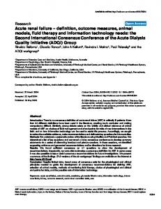
Investigations showed a hemoglobin level of 7.6 g/dl, leukocyte count 16x10 9 /L, 63% polymorphs, 32% lymphocytes, 1% monocytes and 4% eosinophils; the platelet count was 310x10 9/L. The peripheral smear showed macrocytic anemia. Blood urea was 246 mg/dl, creatinine 6.4 mg/dl, sodium 141 mEq/L, potassium 6 mEq/L, cholesterol 197 mg/dl, albumin 4 g/dl, calcium 9.9 mg/dl, phosphate 4 mg/dl, pH 7.35, bicarbonate 19.9 mEq/L. Urinalysis showed 3-4 white blood cells and 5-6 red blood cells/HPF with no casts, proteinuria 2+; urine culture was sterile. C3 level was 96 mg/dl. Blood culture grew Escherichia coli at 48 hours. Renal ultrasonography showed enlargement of both kidneys, with no evidence of hydronephrosis; urinary bladder was normal and post void residue minimal. A renal biopsy, done in view of rapidly worsening kidney function, showed a dense lymphoplasmacytic infiltrate and neutrophils with focal areas of necrosis in the interstitium (Fig. 1a and 1b).
An 8- yr- old boy presented with complaints of low grade fever, malaise and decreased appetite for 15 days and decreased urine output for 2 days. There was no history of frequency, dysuria, flank pain, rash, pyoderma or hematuria. There was no history of consumption of drugs and no past history of urinary tract infections. On examination, he was pale with periorbital and pedal edema. There was no icterus, lymphadenopathy or rash. Blood pressure was 130/88 mm Hg (more than 95th centile for age). Chest, cardiovascular and abdominal examinations were normal.
Correspondence and Reprint requests : Dr. Pankaj Hari, Associate Professor, Department of Pediatrics, All India Institute of Medical Sciences, New Delhi. [Received October 12, 2007; Accepted February 18, 2008]
Indian Journal of Pediatrics, Volume 75—September, 2008
Fig 1(a). Photomicrograph of renal biopsy showing marked interstitial inflammation. A dilated tubule containing a red cell case is visible. (Hematoxylin & Eosin X200)
961
S. Krishnamurthy et al
Fig 1(b): Renal biopsy showing an epitheloid cell granuloma and surrounding dense lymphoplasmacytic infiltrate. (silver methenamine X 200)
Staining for acid fast bacilli was negative. Glomeruli were normal. The renal histology was suggestive of acute pyelonephritis. Repeat ultrasonography 2 weeks later, showed persistently enlarged kidneys with loss of corticomedullary differentiation. Two hypoechoic focal lesions (1.6 cm and 1.2 cm diameter) at the inferior pole of the right kidney and an anechoic lesion (8 mm) were seen at the superior pole of the left kidney. A gadolinium contrast MRI showed 3 lesions in the right and 2 in the left kidney; these were nodular with size ranging from 0.8-2 cm in size, suggestive of multiple abscesses (Fig.2a and 2b). Serological tests for HIV, and hepatitis B and C were negative. Immunoglobulin G, A and M levels and nitroblue tetrazolium (NBT) test were normal.
Fig 2(b).Contrast enhanced T1W axial image shows peripheral rim enhancement after contrast administration. The MR features are suggestive of abscesses.
underwent repeated sessions of haemodialysis following which the urine output normalised and serum creatinine decreased to 3.5 mg/dl. Hypertension was controlled on amlodipine. He became afebrile and was discharged on oral antibiotics. After 2 weeks of discharge from the hospital, ultrasonography showed 2 small renal abscesses in the inferior pole of the right kidney and a small renal abscess in the inferior pole of the left kidney. Aspiration of the abscesses was done; culture of the aspirate was sterile. 4 weeks later, a DMSA renal scan showed bilateral scarred kidneys with impaired function; MCU was normal. 4 months after discharge from the hospital, blood urea and serum creatinine levels were 47mg/dl and 1.57 mg/dl respectively. DISCUSSION
Fig 2(a). Fat suppressed T2W axial image through the kidneys showing well defined, T2 hyperintense lesions (arrows) in both kidneys. Mildly dilated left superior calyx is also noted (star).
Pyelonephritis is an unusual cause of ARF requiring dialysis in children with an anatomically normal urinary tract and no other predisposing conditions.1 ARF due to acute pyelonephritis has been reported in adults, and is often associated with pregnancy, solitary kidney and indwelling catheters. 3,7-9 The incidence of pyelonephritis in adult patients with ARF has been shown to be 2-3% in one study ,10 while another has reported it as 0.7%.11 To the best of our knowledge, only 5 cases of ARF due to pyelonephritis have been described in children in the literature, 1, 2,4-6 and that too, not in recent years. The present report serves as a timely reminder that acute pyelonephritis in children can lead to ARF.
The patient was treated initially with intravenous ceftriaxone and later ciprofloxacin. On Day 2 of admission, serum creatinine increased to 7.5 mg/dl. He
Most of the cases of ARF with pyelonephritis described in literature are due to Escherichia coli.1,7-9 Pyelonephritis has been reported in children with obstructing urethral diverticulum5 and solitary kidney.6 Others 1, 2, 4 however, have reported patients with normal urinary tract
962
Indian Journal of Pediatrics, Volume 75—September, 2008
Pyelonephritis Presenting with Severe Acute Renal Failure anatomy. Hemodialysis1, 6 and peritoneal dialysis 2, 4, 5 have been used for management of these cases. The clinical outcomes vary from complete recovery1, 5 to residual renal injury 2, 6 or death 4. Disruption of tubular function by interstitial infiltrates of neutrophils and phagocytes, interstitial edema, tubular obstruction by cellular debris and intrarenal vasoconstriction are suggested as possible etiologies for the occurrence of ARF in pyelonephritis.1,7 Although the present case had fever, diagnosis of pyelonephritis was not considered initially, due to absence of urinary symptoms and sterile urine culture. The presence of azotemia, oliguria, microscopic hematuria, 2+ proteinuria and hypertension prompted us to perform a kidney biopsy, considering rapidly progressive glomerulonephritis (RPGN) as a possibility. Since acute non-obstructive pyelonephritis may result in severe reversible renal failure, this diagnosis must be considered in patients presenting with acute uremia. Prompt diagnosis and intervention may avoid residual renal injury. Although uncomplicated pyelonephritis is a clinical diagnosis that does not require imaging, the clinical features may occasionally be equivocal. While renal ultrasonography could detect few abscesses, MRI detected additional lesions and confirmed the diagnosis of pyelonephritis with abscess formation. MRI has been shown to be superior to conventional procedures such as ultrasonography and nuclear scintigraphy for the diagnosis of pyelonephritis.12 Caution must be exercised when utilizing gadolinium as a contrast agent in patients with kidney disease.13 Recently, gadolinium has been associated with both renal and extra-renal complications such as ARF, pancreatitis, and nephrogenic systemic fibrosis (NSF) in patients with renal failure. Although cause and effect have not been proven for the NSF-gadolinium link, the impaired renal elimination of gadolinium in patients with kidney disease is suggested to result in this fibrosing disorder.
Indian Journal of Pediatrics, Volume 75—September, 2008
Key Messages Although rare, acute pyelonephritis may cause severe acute renal failure. REFERENCES 1. Turner ME, Weinstein J, Kher K. Acute renal failure secondary to pyelonephritis. Pediatrics 1996; 97 : 742-743. 2. Greenhill AH, Norman ME, Cornfeld D, Chatten J, Buck B, Witzleben CL. Acute renal failure secondary to acute pyelonephritis. Clin Nephrol 1977; 8 : 400-403. 3. Nahar A, Akom M, Hanes D, Briglia A, Drachenberg CB, Weinman EJ. Pyelonephritis and acute renal failure. Am J Med Sci 2004; 328 : 121-123. 4. Söylemezoðlu O, Kale G, Saatçi U, Akçaören Z. Acute renal failure due to acute pyelonephritis. Int Urol Nephrol 1995; 27: 137-139. 5. Lorentz WB, Iskandar S, Browning MC, Reynolds GD. Acute renal failure due to pyelonephritis. Nephron 1990; 54 : 256-258. 6. Noah M, Schoeneman MJ, Haycock G, Levitt SB, Bennett B, Greifer I. Xanthogranulomatous pyelonephritis-presentation as acute renal failure in a child with a solitary kidney. Nephron 1978; 21 : 161-164. 7. Weinstein T, Zevin D, Gafter U, Chagnac A, Levi J. Acute renal failure in a solitary kidney due to bacterial pyelonephritis. J Urol 1986; 136 : 1290-1291. 8. Thompson C, Verani R, Evanoff G, Weinman E. Suppurative bacterial pyelonephritis as a cause of acute renal failure. Am J Kidney Dis 1986; 8 : 271-273. 9. Creyghton WM, Lobatto S,Weening JJ. Acute renal failure caused by Klebsiella pneumoniae pyelonephritis. Clin Nephrol 2001; 56 : 391-393 . 10. Baker LR, Cattell WR, Fry IK, Mallinson WJ. Acute renal failure due to bacterial pyelonephritis. Q J Med 1979; 48 : 603612. 11. Al-Rohani M. Renal failure in Yemen. Transplant Proc 2004; 36: 1777-1779. 12. Kirsch AJ, Grattan-Smith JD, Molitierno JA. The role of magnetic resonance imaging in pediatric urology. Curr Opin Urol 2006; 16 : 283-290. 13. Perazella MA, Rodby RA. Gadolinium use in patients with kidney disease: a cause for concern. Semin Dial 2007; 20 : 179185.
963