Quantum entanglement of K+ ions, multiple channel states and the role of noise in the brain Gustav Bernroider*a and Sisir Roy b a FB Organismic Biology, Neurodynamics and Neurosignaling Unit, University of Salzburg, A-5020 Salzburg, Austria b Physics and Applied Mathematics Unit, Indian Statistical Institute, Kolkatta –700 035, India ABSTRACT We propose a quantum information scheme that builds on the interference properties of entangled ion states that are transiently confined by local potentials within the permeation path of voltage-gated, ion-conducting membrane proteins. We show, that the sub-molecular organization of parts of the protein, as revealed by the recent progress in highresolution atomic-level spectroscopy and accompaning molecular dynamics simulations, carries a logical coding potency that goes beyond the pure catalytic function of the channel, subserving the transmembrane crossing of an electrodiffusive barrier. As we argue that ‘within channel states’ can become super-correlated with the environment , this also sheds new light on the role of noise in controlling the access of ions to voltage-gated ion channels (‘channel noise’). Key words: entanglement, ion channels , quantum computing, channel noise, neural coding, quantum neurophysics, molecular dynamics simulations, Fluid-Mosaic Model, Liquid-Crystal-Model, natural computing, neural quantum computing.
1. INTRODUCTION Although the classical view of the brain as a computational device has been very successful in explaining the major physical aspects behind neural information processes and has encouraged the development of complex artificial networks, the phenomenal aspects behind information in the brain, i.e ‘computing’ the meaning of a message, is not understood. In the neuralistic view complexity and quantity substitute for ‘meaning’. Albeit of historical dimension (e.g. the Chomsky-Shannon debate on the statistical structure of language as discussed by W. Bialek 1 ), such an assumption has never been proven and seems to be incompatible with emerging insight into evolutionary and energetical constraints governing ‘natural computing’ in biological organisms 2,3,4. Beside the ability to adaptively reconfigure in response to novelty, all nervous systems (NS), ranging from one cell type systems of sea anemones to all higher level NS engaging sensory, intermediate and motory cell types 5, are capable of elaborating some degree of self-cognizance, i.e. reflecting upon their own experience in the light of previous responses 6. This involves varying degrees of ‘perceptual consciousness’ or ‘body-ness’, where the role of a classical computation behind these abilities is completely obscure. The explanatory value of the usual association of the integrate and fire status of neurons with the stimulus that triggers this status, as seen from the view of an external observer, has reached it’s limitation. What would be required is a closed description of observation processes 7, i.e. a description that figures and includes the role of an observer. This includes the decisive question on how the brain gains understanding about it’s own internal states (‘conscious experience’). Furthermore, the composed neural system is surprisingly resistant to a description in terms of it’s constituent parts and in sensory systems, possible computations seem to run on all input states at the same time. Taken together, there is a whole list of arguments that call in question the competence of the classical engineering approach behind information theory in explaining the decisive secrets of the brain. •
[email protected]; ++43-662-8044-5604,
[email protected]
Despite the limitation and possible inadequacy of classical concepts in brain science have been expressed more than a decade ago 8,9 , alternatives have not been established in agreement. However, several steps have been made along this line. Striking formal analogies of cognition models with the operator algebra known in quantum mechanics (QM) have been suggested 7, 10, 11. Neural state and propagation models have been successfully modeled using complex wave functions similar to QM postulates 12 and some quite specific candidate realizations employing quantum computation have been forwarded, such as the Penrose & Hameroff microtubule protein hypothesis 13, neural electron spin networks14 and ion trapping within voltage-gated ion channels by one of the present authors 15. At least three challenging questions have become pressing, that need to be plausibly answered should quantum concepts in brain science become generally accepted: (i) Can we identify the unique quantum physical properties such as within system and between system superpositions (entanglement) of some brain-states and (ii) if so, are these states indispensable for the description of some essential features of brain function? Finally (iii), how would these quantum phenomena relate to the classical signals that (at least) correlate with higher level brain functions ? Answers to these questions have been subject of a series of controversial contributions 16,17 and remain open at present. Now, with the recent progress in high-resolution atomic-level spectroscopy of channel forming proteins 18,19 and the accompaning molecular dynamics simulations 20, the identification of quantum states relating to brain signaling has become feasible. We have previously given a summary of candidate states mediating a quantum-classical correspondence in the brain based on these findings 21. Here, we propose a quantum information scheme that builds on the interference properties of entangled ion states that are transiently confined by local potentials within the permeation path of ion-conducting integral membrane proteins. We argue that the underlaying quantum-type interaction has a ‘coding-potency’ as such, something that goes beyond the pure constructing mechanism leading to classical brain signals (action potentials).
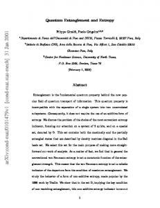
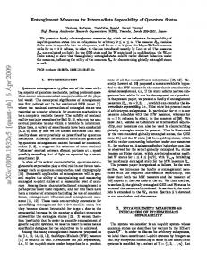
2. ION-CHANNEL CONFIGURATIONS The classical determinant of communication and computation in the brain is provided by the organization and cooperation of ion conducting proteins, inserted into the lipid double layer of the neural plasma membrane. For a membrane transiently shifted from thermodynamic equilibrium (by energy consuming charge separation across the lipid layer) and a given membrane capacitance, the coupled dynamics of these ‘channel proteins’ and voltage is described by a set of equations of motions as originally suggested by Hodgkin and Huxley 1952 22. Congruent with the Singer and Nicolson fluid-mosaic model of the membrane (FMM) 23, these equations of motion provide the basis of electrical excitability and signaling in the nervous system. HH-type non-linear dynamics selects a propagating pulse (the action potential) at the cellular level. In the electric circuit equivalent the inserted membrane proteins thereby play the role of field-effect transistors, with a voltage imposed across the cell membrane ‘gating’ the transfer of ion bound charges through the membrane. Two different aspects characterize channel function: ion-selective permeation and ‘gating’, i.e. control of access of ions to the permeation pathway 24. We will base the subsequent concept on potassium channels, employing the crystal structure of the KcsA and KvAP channels at a resolution ranging from 1.9x 10⁻¹⁰ m to 3.2 x 10⁻¹⁰ m, as revealed by the work of MacKinnon’s group. The channel structure is basically conserved among all potassium channels 25 with some differences relating to gating characteristics rather than ionic selectivity 26. In the open gate configuration the protein selects the permeation of K+ ions against other ions in the ‘selectivity filter‘and can still allow ion permeation rates near the diffusion limit 27. In the view of HH-type models of membrane potentials , K+ permeation stabilizes the membrane potential, resetting it from firing threshold values to resting conditions 28. The atomic level reconstruction of parts of the channel and accompanying molecular dynamics simulations (MD) at the 10 –12 sec resolution 29 have changed the picture behind ion permeation: the channel protein can transiently stabilize three K+ states, two within the permeation pathway and one within the ‘water-cavity’ located towards the intracellular side of the permeation path. The overall state structure can be summarized as seen in the following Figure 1.
Inside
water-cavity
Gate Closed
permeation path (a)
W4 -- K+ -- W4 (b)
Open
W 4 − K+ − W4
outside
O4 − K+ − O4 W
O4 − K+ − O4 W O4
4
2
O4
3 W
1
0
O 4 – K + − O 4 W O 4 – K+ − O 4
W4 − K+ − O4K+ − W –
K+ O 4 –
W –
K+O4 – W
Figure 1: K+ channel state configurations, based on the crystallographic X-ray structure from MacKinnon. The channel is shown in the three ion mode, consistent with it’s proposed ‘functional state’ 26, 30 and is schematically aligned along one (z) axis (longitudinal section of the protein core), with the cells interior (cytoplasmatic) site to the left. The gate states are either closed (above) or open (below). Ions are coordinated by their Coloumb interaction with either carbonyl derived oxygens in the permeation path (Oi − K+) or water (W) - ion interaction (Wi – K+). The subscript i denotes the ‘in-plane’ number, along the perpendicular x,y plane of molecules (water) or atoms (oxygens) coordinating the ion. The interior of the permeation pore is delineated and the Coloumb domains giving rise to 8-fold coordination states under the closed gate conditions (a,b) are shaded. Note: with double ion-occupancy of the permeation path , there are two ‘confined’ ion configurations in the closed gate state. Using the originally proposed z-coordinate number 31 (numbers 0-4), these are the configurations 2,4 (a) and 1,3 (b). Only short range electrostatic interaction terms are given, leaving out i) water-damped dipole interactions from surrounding short pore helices in the water cavity and ii) side chain interaction terms from the ionizable residues near the selectivity filter 32 .
The essential feature behind the channel structure relating to the present work is provided by the closed gate, low-K+ state of the permeation pore of the protein, to which the original crystallographic image refers to (a,b in Figure 1). Whereas in the open gate state, with the cavity exposed to the high intracellular K+ concentration, the interior of the protein represents an almost barrierless pathway for selected ion flow, the closed gate state represents a stable ionprotein conformation. In this state two ions can become transiently ‘trapped’ in the selectivity filter by confining potentials in the 1,3 and 2,4 configuration. Depending on the total ‘loading state’ of the channel, i.e. whether there are 1,2 or 3 ions present and depending on the filter radius and imposed electrical field strength, the confining energy scales between 10 kT and 60 kT at 300K 30,33. In Figure 1 the ‘closed’ three ion configuration may already reflect a transition state between the ‘closed’ to the ‘open’ configuration by the presence of a fully hydrated K+ ion (W4 − K+ − W4) in the cavity. When this ion becomes partially dehydrated , carbonyl oxygens at position 4 of the filter compensate for dehydration energies of the ion, taking this ion into a (W4− K+ − O4) configuration. The resulting Coulomb interaction with the innermost filter-ion (corresponding to a field strength of up to 9x10⁹ Vm-1) 30 destabilizes the carbonyl binding pockets within the filter configuration, eliminating the ‘locality’ of energy profiles and finally leading to the extrusion of the outermost ion from the channel. The ability of the channel protein to provide an optimal ‘bulk-like solvation environment’ for one ion species (e.g K+) compared to other ions (e.g. Na+) during the cavity-filter transition seems to be the essential mechanism behind ‘selective permeation’. Also the view on gating, the second aspect characterizing channel dynamics, has to be changed with respect to the recently analysed high-resolution structure 34,35. In voltage controlled channels, changes in the imposed membrane
field induce a charge transfer transition (12-14 electron charges in K+ channels), carried by amino acids called voltage sensors 36. The transition moment in gating correlates with the open probability of the channel. In the conventional model the four helical screws carrying the ‘sensing charges’ move quite independently from other parts of the protein through ‘gating pores’, whereas in the recent ‘paddle-model’ the transition moment couples with the neighbouring lipids of the channel protein that in turn changes the gating state of the channel. This latter view, as suggested by MacKinnon’s group, gains strong recent support from an increasing number of studies demonstrating that protein surrounding lipids engage in the control of voltage gated Kv channels 37. As the main source of noise in neurons has traditionally been attributed to the probabilistic nature of gating, a reconsideration of channel noise in the new context of atomic resolution dynamics seems to become unavoidable. The conventional FMM type ‘isolated-dipole field-response’ and ‘permeation’ feedback relationship that gave rise to the successful description of action potentials in the squid axon, should become gradually extended into a liquid-crystal type ‘charge-transfer’ model, involving higher moments, e.g. electric quadrupole moments, for the description of channel responses to electrical field changes across the membrane. To what extent this type of membrane model might host computational abilites instantiated at the atomic level, as suggested previously 13,14,15, 38,39 will be addressed in the following chapter.
3. ION CHANNELS AS ARRAYS OF MICROSCOPIC ION TRAPS The present view on ion-channel-membrane configurations allows the identifcation of state-dependent interaction terms between ions and membrane components that could establish a resource for mappings between different subsystems engaged in ion-conduction across the membrane. Together with the identification of ‘logical states’ encoded by atoms or molecules, the identification of conditional dynamics between these sub-systems seems to be a prerequisite to attribute ‘computational’ properties to the physics of membrane signaling at the atomic scale. This view implies a strong analogy with two-level logical gate operations in quantum computing as proposed by DiVincenzo, Cirac and Zoller 40, 41. However, intentional aspects are on opposite course: whereas in quantum computing the challenge is to design operating conditions that implement pregiven computational tasks 46, 47, the main intention behind the search for neuro-molecular computing is to identify unique logical states that reflect the ‘closed observational’ states that characterize the brain. It may finally turn out that these physical states are quite different from artificial quantum computational structures and their logical composition may not be based on the universal set (‘and, or, not and idendity’) that characterizes all Turing type computations. Here we identify some key electric properties of channel proteins based on the above summary of experimental findings and relate these properties to previously suggested quantum-computation schemes, e.g. as reviewed by C. Monroe42 or Milburn et al 43 . In particular, we will draw comparisons to the Cirac & Zoller ion trap architecture, based on electrostatic interactions between ions transiently kept in arrays of microscopic traps 41, 44, 45.
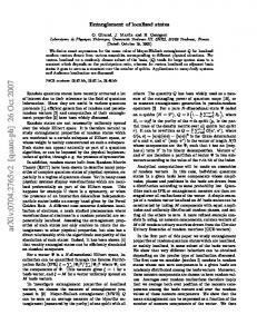
3.1 Operational structures The frame is set by the question how dipoles in the interior of a molecule giving rise to ‘local fields’ relate to the electric field applied at the surface of a membrane inserted protein. As available from the 3-ion-closed gate state of KvA channels in Figure 1 (a,b), two K+ ions are transiently trapped in the permeation path of the protein if the channel gate is closed. The two ions are trapped within the carbonyl binding pockets via their eight-coordinated oxygen electrostatic interaction. The situation is depicted in an idealized outline in Figure 2. In this figure the ion-oxygen coordination structure within the filter region along a longitudinal extension of 1.2 nm is given. The ‘filter’ accounts for roughly 1/6 of the total trans-membrane length of the channel protein. Congruent with the crystallographic K+ electron density profiles 31, adjacent K+ locations are separated by ∆S ∼ 0.3 nm in their S1-S3 and S2-S4 configurations. In Figure 2 the ion diameter of 0.27 nm is graphically reduced, compared to the (K+ - O) coordination distances ranging between 0.27(short) to 0.308 nm (long) 29. The energy depth provided by the confinement potentials V along the z-axis of the filter is in the order of 10 kT (300K) and approximated by harmonic potentials, assuming a simplified isotropic and homogeneous structure. Both ions interact by electrostatic repulsion and are exposed to a homogeneous external transmembrane field E of magnitude 107 V/m giving rise to a ∆V ∼ 100mV. Results from Brownian Dynamics Simulations (BD) provide measures about the dynamics of local electric potential profiles confining the ions 33 and are similar to those found in Poisson-Boltzmann solutions 48. The potentials inside the pore are found to fluctuate due to charges on the surrounding protein and the presence of ions in the range of 100mV amplitudes. MD simulations at the 10-12 sec
timescale reveal transition times between the S1 → S2 ion states of 6x10-11 sec 29. Thus in the low-K+ condition and closed channel gate, the ions may oscillate between the S1-S3 and S2-S4 locations many times, as originally proposed by Morais-Cabral 31. As gate changes occur at the 10-3 sec scale, we can expect 1/6 x 108 transitions of the trapping potential before the channel switches to it’s open state (which is equivalent to a trapping frequency in the order of 10-100 GHz).
Axial position in nm 1.2
K+
O
1.2
S0
0.9
0.9
S1 0.6
0.6
S2 S3
0.3
∆d ∆S
0.3
S4 100 mV
0
0
1,3
2,4
Potential (V)
100mV Potential (V)
Figure 2: Ion trapping within the selectivity filter of K+ channels. The filter region of the channel is shown as five sets of four carbonyl oxygens with the K+ ion bound by eight-oxygen coordinated, electrostatic interactions. Site S0 is outside and site S4 towards the intracellular domain of the channel. The confining potentials are modelled as homogeneous harmonics to the left (relating to the 1,3) and to the right side (relating to the 2,4 configurati on). The filter is hosting 2 K+ ions (grey spheres) and 2 intervening water molecules. During the closed gate state ions can be modelled as being confined to two moving potentials, shifted along the z-axis by distance ∆d. Measures on the potential, ion distances and temporal characterisitics are given in the text.
3.2 Energetical considerations According to the ‘energetic optimization hypothesis of ion channels’ 31 , stating that ‘conspecific’ ions in the channel are energetically balanced, whereas ‘heterospecific ions’ such as Rb+ ions in a K+ channel, are energetically imbalanced, the states of the joint two K+ ion configurations (1,3) and (2,4) may be assumed to be degenerate, i.e. their energy difference ∆E = 0. If we are interested in finding the system in one of these states as a function of time, we can apply time dependent perturbation concepts, in particular Rabis formula, which allows us to estimate the probability of occupying e.g. state (2,4) = P2 , if a constant perturbation is swiched on at t = 0 and the system sets out from state 1 (1,3 configuration): P2 = |a2| 2 = [(4V2)/(ω02 + 4V2)] . sin2 ½ (ω02 + 4V2)1/2 t
(1)
For the degenerate case ∆E = 0, we also have ω0 = 0 and (1) simplifies to P2 = sin2 Vt. This predicts a complete state transition for degenerate systems, with P → 1 after a time t = π /2V. As we know the time t from MD simulations (chapter 2.1) to scale along 6.10-11 sec, we can estimate the perturbation strength V ≈ π/2 . 1011 s-1 . So the ‘trapping frequencies’ above can be recovered from the view of time-dependent perturbation theory. This sheds new light on the meaning of ion selection by channel proteins. Non-selected ions (e.g. Na+ in K+ channels), leading to a separation of energy levels between the preferred filter positions, would still allow state oscillations to occur , but with maximum levels for P in the range of 4V2/ω02 < 1. Thus the effect of the external membrane field, perturbing the local fields within the selectivity pore of the channel, resides within the relative off-set between the energy separation of ion-channel states and the strength of the external field. In this way it is guaranteed that in the co-evolved ion-channel configuration, even very week perturbations caused by external field variations can drive the system completely between the degenerate ionchannel coordination states. In addition, these findings suggest that within the selectivity filter, under the nonconducting, low ion condition and under the influence of a constant perturbation, pure ion states can evolve that are delicately balanced with the ion, channel and overall electric membrane characteristics at a given time. The ‘trapped’ ion states under the non-conducting closed gate state seem to be particularly important
4. CORRELATIONS AND LOGICAL STATES 4.1 Quantum interactions The physical action behind ion permeation through protein pores requires action orders at the quantum scale. We have previously estimated the action distance between ion states in the K+ channel to scale along ≈ π. 10-33 Lagrangian units (L). A propagating action potential would require 1.8 x 10⁻¹⁵ L . The brain actually spans at least 20 orders of magnitudes of physical action with physiologically significant signal properties 21. Table 1 below provides a summary of relevant action orders in the brain. Source/Action
Mechanical Units (A = M.L2. T-1) (kg m2 s-1)
Energy/Time (A = E. T)
Electrical Units (A = C.V.T)
Cell / Action Potential
1,8 x 10⁻¹⁵ L (Js) (1)
6. 10-16 L (Js) (2)
2,1 . 10-16 L (Js) (2)
Molecular / Ion permeation
0,48 . 10-34 L (Js)
6,95 x 10⁻³³ L (Js) (3)
7 x 10-33 L (Js) (3)
Table 1: Action orders in the brain, 1 time at the scale of spiking events (10-3) sec, 2 time T to be close to 10-8 sec for conduction rates of ions, 3 time T to scale at 10-12 sec , the min transition time for K+ ions in the permeation path (from Bernroider & Roy 21) At the Planck scale of ≈ 10-34 L, a pre-decohered quantum system hosts all information needed for the description of the system in a linear superposition of it’s basis states. Decoherence of superposition states establishes a qualitatively new interaction term between the subsystems under consideration (e.g. a system and it’s environment). The question holds whether this ‘new’ interaction term is of any significance for i) within-system correlations of ion channel states and/or ii) between system correlations (entanglement) of ion-channel states. From a physical point of view, all short length Colombic interaction terms as seen in Fig.1 and Fig.2 should also carry quantum mechanical effects, because near or overlapping electronic wavefunctions would for example be subject to Pauli’s exclusion principle. Quantum concepts are also contained in the ab initio MD simulations, if one intends to trace the motion of individual ions through channel proteins involving the electronic degrees of freedom 20,29. Here we want to briefly touch upon the question whether we can associate ‘logic states’ to the model emerging for atomic-scale ion channel states.
One way to envisage a superpositon of ion states in the permeation filter of channels is to ‘conceptually’ close the filter from it’s environment and describe the state of K+ ions by a spatial superposition according: ψ〉 = k1,3〉 〈 k1,3ψ〉 + k2,4〉 〈 k2,4ψ〉
(2)
where Ψ〉 denotes the ions state vector for the low-K+ structure and ki,j〉 the ket-vector of either (i=1,j=3), or ( i =2, j=4) ion locations according to the outline in Fig. 2. If the expansion (2) is written using complex coefficients α, β for the components