Jul 19, 1989 - No good match with the E.coli d7° promoter consensus sequence is ..... that has a cat cassette inserted into the NruI site in recO. pNC 105 ...
The EMBO Journal vol.8 no. 1 1 pp. 3401 - 3407, 1 989
Autoregulation of RNase
III
operon
James C.A.Bardwell, Philippe Regnier', Su-Min Chen, Yoshikazu Nakamura2, Marianne Grunberg-Manago1 and Donald L.Court Laboratory of Chromosome Biology, BRI-Basic Research Program, National Cancer Institute, Frederick Cancer Research Facility, Frederick, MD 21701, USA, 'Institut de Biologie Physico-Chimique, 13 rue Pierre et Marie Curie, 75005 Paris, France and 2Department of Tumor Biology, The Institute of Medical Science, University of Tokyo, Tokyo 108, Japan
Communicated by M.Grunberg-Manago
RNase III has been implicated in the control of gene expression by the processing of mRNA. We have found that the mc operon is autoregulated; rnc- mutant strains oversynthesize the operon's mRNA and protein products. A site in the 5'-noncoding region of the operon's message is cleaved by RNase III. This sitespecific cleavage appears to be the initial step in the functional inactivation of the message, since the half-life of the cut message is dramatically shorter than that of the uncut message. Key words: ribonuclease/RNA half-life/RNA secondary structure/transcript stability
Introduction The level of gene expression is primarily determined by three factors: transcription rates, efficiency of translation and message stability. The factors that influence transcription and translation rates have been the subject of intense scrutiny. However, regulation of mRNA stability is still poorly understood, even though the synthesis rate of many proteins is profoundly influenced by rates of mRNA decay. It has been recognized since about 1961 that mRNA is inherently unstable in vivo (Brenner et al., 1961; Gros et al., 1961). Rates of message decay differ widely; in Escherichia coli, message half-lives vary over at least a 50-fold range from seconds to 25 min (Nilsson et al., 1984). In eukaryotes, polyadenylated message half-lives vary from a few minutes to a few weeks (Brock and Shapiro, 1983; Greenberg and Ziff, 1984). It appears that the decay of a transcript is often controlled by a rate-limiting first cleavage step followed by rapid degradation (Blundell and Kennell, 1974; Schmeissner et al., 1984; Belasco et al., 1985; Portier et al., 1987; Melefors and von Gabain, 1988). In several cases the endoribonuclease RNase III is implicated in the rate-limiting cleavage step. There is an RNase III-sensitive site in phage X mRNA 3' to the int gene. Cleavage by RNase III triggers int mRNA degradation, thus negatively regulating int gene expression (Schmeissner et al., 1984). RNase III cutting in the 5' noncoding region of the mRNA for E. coli polynucleotide phosphorylase is apparently )IRL Press
by mRNA processing
rate limiting in mRNA decay. In the absence of processing, the pnp mRNA is more stable, resulting in an accumulation of the message and an oversynthesis of polynucleotide phosphorylase (Portier et al., 1987). In addition to triggering mRNA decay, RNase [H has been implicated in the processing of rRNA, tRNA and mRNA of E. coli (Young and Steitz, 1978; Bram et al., 1980; Barry et al., 1980; Saito and Richardson, 1981; Downing and Dennis, 1987). RNase III cleavage 5' to the phage T7 0.3 gene mRNA positively regulates gene expression by allowing increased translation (King et al., 1986). The expression of the cII and cIII genes of phage lambda is also dependent on RNase III (Krinke and Wulff, 1987; Altuvia et al., 1987). Finally, 10% of the proteins examined on two-dimensional gels of total E. coli protein extracts are either under- or overproduced in rnclOS mutant strains, implying a broad function of RNase III in gene expression (Gitelman and Apirion, 1980; Takata et al., 1987). Since there is little information on how nucleases involved in the initial stages of mRNA degradation are regulated and given the wide role of RNase III in gene expression, it is important to understand how RNase III itself is regulated. The mc gene, encoding RNase III, is located at 55 min on the E.coli chromosome (Studier, 1975) upstream of the adjacent era gene, which encodes a GTP-binding protein called Era (Ahnn et al., 1986). We demonstrate here that mc and era belong to the same transcriptional unit and that RNase III controls the expression of both genes by mRNA processing. -
Results Co-transcription of mc and era We have used S1 nuclease mapping to characterize mc -era transcripts. In order to determine the 5' end of this mRNA, p-
p RI
r-
RIII
-It r-jm.
SspmPstHicDdeI
r Cla
era RV
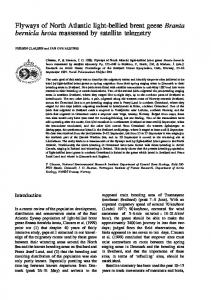
Si probes* 1kb Fig. 1. Transcription and maturation of the mnc-era operon. The results of SI mapping experiments of Figures 2 and 3 are summarized over the map of the E.coli chromosome showing the mc, era and recO genes. Transcripts identified by S1 mapping are shown by horizontal arrows of a width proportional to abundance. DNA probes are represented by solid bars. Labeling of 5' ends is shown by a star. The promoter (P) and RNase III (RIII) maturation sites deduced from the S1 nuclease experiments are indicated on the genetic map. Restriction sites are RI, EcoRI; Ssp, SspI; Pst, PstI; Hinc, HincIl; RV, EcoRV.
3401
J.C.A.Bardwell et al.
,f" .." %L.
:i
--. ...
-4
-.
v
e'.
.... BSS
:*;.:ijf.' ::
.::::
*.:.
:....:....::::
:. ..:
.:
:.:
*:::.:::.: .:.
*:
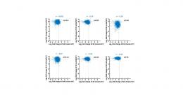
Fig. 2. Quantitative SI nuclease mapping of the mc-era transcript in rnclOS and mnc+ strains. The probe was a 1.7 kb double-stranded EcoRI-EcoRV DNA fragment labeled at each 5' end with 32p. Excess of the probe was hybridized overnight at 52°C with 12.4 yg (lane 1) or 18.6 Ag (lane 2) of isolated RNA from BL322 (rnc+), and 18.8 itg (lane 3) or 28.8 Ag (lane 4) of isolated RNA from BL321 (mc1O5). After S1 nuclease digestion at 37°C, the protected DNA fragments were glyoxylated and subjected to electrophoresis in a 1.2% agarose gel. Experimental procedures and conditions are as described in Materials and methods. In lane 5, mRNA was replaced with 30 yg tRNA. Size markers in lane 6 are 5'-end-labeled fragments of HindIII/EcoRI digests of lambda DNA. The major bands at 1300 and 1150 correspond to initiated and RNase Ill processed ends respectively (see text). The weak band at the nucleotide 1003 position corresponds to an SI digestion product at the single base mismatch formed by the hybrid between the mclO5 mutant RNA and the wild-type probe DNA. Other minor bands may correspond to minor promoters, degradation products, or transcripts hybridizing to the EcoRI-labeled strand.
764
i_
*_
;;
:...::. :: .::::
..
_F
'':: :.:
.:
*::
:::
............. i} .::::: ...; ....
:.!. .:i: ::..::
:.:.: *: ..:: .. ::i'.:: .:
'
.::
*: .:
.:.
:.
..
..
..
*: ::: .:
:..
:.:::
U.J,
16
..
..
:.
i: :.:
... :.:
.. .;
.. ... .: ..
..
..
0o
_
a 1.7 kb DNA fragment that extends from the EcoRI site upstream of the ATG in rnc to the EcoRV site in era was labeled at the 5' ends, hybridized to total E. coli RNA, treated with SI nuclease and analyzed by agarose gel electrophoresis. A 1. 15 kb region was protected from SI digestion (Figures 1 and 2), which indicates that mc is co-transcribed with era. This is consistent with the following observations suggesting that mc and era belong to the same operon with mc being promoter proximal. First, insertion mutants in mc are polar on era; second the UGA termination codon of mc overlaps the two last nucleotides of the AUG initiation codon for the Era protein; and third, cloning of a DNA fragment containing the two genes under control of the XPL and lppP5 promoters leads to overexpression of both RNase Im and Era (Ahnn et al., 1986; Takiff et al., 1989; S.-M.Chen, H.E.Takiff, G.G.Dubois and D.L.Court, in preparation). Downstream from era lies a third gene called recO. Twenty five base pairs lie between the terminal codon for era and the most likely initiation codon for recO (Morrison et al., 1989). Genetic evidence suggests that it is also part of the same operon, since insertions in era are polar on recO (Takiff et al., 1989). The recO gene product is required for recombination of circular plasmids and repair of UV damage to DNA (Morrison et al., 1989).
3402
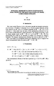
Fig. 3. High resolution S1 nuclease mapping of the 5' ends of the mc-era transcript. The probe was a 727 bp EcoRl-DdeI fragment 5'-end-labeled at the DdeI site. Excess of probe was hybridized for 3 h at 45°C with either 100 Ag tRNA (lane 1), 70 Ag tRNA plus 30 jig E.coli RNA isolated from NC124 pNC105 (mclO5) (lane 2) or 70 jig tRNA plus 30 ltg RNA from NC124 pKKFI (mc+) grown at 32'C (lane 3). Lanes 6 and 7 are identical to lanes 2 and 3 respectively except that the RNA was isolated from cells shifted to 10°C 1 h prior to harvesting, which resulted in overexpression of plasmid-encoded transcripts including mc-era. After S1 nuclease digestion at 37°C, the protected fragments were analyzed on a 7% acrylamide denaturing gel together with sequencing ladders obtained by chemical cleavage of the probe at G+A (lane 4) and C+T (lane 5). For lane 9, 8 ytg of mclOS RNA were treated with 40 ng of RNase III for 10 min at 34WC in RNase III buffer (Steege et al., 1987). After phenol extraction, the RNA was hybridized to the EcoRI-DdeI probe and submitted to SI nuclease digestion. Lane 8 is identical to lane 7 except that less RNA and probe of a lower specific activity were used.
Maturation of the mc -era transcripts by RNase 111 No good match with the E. coli d7° promoter consensus sequence is present in the DNA sequence just upstream from the 5' RNA end identified in the above S1 mapping, so this
RNase III processes its own message
PRIFPER
A
1
PR M El
I
a. -0 .
4m4w,AlMmllLAa&AM
-9
.....
ft
.illli
W
'm
4w
B 'I
R 1l F *
A
I
"
R
Fig. 4. (A) Primer extensions to define RNase III cleavage sites. RNA of the mc 5' leader region was made in vitro by transcribing pAR2526 with T7 RNA polymerase. It was then incubated for 10 min at 37°C either with (+) or without (-) RNase III in RNase III buffer (Steege et al., 1987), and 1 pmol of RNA was annealed to 70 ng of primer and extended using reverse transcriptase as described in Materials and methods. Two primers were used: primer 1, 5'd(TTTCAGTTTACCGGTAT)3'; and primer 2, 5'd(CACTATCGACTACGCGATCA)3'. Using the same primers, an M 13 clone of the mc leader region was sequenced (the lanes marked GATC). The primer extension products found in RNase Ill-treated samples are shown with arrows marked RI,, and are indicated in the schematic below. (B) Predicted secondary structure in the mc transcript recognized by RNase III. The RNase III cleavage sites as determined by reverse transcriptase are shown by arrows. The sequence is numbered backward from the translation start site. This ends of the region of sequence was manipulated with the Zuker computer folding program, which predicts optimal RNA secondary structures. The folding energy of the structure shown is 40.3 kcal (Zuker and Stiegler, 1981).
end may not correspond to a transcription initiation site. Moreover, a hairpin loop structure that might be an RNase III site can be drawn in this region (March et al., 1985). To determine whether the 5' end identified above is generated by RNase III processing, RNA was prepared from a strain deficient for RNase III [BL321 (rnclOS)] and analyzed by SI nuclease mapping using the 5'-labeled EcoRI-EcoRV DNA from the previous experiment. A 1.3 kb fragment was protected in this rnc- strain (Figure 2), locating the 5' end of the mcc-era message in the vicinity of a putative promoter that was previously predicted by sequence analysis (March et al., 1985; Nashimoto and Uchida, 1985). These results suggest that the RNA observed in mc+ strains is a result of RNase III cleavage and that the mnc-era polycistronic transcripts are cleaved by RNase III in the 5' sequence upstream from the mc coding region. Moreover, the difference of the amounts of protected DNA indicates that the intracellular concentration of the mnc-era RNA is higher in the ncthan in the mc+ strain. More precise localization of the 5' extremities of mc- era transcripts was achieved by fine-structure SI mapping and
C.
Fig. 5. Decay of the mnc-era mRNA in mc+ and nc- strains. (A) Total RNA was prepared from aliquots of cultures of BL322 (mc+) and BL321 (mc-) withdrawn at 0 min (lanes 1 and 8), 0.5 min (lanes 2 and 9), 1 min (lanes 3 and 10), 1.5 min (lanes 4 and 11), 2 min (lanes 5 and 12), 5 min (lanes 6 and 13) and 10 min (lane 14) after addition of rifampicin. Fifty micrograms of mc+ RNA (lanes 1-6), 20 ,ug of mc- RNA (lanes 8-14) and 50 ytg of tRNA (lane 15) were hybridized to an excess of the EcoRI-EcoRV probe described in Figure 2 and processed as described in Materials and methods. Lane 7 contains the same size markers as in Figure 2. (B) Relative amount of mnc-era mRNA in the samples described above were estimated by scanning the autoradiograph of A and are presented as percent of the intracellular concentration of mnc-era mRNA at the time of rifampicin addition (0, mc+; 0 mc-).
primer extension analysis (Figures 3 and 4). In mcstrains, the 5' ends of the message map just downstream from the TAGAAT-10 region of the potential promoter previously suggested (March et al., 1985; Nashimoto and Uchida, 1985) (Figure 3). These ends probably correspond to transcription initiation sites of the operon. Two 5' ends were observed; multiple initiation sites had been previously observed with other promoters (Cowing et al., 1985). In Mc+ strains the most predominant mRNA 5' end was at approximately -40 from the mc initiation codon with a less abundant transcript found ending at approximately 125 (Figure 3, lanes 3 and 7). These ends lie on either side and near the bottom of an extensive stem-loop structure that can be drawn in the RNA (Figure 4). -
3403
J.C.A.Bardwell et al.
In order to demonstrate that RNase III is directly responsible for the processing of its own transcript, uncut RNA isolated from the rncl05 strain was treated with highly purified RNase III in vitro and the 5' ends of mc transcripts were identified by high-resolution SI nuclease mapping. The mc mRNA is cut to exactly the same length in vitro and in vivo, showing that the 5' ends observed in mc+ strains are the direct result of RNase III cleavage (Figure 3, lanes 8 and 9).
Precise localization of RNase 111 processing sites The SI nuclease digestion left a cluster of ends, making it difficult to assign the exact RNase III cleavage sites. This heterogeneity is presumably due to fraying inherent in the S1 mapping technique (Aiba et al., 1981). To precisely localize the cut sites, mc RNA was made in vitro, cut to completion with RNase m and used as a template for reverse transcription. An SspI-HinclI fragment containing the stem -loop region was cloned into the BamHI site of pET I to create pAR2656, placing this fragment under control of the T7 10 promoter (Rosenberg et al., 1987). RNA was synthesized in vitro using T7 RNA polymerase and treated with RNase III. Two primers were used in separate reverse transcription experiments. One hybridized within the stem-loop region of the RNA and was used to determine the cut site near -125; the other hybridized to RNA distal to the cut site near -40. Single bands corresponding to 5' ends at positions -120 and -38 were found (Figure 4). RNA that had been partially cleaved by RNase III was examined using the more distal primer. Three run-off bands were seen, one corresponding to each of the cleaved sites at -120 and -38, and one corresponding to the T7 transcription start site. This indicates that RNase III cuts the stem -loop only at the two sites shown by the arrows in Figure 4. This experiment also demonstrates that the 144 bp SspI -HinclI fragment, which extends 39 nucleotides upstream and seven nucleotides downstream of the base of the stem, contains all the information necessary for the RNase III cleavage. A PstI - ClaI fragment containing half of the stem -loop and all of the mc gene was also cloned into pET-1. RNA made from the resulting clone, called pAR2637, was not cleaved by RNase III, indicating that sequences upstream from the PstI site are required for the -38 RNase III cleavage (A.Rosenberg and W.F.Studier, personal communication).
Steady-state levels and relative rates of decay of mc - era mRNA in mc + and mc - strains In addition to generating somewhat longer transcripts, the mc 105 mutation has a striking effect on the amount of mc -era mRNA deduced from the efficiency of the
protection of the DNA probe against Sl nuclease attack (Figures 2 and 3). The relative intracellular concentration of uncut mRNA in the rnc- strain was about six times higher than that of the processed mRNA in the wild-type strain. To determine whether the higher concentration of the inc-era transcripts in the mc- strain is due to the greater stability of the uncut message, the rates of decrease in the intracellular concentration of this mRNA in the mc+ and mc7 strains were measured (Figure Sb). The half-life of the inc-era transcripts in the mrclO5 strain (4.8 min) was much greater than the half-life in the mc' strain (0.75 min). The large difference in half-life observed is sufficient to explain
3404
the 6-fold difference in steady-state mRNA levels observed. Previous investigations demonstrated that this is not the consequence of a general increase in mRNA stability in mc bacteria. In fact, the mc105 mutation did not appreciably affect the rate of degradation of total mRNA (Apirion and Gitelman, 1980) or of some specific mRNAs, i.e. for the thrS and rpsO genes (Portier et al., 1987).
Oversynthesis of RNase 1/l and Era in mc 105 strain We next asked whether the mcc-era mRNA accumulated in the mc- strain is capable of being translated into RNase III and Era proteins. For this purpose, 35S_pulse-labeled proteins were immunoprecipitated from mc+ and mcbacteria by specific anti-Era and anti-RNase III antisera and the amounts of these proteins precipitated were quantified by scanning autoradiographs of denaturing polyacrylamide gels. RNase III and Era were synthesized 3-5 times more rapidly in the mc105 strain than in the wild-type (Figure 6). This coordinate overproduction of the two proteins in the mutant was found when the mc operon was present in single copy on the chromosome or in multiple copies on a plasmid. [S]methionine was incorporated into Era 3.8 0.9 fold more than into RNase III. Since the Era protein has three times more methionines than RNase III, we conclude that the ratio of Era to RNase III synthesis is - 1: 1. Discussion We have found that RNase III processes its own message. The RNase III cleavage occurs upstream of the mc coding region in the stem of a hairpin loop. The inc-era mRNA that has been processed by RNase HI was discovered to have a shorter half-life than the uncut transcript. The stem loop of the RNase III cleavage site or some other feature of the 122-nucleotide mRNA leader that is removed by RNase III may confer greater stability to the uncut message. Stem -loop structures have been implicated in determining mRNA stability in a number of different systems. It appears that unprotected ends of mRNA are sensitive to degradation. In support of this idea, circular endless mRNA injected in Xenopus embryos is exceptionally stable (Harland and Misher, 1988). Evidence from several organisms suggests that hairpins at the 3' ends of messages can protect upstream mRNA from degradation by 3' exonucleases (Hayashi and Hayashi, 1985; Mott et al., 1985; Newbury et al., 1987; Chen et al., 1988; Plunkett and Echols, 1989). Sequences at the 5' ends of transcripts that are known to stabilize mRNA may or may not form secondary structures (Gorski et al., 1985; Belasco et al., 1985; Bechhofer and Dubnau, 1987; Mackie, 1987). How these 5' sequences protect the downstream message is unclear because no 5'-exonucleases have been found in bacteria (Deutscher 1985). There are several mechanisms by which the removal of the 5' fragment by RNase Il endonucleolytic cleavage could initiate degradation of mc mRNA. One possibility is that this cleavage could provide an entry site for a wave of processive endonuclease attacks that degrade the message (Melefors and von Gabain, 1988). Close examination of Figure 3 (lanes 3 and 7) reveals minor 5' ends located just 3' to the major RNase III cleavage site at -38 from the mc initiation codon. These may represent decay intermediates. They are not RNase III cleavage sites because they are present in RNA from both mc- and rnc+ strains and they
RNase
A 1
Era
2 3 4 5
6 7 8
1011 12
-
B 1 2
3 4 5
6 7
Fig. 6. Immunoprecipitations of Era and RNase III proteins in mc+ and mc- strains. Cells were grown to log phase and pulse-labeled with [35S]methionine. (A) A lysate was prepared and immunoprecipitated using either pre-immune serum (lanes 1,4,7,10), antiserum raised against purified Era protein (Chen et al., in preparation) (lanes 2,5,8,11) or anti-Era antiserum plus 10 J4g of purified Era protein as competitor (lanes 3,6,9,12). The following bacterial strains were used: lanes 1-3, W3110 (mnc+): lanes 4-6, NC124 (mc 105); lanes 7-9, W3110 (pACSl); lanes 10-12, NC124 (pACS 105). (B) Immunoprecipitations using anti-RNase III antiserum. An E.coli cell lysate was either directly loaded (lane 4) or immunoprecipitated using pre-immune serum (lanes 1 and 5) antiserum raised against purified RNase III (lanes 2 and 6), or anti-RNase III antiserum plus 10 ug purified RNase III protein as competitor (lanes 3 and 7). The following bacterial strains were used: lanes 1-3, W3110 (pACS1); lanes 5-7, NC124 (pACS105). Pelleted proteins were analyzed on an SDS-polyacrylamide gel and autoradiographed. Quantification was achieved by scanning the autoradiofraph. Three independent experiments allowed the calculation that [V5S]methionine is incorporated 3.8 ± 9-fold more rapidly in Era than in RNase III.
are not sites cleaved by RNase III in vitro. They are more abundant in mc+ strains than in mc- strains, which is consistent with the idea that the RNase III-cleaved message is more susceptible to nuclease attack. Another possibility is that cleavage by RNase III reduces the translational efficiency of the mc message by disrupting the loading of ribosomes. Transcripts free of ribosomes may be more sensitive to attack by secondary endo- and exonucleases (Schneider et al., 1978). However, recent experiments suggest that the effects of translation on mRNA stability cannot be easily explained in terms of physical masking of the mRNA by ribosomes (Nilsson et al., 1987; Stanssens et al., 1986). The exact mechanism by which RNase III endonucleolytic attack promotes the degradation of mc mRNA remains to be determined. In cells deficient in RNase III, the hairpin-loop structure in the 5' leader of the mc mRNA fails to be cleaved, the stable uncut message accumulates and more RNase III is produced. This may be the first example of any RNase controlling its own expression. Several other RNAs could compete with mc transcripts for processing by RNase III. The feedback loop of regulation described above enables us to predict that increasing the intracellular concentration of competitor RNAs could lead to an accumulation of uncut mcc-era mRNAs and an enhancement of RNase Im
IlIl
processes its own message
synthesis. Ribosomal RNA precursors are the most abundant RNA substrates for RNase HII in the cell. Depending on growth conditions, they can represent 41-85 % of the total RNA synthesized (Bremer and Dennis, 1987). The autogenous regulatory mechanism described above would ensure an efficient processing of rRNA precursors by adapting the rate of RNase III synthesis to the intracellular concentration of these precursors. S 1 and primer extension experiments show that in the stem -loop structure of the 5'-non-coding region of mc RNA both strands are cleaved. All sites known to be cut by RNase HI have the potential to form stem-loop structures with long stretches of double-stranded RNA. (Young and Steitz, 1978; Bram et al., 1980; Saito and Richardson, 1981; Schmeissner et al., 1984; Downing and Dennis, 1987; Regnier and Portier, 1986; Dunn and Studier, 1983). Our deletion analysis suggests that both sides of the mc stem are required for cleavage. In a survey of 18 RNase III sites, a consensus 5'AAG/CUU 3' base-paired sequence for RNase III cleavage was derived, with the cuts being one nucleotide to the 5' side of the AAG sequence and/or three bases to the 3' side of the CUU sequence (Daniels et al., 1988). The mc RNase III cleavage site has two of these three base pairs conserved and with a double strand cut leaving a 2-base 3' overhang (Figure 4). It seems unlikely, however, that the consensus sequence alone is sufficient to direct RNase III cleavage. The results presented here demonstrate that the mc and era genes, encoding respectively RNase III and the GTPbinding protein Era, comprise a single operon transcribed into a polycistronic mRNA, which is processed by RNase III. The potential promoter visible in the sequence just upstream of the 5' end of the uncut RNA probably directs the transcription of the operon. SI mapping of 3' ends (not shown) reveals a major 3' end -85 bases beyond the termination codon for Era in both mc+ and mc- strains. However, this putative terminator appears to be < 100% efficient and genetic analysis revals that recO, which lies just downstream from era is also part of the mc operon (Takiff et al., 1989). The following observations suggest that mc and era are translationally coupled: equivalent amounts of Era and RNase III are synthesized in the cell; the mclOS mutation induces a parallel increase of RNase III and Era; the initiation codon for Era overlaps the termination codon for mc; and the era gene lacks a functional ribosome binding site sequence (Chen et al., in preparation). Era is a GTP-binding protein that hydrolyses GTP, but its function is not known (Chen et al., in preparation). Era has been reported to have homology to eukaryotic Ras proteins (Ahnn et al., 1986). However, our computer analysis has shown that Era is no more closely related to eukaryotic Ras proteins than are other GTP-binding proteins such as EFTu (unpublished data). Guanosine nucleotides, e.g. GTP, ppGpp and pppGpp, play a central role in macromolecular synthesis pathways as energy sources and regulatory factors. Since the expression of Era appears to be closely coordinated with that of RNase III, therefore there may be a direct correlation between the rate of Era synthesis and both the growth rate and intensity of macromolecular
synthesis.
RNase HI, in addition to being autoregulatory, is involved in the expression of polynucleotide phosphorylase, a 3'-processive exonuclease. In both cases, RNase m cleavage upstream of the coding sequence destabilizes the mRNA
3405
J.C.A.Bardwell et al.
(Regnier and Portier, 1986; Portier et al., 1987). Therefore, synthesis of the two RNases may be controlled by a common mechanism, including the RNase III feedback loop. It is possible that other genes or operons also belong to what can be called the RNase III regulon. An intriguing question is whether other RNases are involved in this regulatory network.
Materials and methods Bacterial strains and plasmids Bacterial strains (E. coli K-12) were BL322 (mc+), BL321 (mclO5) which were a gift of F.W.Studier and W3110 and NC124 (W3110 mclO5 glyA:: TnS) (Chen et al., in preparation). Plasmids: pASCI is a derivative of pBR322 that carries the mc gene on a 4.3 kb EcoRI fragment; pACS105 is a derivative of pACSI that carries the rnclOS mutation (Takiff et al., 1989); pKKFl, which was a gift of K.Kawakami, is a derivative of pACS1 that has a cat cassette inserted into the NruI site in recO. pNC 105, which was a gift of N.Costantino, is a mclO5 derivative of pKKFI. pAR2656 and pAR2636, which respectively contain the SspI-HincII and PstI-ClaI fragments from PACS1 cloned into the pETI T7 expression vector, were gifts from A. Rosenberg.
Si nuclease protection experiments For low-resolutionS1 experiments, RNAs were extracted from cells of the BL321(mclO5) and BL322(mc+) strains as described (Regnier and Portier, 1986). These RNAs were mixed with 5'-end-labeled DNA probe in hybridization buffer, incubated overnight at 52°C and digested with Sl nuclease at 37°C for 1 h as described (Regnier and Portier, 1986). Samples were glyoxylated and analyzed by electrophoresis in 1.2% agarose gels. The DNA probe was added in a large excess to the hybridization mixture so that the amount of the protected fragment was proportional to the amount of the E. coli mRNA. Relative amounts of mRNA were estimated from densitometric analysis of the autoradiographs. High-resolution S1 experiments were performed in a similar manner, except RNA that was extracted from NC 124 (pKKF1) and NC 124 (pNC 105) using the method described by Sarmientos et al. (1983). Hybridization was for 3-4 h at 45°C and samples were analyzed on 7% acrylamide sequencing gels. Dideoxy sequencing and primer extensions Dideoxy sequencing of DNA was performed using Sequenase and reagents supplied by United States Biochemical Corp. under conditions recommended by the manufacturer. Primer extension reactions were performed using reverse transcriptase, esssentially as recommended by International Biotechnologies, Inc. for DNA sequencing except that all four deoxynucleotide triphosphates (0.25 mM each) were used in place of a dideoxynucleotide-containing mixture.
Determination of mRNA decay Cells (BL321 and BL322) were grown at 37°C in MOPS-Tricine medium supplemented with 1 tg/ml thiamine, 0.4% glucose, 2 mM potassium phosphate and 0.4 mM each of the 20 amino acids. Exponentially growing cultures were treated with rifampicin (500 jig/mI) and 10-mil aliquots were withdrawn at different times, followed by quick lysis by adding 2 ml of lysis buffer [5% sodium dodecyl sulfate, 1.5 M sodium acetate (pH 5.2), and 1 mM EDTA] and heating at 100°C for 2-3 mmn. RNAs were extracted once with hot phenol at 65°C and with chloroform, then precipitated with ethanol and used for quantitative S1 mapping.
Immunoprecipitations Bacteria to be labeled were grown at 37°C in M56 medium supplemented with 0.2% glucose, 0.01% thiamine, 0.003% biotin and 0.1% of each of 18 amino acids (excluding methionine and cysteine). At log phase (A60 0.3-0.55) the cells were pulse-labeled with 50 pCi of 400 Ci/mmol [35S]methionine for 3 min and then mixed with a equal volume of ice-cold M56 salts buffer supplemented with 0.5% sodium azide and 0.05% methionine. Cells were pelleted and then lysed by resuspension in 1% SDS- 10 mM Tris (pH 8) and heated at 100°C for 5 min. The lysate was centrifuged for 5 min at 13 000 g. The supernatant was diluted 10-fold in IP buffer [10 mM Tris (pH 7.5), 1% NP40, 2 mM EDTA, 0.15 M NaCl, 0.02 % methionine] and adsorbed with pre-immune serum for 1 h at 4°C. The immune complexes were removed from solution by incubating at 4°C for 3 h with protein A -Sepharose CL-4B (Pharmacia LKB Biotechnology, Inc.), followed by centrifugation. The supernatant was then reacted with antiserum to RNase III or to Era at 48°C overnight, with or without excess
3406
purified competitor protein (RNase HI or Era). The immune complexes were adsorbed to protein A - Sepharose for 30 min at 4'C. The immune complexes were removed from solution by centrifugation and washed once in IP buffer plus 0.1 % bovine serum albumin and then three times in IP buffer alone. The pellet was resuspended in SDS loading buffer, heated at 100°C for 2 min and loaded onto a 12% SDS-polyacrylamide gel.
Acknowledgements We thank A.Rosenberg for the observation that RNase III cleaves RNA made from pAR2656, helpful discussions and for providing pAR2656 and pAR2637. We also thank H.Takiff, K.Kawakami, and N.Costantino for providing mc clones. This work was partially supported by the National Cancer Institute, DHHS, under contract NO. NO1-CO-74101 with BRI and by grants from the CNRS (U.A. 1139), from the University Paris 7, from ARC, from La Fondation Pour La Recherche Medicale, from E.I. Dupont de Nemours, from the INSERM and the EMBO (short-term fellowships to Y.N.) and from the US-Japan Cooperative Science Program. Ph. R. is a fellow of University Paris 7. The contents of this publication do not necessarily reflect the views or policies of the Department of Health and Human Services, nor does mention of trade names, commercial products, or organizations imply endorsement by the US Government. By acceptance of this article, the publisher or recipient acknowledges the right of the US Government to retain a nonexclusive royalty-free license in and to any copyright covering the article.
References Ahnn,J., March,P.E., Takiff,H.E. and Inouye,M. (1986) Proc. Natl. Acad. Sci. USA, 83, 8849-8853. Aiba,H., Adyha,S. and de Crombrugghe,B. (1981) J. Biol. Chem., 256, 11905-11910. Altuvia,S., Locker-Giladi,H., Koby,S., Ben-Nun,O. and Oppenheim,A.B. (1987) Proc. Natl. Acad. Sci. USA, 84, 6511-6515. Apirion,D. and Gitelman,D.R. (1980) Mol. Gen. Genet., 177, 339-343. Barry,G., Squires,C. and Squires,C.L. (1980) Proc. Nati. Acad. Sci. USA, 77, 3331-3335. Bechhofer,D.H. and Dubnau,D. (1987) Proc. Natl. Acad. Sci. USA, 84, 498-502. Belasco,J.G., Beatty,J.T., Adams,C.W., von Gabain,A. and Cohen,S.N. (1985) Cell, 40, 171-181. Blundell,M. and Kennell,D. (1974) J. Mol. Biol., 83, 143-161. Bram,R.J., Young,R.A. and Steitz,J.A. (1980) Cell, 19, 393-401. Bremer,H. and Dennis,P.P. (1987) In Neidhardt,F.C. (ed.), Escherichia coli and Salmonella typhimurium. American Society of Microbiology, Washington, Vol. 2, pp. 1527-1542. Brock,M.L. and Shapiro,D.J. (1983) Cell, 34, 207-214. Chen,C.-Y.A., Beatty,J.T., Cohen,S.N. and Belasco,J.G. (1988) Cell, 52, 609-619. Cowing,D.W., Bardwell,J.C.A., Craig,E.A., Woolford,C., Hendrix,R.W. and Gross,C.A. (1985) Proc. Natl. Acad. Sci. USA, 82, 2679-2683. Daniels,D.L., Subbarao,M.N., Blattner,F.R. and Lozeron,H.A. (1988) Virology, 167, 568-577. Deutscher,M.P. (1985) Cell, 40, 731-732. Downing,W.L. and Dennis,P.P. (1987) J. Mol. Biol., 194, 609-620. Dunn,J.J. and Studier,F.W. (1983) J. Mol. Biol., 166, 477-535. Gitelman,D.R. and Apirion,D. (1980) Biochem. Biophys. Res. Commun., 96, 1063-1070. Gorski,K., Roch,J.-M., Prentki,P. and Krisch,H.M. (1985) Cell, 43, 461-469. Greenberg,M.E. and Ziff,E.B. (1984) Nature, 311, 433-438. Gros,F., Hiatt,H., Gilbert,W., Kurland,C.G., Riseborough,R.W. and Watson,J.D. (1961) Nature, 190, 581-585. Harland,R. and Misher,L. (1988) Development, 102, 837-852. Hayashi,M.N. and Hayashi,M. (1985) Nucleic Acids Res., 13, 5937 -5948. King,T.C., Sirdeskmukh,R. and Schlessinger,D. (1986) Microbiol. Rev., 50, 428-451. Krinke,L. and Wulff,D.L. (1987) Genes Dev., 1, 1005-1013. Mackie,G.A. (1987)
J. Bacteriol., 169, 2697-2701.
March,P.E., Ahnn,J. and Inouye,M. (1985) Nucleic Acids Res., 13, 4677-4685. Melefors,O. and von Gabain,A. (1988) Cell, 52, 893-901. Morrison,P.T., Lovett,S.T., Gilson,L. and Kolodner,R. (1989) J. Bacteriol, 171, 3641-3649. Mott,J.E., Galloway,J.L. and Platt,T. (1985) EMBO J., 4, 1887-1891. Nashimoto,H. and Uchida,H. (1985) Mol. Gen. Genet., 201, 25-29.
RNase III processes its own message
Newbury,S.F., Smith,N.H. and Higgins,C.F. (1987) Cell, 51, 1131-1143. Nilsson,G., Belasco,J.G., Cohen,S.N. and von Gabain,A. (1984) Nature, 312, 75-77. Nilsson,G., Belasco,J.G., Cohen,S.N. and von Gabain,A. (1987) Proc. Natl. Acad. Sci. USA, 84, 4890-4894. Plunkett,G. and Echols,H. (1989) J. Bacteriol., 171, 588-592. Portier,C., Dondon,L., Grunberg-Manago,M. and Regnier,P. (1987) EMBO J., 6, 2165-2170. Regnier,P. and Portier,C. (1986) J. Mol. Biol., 187, 23-32. Rosenberg,A.H., Lade,B.N., Chui,D., Lin,S.-W., Dunn,J.J. and Studier,F.W. (1987) Gene, 56, 125-135. Saito,H. and Richardson,C.C. (1981) Cell, 27, 533-542. Sarmientos,P., Sylverster,J.E., Contente,S. and Cashel,M. (1983) Cell, 32, 1337- 1346. Schmeissner,U., McKenney,K., Rosenberg,M. and Court,D. (1984) J. Mol. Biol., 176, 39-53. Schneider,E., Blundell,M. and Kennell,D. (1978) Mol. Gen. Genet., 160, 121- 129. Stanssens,P., Remaut,E. and Fiers,W. (1986) Cell, 44, 711 -718. Steege,D.A., Cone,K.C., Queen,C. and Rosenberg,M. (1987) J. Biol. Chem., 262, 17651-17658. Studier,F.W. (1975) J. Bacteriol., 124, 307-316. Takata,R., Mukai,T. and Hori,K. (1987) Mol. Gen. Genet., 209, 28-32. Takiff,H.E., Chen,S.-M. and Court,D.L. (1989) J. Bacteriol., 171, 2581 -2590. Young,R. and Steitz,J. (1978) Proc. Natl. Acad. Sci. USA, 75, 3593 -3597. Zuker,M. and Stiegler,P. (1981) Nucleic Acids Res., 9, 133-148. Received on Mav 29, 1989; revised on July 19, 1989
3407


![AC2[R+,R] - Hindawi](https://m.moam.info/img/260x300/ac2rr-hindawi_5b5b550f097c47ff718b4589.jpg)

!['[--)][' R·-E···' CT()R](https://m.moam.info/img/260x300/-r-e-ctr_5a2f16371723dddcaca8f531.jpg)


