ANTICANCER RESEARCH 35: 2857-2860 (2015)
Radiation-induced Chondrosarcoma of the Bladder. Case Report and Review of Literature NORBERT SULE1, BO XU1, DIMA EL ZEIN2, KINGA SZIGETI3, SABY GEORGE4, JOHN M. KANE5 and RICHARD CHENEY1
Departments of 1Pathology, 4Medical Oncology and 5Surgical Oncology, Roswell Park Cancer Institute, Buffalo, NY, U.S.A.; Departments of 2Pathology and Anatomical Sciences and 3Neurology, University at Buffalo, Buffalo, NY, U.S.A.
Abstract. Background: Chondrosarcoma of the bladder is an extremely rare disease. Only five previously described cases are known in the medical literature. Patients and Methods: We present a chondrosarcoma developed 19 years after radiation treatment in a 73-year-old patient. A literature search of articles published from 1984 to 2014 was performed. Results: This is the first reported case of post-radiation bladder chondrosarcoma. We compared the clinicopathological features of the previously reported cases and reviewed the medical literature of the bladder sarcomas and post-radiation sarcomas. Conclusion: The primary treatment for bladder mesenchymal neoplasms is surgical, preferably radical cystectomy with or without chemotherapy. Positive surgical margin is one of the most important factors negatively affecting disease-specific, recurrence-free and overall survival rates. Soft tissue sarcomas are relatively rare in the genitourinary (GU) system representing only 2.1 % of all soft tissue sarcomas (1). Extraosseous chondrosarcoma is a rare neoplasm. Chondrosarcomas are reported in multiple locations, including the larynx, lung, ovary, kidney, ureter and spermatic cord. The differential diagnosis of extra-skeletal soft tissue neoplasms that show chondroid differentiation is limited and includes (i) true sarcoma (extraskeletal myxoid chondrosarcoma, mesenchymal sarcoma), (ii) carcinomasarcoma with chondroid differentiation and (iii) malignant neoplasm with chondroid metaplasia.
This article is freely accessible online. Correspondence to: Norbert Sule M.D., Ph.D., Department of Pathology, Roswell Park Cancer Institute, Elm & Carlton Street, Buffalo, NY 14263, U.S.A. Tel: +1 7168451765, Fax: +1 7168453427, e-mail:
[email protected] Key Words: Bladder chondrosarcoma, postradiation sarcoma.
0250-7005/2015 $2.00+.40
Advances in cancer treatment have resulted in multimodal therapeutic approaches, which include radiation therapy. Therapeutic radiation can result in a slightly increased risk for second primary tumors.
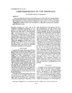
Case presentation History. The patient is a 73-year-old white female with a prior history of transformed non-Hodgkin's lymphoma. The patient underwent an autologous bone marrow transplant in 1993 followed by consolidation radiation to the pelvic and periaortic nodes in 1994. Beginning in April 2013, she developed urinary symptoms and hematuria. Computed tomography (CT) scan of the abdomen and pelvis in June, 2013, showed a 4.2 cm mass in the bladder with bilateral hydronephrosis. The patient underwent transurethral resection of bladder tumor (TURBT) and the final pathology finding was consistent with a high-grade sarcoma. A preoperative restaging CT scan performed in August showed a rapidly enlarging mass measuring 7.5 cm with persistent bilateral hydronephrosis (Figure 1). Resection of the sarcoma was performed, including en block total cystectomy, ureterectomy, total abdominal hysterectomy with bilateral salpingo-oophorectomy combined with intraoperative brachytherapy radiation. Gross description. The bladder is opened to reveal a polypoid, smooth surfaced, tan-pink lesion measuring 9.5×9.0×6.0 cm filling up most of the bladder cavity. The mass is attached to the trigone and inferior posterior portion of the bladder wall (Figure 1). Sections show heterogeneous, tan-pink, focally necrotic cut surface with focal areas of cystic degeneration and hemorrhage. The mass centers in the bladder wall with gross involvement of the perivesical fat. The tumor grossly approaches the inked surgical margin. The urothelium covering the bladder and the urethra is slightly hyperemic, otherwise grossly unremarkable with no other lesions noted.
2857
ANTICANCER RESEARCH 35: 2857-2860 (2015) Table I. Clinicopathological characteristics of the reported cases. Case
Sex
Age
History of radiation
Location
Size
Grade
Patient presentation
Patient follow-up
Reported by (year)
#1
male
81
no
Trigone
5 cm
High
Dysuria, frequency, hematuria
Local recurrence 10 weeks, DOD
Goertchen et al. (1984)
#2
male
65
no
Left wall
9 cm
High
Dysuria, frequency, hematuria
Free of disease 12 month , died of unrelated cause
Suster et al. (1987)
#3
female
73
no
Right wall
7 cm
High
Dysuria, frequency, hematuria
Local recurrence 4 month, DOD
Torenbek et al. (1993)
#4
male
66
no
Trigone
Not described
High
No clinical history given
#5
male
73
no
Posterior
3 cm
High
Macroscopic hematuria
DOD, 6 month, metastatic pleural effusion
Ikemoto et al. (2004)
#6
female
73
yes
Posterior wall trigone
9.5
High
Urinary symptoms, hematuria
Alive, no residual disease
Current
Kingsley et al. (1997)
DOD, Died of disease.
Microscopic description. Histological examination of the representative tumor sections showed a poorly differentiated neoplasm infiltrating through the muscularis propria and involving the perivesical adipose tissue. The neoplastic cells showed predominantly spindle cell morphology with prominent leucocytic infiltrate at the edges (Figure 2). The urothelial mucosal surface covering the polypoid lesion was ulcerated. The adjacent preserved bladder mucosa did not showed neoplastic change. Focal areas of myxoid change and chondroid matrix production were observed in the center of the lesion (Figure 3). Immunohistochemical studies of the poorly differentiated portion showed only vimentin positivity. No cytokeratin or S100 reactivity was observed (Figure 2). The area with chondroid differentiation was positive for vimentin and S100 (Figure 3).
Discussion The paratesticular soft tissue and kidney represent most common GU site for sarcomas (44% and 20%, respectively). Prostate and bladder, as primary location, are less frequent and occur in 16% and 15% of cases. Leiomyosarcoma is reported as being the most common type of bladder sarcoma accounting for 60-73% (2,3) of all bladder sarcomas. Other, less common variants of malignant soft tissue tumors of the bladder are rhabdomyosarcoma and angiosarcoma. Rhabdomyosarcoma was diagnosed in 20% of cases in the Memorial Sloan Kettering study (3), while, in M. D. Anderson Cancer Center’s case series, the second most common neoplasm was the angiosarcoma, reported in 15% of the study cases(2).
2858
Observations and experiences of the various effects of radioactivity, including radiation-induced neoplasm, came from laboratory experiments, nuclear catastrophe survivors and therapeutic, diagnostic and occupational exposure to radiation. Multiple factors influence the development of neoplasm, including dose, age, gender, anatomical site and post-radiation latency period. An increased relative risk (compared to general population) for radiation-induced bladder cancer was observed in patients who received radiation treatment for carcinoma of cervix, seminoma or prostate cancer. Above the 2 Gy radiation dose, the relative risk to develop sarcoma increases significantly (4). Post-radiation sarcomas are rare secondary neoplasms of the affected irradiated field and they can occur in bone and soft tissue. There are no definite criteria to ascertain the diagnosis of postradiation neoplasm but, after exclusion of patients with cancer syndromes, a number of factors could favor the diagnosis, including (i) history of tumor developing in the field of irradiation treatment, (ii) unusual tumor site for sarcoma and (iii) unusual patient age for sarcoma (5). The latency period to develop post-radiation sarcoma is variable with a minimum of 3-4 years since the radiation. The majority of the post-radiation sarcomas develop in the bones of the affected areas, only 8.1% of cases occurred in the soft tissue. The majority of these neoplasms were osteosarcomas (61.5%). In less proportion, fibrosarcoma (23.7%), malignant fibrous histiocytoma (9.6%) and chondrosarcoma (3.7%) were also observed. The latency period for post-irradiation sarcoma ranges from 4-55 years.
Sule et al: Radiation-induced Chondrosarcoma of the Bladder
Figure 2. The majority of the lesion showed a poorly differentiated neoplasm showing infiltrating the muscularis propria and perivesical adipose tissue. The tumor cells were reactive only to vimentin, no S100 positivity was detected (inlets).
Figure 1. A. CT scan with contrast showing an enhancing, irregular bladder tumor. The mass abuts the anterior, left (shown in image) and posterior walls of the bladder. B. Large polypoid mass protruding into the bladder cavity. The surrounding bladder mucosa is unremarkable.
The mean age at the diagnosis was 47.9 years with a peak incidence in the sixth decade of life (5). We present a unique case of post-radiation chondrosarcoma of the bladder. Only six, including the current tumor, isolated cases of bladder chondrosarcoma are documented in the English medical literature (6-10). No history of radiation treatment was mentioned in previous reports. The age of presentation ranges from 65 to 81 years (mean=71.83). There is a male predominance in reported cases with a ratio of 2 males: 1 female. The presenting clinical symptoms of this rare tumor resemble other traditional bladder epithelial tumors with hematuria, dysuria and frequency described for the majority of cases. The tumor size ranges from 3 to 9.5 cm (mean=6.7 cm) with no site predilection noted within the bladder. All tumors were surgically managed with at least a cystectomy. Histologically, they all exhibited high-grade morphology. Three of the cases had a dismal prognosis and died of their disease within 10 weeks to 6 months of presentation. One case was disease-free but died of unrelated
Figure 3. Central portion of the tumor showed areas of cartilagenous hyalin matrix deposition. The neoplastic cells display strong vimentin and S100 stainings consistent with chondrocytic differentiation (inlets).
causes (12 months from presentation).The currently described case, similarly to the previously documented cases, developed at late age and presented with urinary symptoms. Our patient remains disease-free and is alive after fifteen months. This is the first post-radiation urinary chondrosarcoma and second documented case in females (Table I). Chromosomal aberrations have been reported in a case of bladder chondrosarcoma, including multiple rearrangements that appeared clonal, being present consistently in 20 analyzed metaphase cells. Due to the multitude of rearrangements, involving chromosomes 2, 3, 4, 5, 6, 8, 10,
2859
ANTICANCER RESEARCH 35: 2857-2860 (2015) 13, 16 and 22 and consisting of translocations, insertions, deletions and inversions, the significance of these changes are unclear. The OCUU-6 cell line established from a chondrosarcoma subject is aneuploid ranging from 98-102 chromosomes and harbors multiple rearrangements as well; the aneuploidy is likely a tissue culture artifact. Chromosome or microarray analysis of rearrangements on a larger sample size of chondrosarcomas is needed to elucidate the significance of these findings (7,8). Bladder sarcomas are often fatal neoplasms. The 5-year disease-specific survival rate in patients with bladder sarcomas varies between 56%-62% (2,3,11). In their study, Spiess et al. specifically analyzed bladder sarcoma and found that positive surgical margin is one of the most important factor negatively affecting disease-specific, recurrence-free and overall survival rates. The presence of lymphovascular invasion and lymph node metastasis also has a significantly negative impact on disease-specific survival. It seemed that the prognosis of bladder sarcoma is independent of their histological type (2). The local recurrence rate is found to fall between 14-16% (2, 11), 34% (3). The recurrence-free five- and ten-survival rate is 67% and 53%, respectively (3). Russo found a remarkable 80% recurrence-free survival rate in bladder soft tissue malignancies. He speculated that early symptoms (hematuria) and detection at this site could contribute to the better survival data. Univariate analysis of multiple factors by Russo showed that localized tumor at presentation, tumor diameter less 5 cm, low histological grade, paratesticular and bladder tumor site and complete tumor resection are favorable prognostic predictors in relapse-free survival of adult urological soft tissue tumors (12). In a most recent article, Dotan showed that only tumor size at presentation was found to have significant effect on predicting the development of local recurrence (3). The available studies in bladder sarcoma described a range of 15-20% in the rate of distant metastasis, mostly developing in the lungs, bone, liver and brain (3, 11). The median survival rate of patients with distant metastasis was 2.9 years (3). Metastasis-free survival was 66% at 5 years and 59% at 10 years (3). Factors predicting metastasis-free survival by univariate and multivariate analyses include the patients’ age and tumor histology. Univariate analysis also found significant association between tumor grade and metastasis-free survival (3). The primary treatment for bladder mesenchymal neoplasms is surgical, preferably radical cystectomy with or without chemotherapy. The use of neoadjuvant or adjuvant
2860
therapy resulted increased disease-specific survival; however, this observation was not statistically significant due to the limited number of patients in the study (2, 3, 5, 11, 12).
References 1 Stojadinovic A, Leung DH, Allen P, Lewis JJ, Jaques DP, Brennan MF: Primary adult soft tissue sarcoma: time-dependent influence of prognostic variables. J Clin Oncol 20(21): 4344-52, 2002. 2 Spiess PE, Kassouf W, Steinberg JR, Tuziak T, Hernandez M, Tibbs RF, Czerniak B, Kamat AM, Dinney CP, Grossman HB: Review of the M.D. Anderson experience in the treatment of bladder sarcoma. Urol Oncol. 25(1): 38-45, 2007. 3 Dotan ZA, Tal R, Golijanin D, Snyder ME, Antonescu C, Brennan MF, Russo P: Adult genitourinary sarcoma: the 25-year Memorial Sloan-Kettering experience. J Urol 176(5): 2033-8; discussion 2038-9, 2006. 4 Suit H, Goldberg S, Niemierko A, Ancukiewicz M, Hall E, Goitein M, Wong W, Paganetti H: Secondary carcinogenesis in patients treated with radiation: a review of data on radiationinduced cancers in human, non-human primate, canine and rodent subjects. Radiat Res 167(1): 12-42, 2007. 5 Inoue YZ, Frassica FJ, Sim FH, Unni KK, Petersen IA, McLeod RA: Clinicopathologic features and treatment of postirradiation sarcoma of bone and soft tissue. J Surg Oncol 75(1): 42-50, 2000. 6 Goertchen R, Dominok G, Mobius G: [Chordoid sarcoma of the urinary bladder]. Zentralbl Allg Pathol 129(4): 379-82, 1984. 7 Ikemoto S, Sugimura K, Yoshida N, Nakatani T: Chondrosarcoma of the urinary bladder and establishment of a human chondrosarcoma cell line (OCUU-6). Hum Cell 17(3): 93-6, 2004. 8 Kingsley KL, Peier AM, Meloni-Ehrig AM, Sandberg AA, Klein EA: Cytogenetic findings in a bladder chondrosarcoma. Cancer Genet Cytogenet 96(2): 183-4, 1997. 9 Suster S, Huszar M, Bubis JJ, Geiger B: Fibrosarcoma of the urinary bladder. Study of a case showing extensive chondroid differentiation. Arch Pathol Lab Med 111(8): 767-70, 1987. 10 Torenbeek R, Blomjous CE, Meijer CJ: Chondrosarcoma of the urinary bladder: report of a case with immunohistochemical and ultrastructural findings and review of the literature. Eur Urol 23(4): 502-5, 1993. 11 Rosser CJ, Slaton JW, Izawa JI, Levy LB, Dinney CP: Clinical presentation and outcome of high-grade urinary bladder leiomyosarcoma in adults. Urology 61(6): 1151-5, 2003. 12 Russo P, Brady MS, Conlon K, Hajdu SI, Fair WR, Herr HW, Brennan MF: Adult urological sarcoma. J Urol 147(4): 1032-6; discussion 1036-7, 1992.
Received January 21, 2015 Revised January 29, 2015 Accepted February 2, 2015