Dentomaxillofacial Radiology (2007) 36, 121–124 q 2007 The British Institute of Radiology http://dmfr.birjournals.org
CASE REPORT
Radiographic assessment of Gardner’s syndrome LC Fonseca1,2, NK Kodama1, FCF Nunes2, PH Maciel3, FA Fonseca4, M Roitberg5, JX de Oliveira1 and MGP Cavalcanti*,1 1 Department of Radiology, College of Dentistry, University of Sa˜o Paulo, Sa˜o Paulo, Brazil; 2College of Dentistry, PUC of Belo Horizonte, Brazil; 3Hospital of Public Employees at the State of Minas Gerais, Brazil; 4Private Practice, Belo Horizonte, Brazil; 5 Federal University of Minas Gerais, Belo Horizonte, Minas Gerais, Brazil
The detection of osteomas in the maxillofacial region may be the initial clinical finding in Gardner’s syndrome (GS). The most common location of osteomas is in the skull, but the lesion can also occur in the jaws. We present a case of a 47 year old male patient with GS who was referred for radiological evaluation. Extraoral examination revealed an epidermoid cyst and the patient had a history of intestinal polyps. A panoramic radiograph demonstrated numerous osteomas and diffuse sclerosis of the mandible, and compound odontomas with impacted teeth. CT scan allowed the localization and extension of the osteomas, and showed other sites in the maxillofacial region as well. CT images also revealed a different behaviour of osteoma, invading the mandibular canal. Dentomaxillofacial Radiology (2007) 36, 121–124. doi: 10.1259/dmfr/18554322 Keywords: Gardner’s syndrome; tomography; X-ray computed; osteoma Case report A 47 year old white male patient was referred for radiological evaluation, with a medical history of Gardner’s syndrome (GS). The patient reported a clinical history of intestinal polyps and an extraoral examination revealed an epidermoid cyst localized in the forehead (Figure 1a) and in the right leg (Figure 1b). He did not complain of any symptoms. A panoramic radiograph (Figure 2) revealed multiple radiopaque masses at the right ascending ramus and at the bilateral angle of the mandible, also into the left maxillary sinus and in the right zygomatic – maxillary process, suggesting osteomas. Diffuse sclerosis was also noted throughout the mandibular body. A radiopaque mass appeared close to the mandibular canal at the left side, displacing the superior cortex. A large radiopaque mass was distinguished at the right ascending ramus, near the condyle and the mandibular foramen. Compound odontomas were observed in both jaws, which were better visualized by periapical radiograph (Figures 3a and b). Moreover, the right superior canine had suffered impaction by a small compound odontoma (Figure 3b). To determine the exact location of the lesions, a CT scan was performed. Axial CT images demonstrated several *Correspondence to: MGP Cavalcanti, Department of Stomatology, College of Dentistry, University of Sa˜o Paulo, Avenida Prof. Lineu Prestes, 2227, Cidade Universita´ria, Sa˜o Paulo 05508-900, SP, Brazil; E-mail:
[email protected] Received 6 October 2005; revised 14 March 2006; accepted 24 March 2006
hyperdense areas in both sides of the mandible (Figure 4a), and also showed an osteoma in the lingual cortex involving the mandibular foramen and invading the right mandibular canal (Figure 4b). The same lesion was detected in coronal reconstructed image in the right ascending ramus, near to the inferior-medial portion of the condyle, invading the medial cortex (Figure 4c), even though the patient did not suffer paraesthesia. Other osteomas were also seen in the left maxillary sinus and in the right zygomatic bone (Figure 5). No treatment was carried out and subsequently the patient was lost to follow-up. Discussion The early detection of GS is extremely important because patients can develop colorectal adenocarcinoma.1 The maxillofacial features of the syndrome can appear many years before the intestinal polyposis, so dentists should be familiar with the significance of GS as a pre-cancerous condition.1 The presence of multiple bone changes of the skull and abnormal dental findings should alert the clinician to initiate further investigation.2 Ida, Nakamura and Utsunomiya3 revealed that patients with more than three osteomas need to have the familial history checked, with an emphasis on intestinal disease, because this is highly suggestive of GS. Utsunomiya and Nakamura4 reported that osteomatous changes in the jaws were more
Gardner’s syndrome LC Fonseca et al.
122
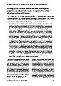
Figure 1 (a) Facial appearance showing an epidermoid cyst in the forehead (arrow). (b) Ulcerated epidermoid cyst in the right leg
frequent than dental abnormalities. Our case showed the classical clinical features associated with GS: multiple osteomas and odontomas, adenomatous polyps, epidermoid cyst and impacted tooth. The understanding of the behaviour of the osteoma is essential, since it can cause clinical problems. Furthermore, it might be the only extracolonic manifestation related to the
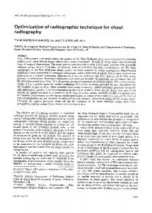
Figure 2 Panoramic radiograph depicting multiple osteomas at the right ascending ramus and at the bilateral angle of the mandible, also into the left maxillary sinus and in the right zygomatic – maxillary process (arrows), and diffuse sclerosis in the mandible Dentomaxillofacial Radiology
Figure 3 Periapical radiography showing (a) compound odontomas in the mandibular premolar region (arrows) and (b) an impaction of right maxillary canine by odontoma (arrow)
disease.4 Lew et al5 reported a case in which an osteoma of the condyle caused a limitation of mouth opening. In addition, Baykul et al6 reported that this lesion can reach a considerable size causing disfigurement. In our case, we did not find a relevant disfigurement and the invasion of the mandibular canal by the osteoma did not cause any clinical alteration. The absence of paraesthesia could be explained because the osteoma in the right ascending ramus was invading the entrance of the mandibular foramen, but did not obstruct the nerve pathway. However, Takeuchi et al7 verified that an increase in the number and size of the jaw lesions, including osteomas, in GS patients can occur gradually even in adulthood. Thus, in our reported case, the osteoma near to the right mandibular canal could enlarge and produce some symptomatology in the future. Consequently, a careful follow-up radiological examination of the jaws must be performed.
Gardner’s syndrome LC Fonseca et al.
123
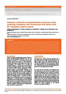
Figure 4 (a) Axial CT image showing several hyperdense areas in the mandible and osteomas (arrows) and (b) an osteoma in the lingual cortex involving the mandibular foramen and invading the right mandibular canal (arrow). (c) Coronal reconstructed image revealing the involvement of medial cortex by the same lesion (arrow)
Skeletal abnormalities appear in approximately 90% of patients with GS, and osteoma is considered to be the most common.5 The frequent sites for these tumours are the outer cortex of the skull, paranasal sinuses and the mandible.5 Supernumerary teeth, compound odontomas and/or impacted teeth were seen in 30% of the patients with this disease.8,9 In a normal population, the incidence of osteosclerosis in the mandible or maxilla is low, whereas in some studies these are detected in more than 70% of cases of familial adenomatosis coli or GS.3,9 In our case there were no supernumerary teeth, but we did find some compound odontomas in the mandible and in the maxilla. The compound odontomas in the maxilla caused the impaction of the right superior canine, and the patient also had diffuse sclerosis of the mandible. Panoramic radiography can be useful for early detection of GS by the dentist, because the components of this entity, like osteoma, odontoma, supernumerary and impacted teeth, can be detected in the routine radiological examination.9 Nevertheless, panoramic radiography is of limited value in evaluation, localization and extension of the tumour mass, considering the superimposition of bony structures and that it is a bidimensional image. DelBalso and Werning10 related that CT is the best imaging modality for the diagnosis of osteoma. Halling et al2 reported a case of GS studied by panoramic radiography that showed multiple widespread osteomas in the mandible associated with composite odontomas. Furthermore, this patient was submitted to a CT scan that demonstrated enostoses in the inner surface of both cortices and the full extent of the osteomas at the angles of the mandible and the ethmoidal sinuses, as well as smaller lesions lingual to the lower right first premolar. Yuasa et al11 reported that the osteomas of the maxilla were described correctly when studied in CT
scans because the panoramic radiography was a less adequate technique for detecting maxillary lesions. CT provides better delineation of the bony structures of the skull base and facial skeleton than conventional radiography. In addition, it allows the visualization of cross-sectional images and it is the only examination that provides details of the extension and localization of the osteomas.11 Owing to this, CT scan has become an important diagnostic tool for maxillofacial lesions,10 but only a few cases of GS have been studied using this technique. In our case report, using CT images we could identify an osteoma in the maxillary sinus and in the zygomatic bone accurately. In addition, CT showed a modified pattern of the bone in the mandible. All of these manifestations in the mandible were more clearly elucidated by CT than panoramic radiography. Also, the localization and extension of the osteoma in the mandible was determined by CT scan, which revealed a particular behaviour of the osteoma that invaded the mandibular canal. This invasion of the mandibular canal is a new finding in GS. Furthermore, a CT examination can be useful to the clinicians in diagnosis of this lesion, the
Figure 5 Coronal CT image of the maxilla demonstrating osteoma in the left maxillary sinus and in the right zygomatic bone (arrows) Dentomaxillofacial Radiology
Gardner’s syndrome LC Fonseca et al.
124
establishment of appropriate treatment plan and follow-up for the patient. The present case illustrated the importance of CT examination in GS with multiple osteomas. CT revealed a new behaviour of osteoma, invading the mandibular canal, but without a clinical manifestation.
In conclusion, dentists must be alert to the presence of osteomas, dental abnormalities, odontomas and osteosclerosis because it can be the first manifestation of GS, and CT is the recommended imaging modality for detecting the extension and localization of osteomas.
References 1. Wesley RK, Cullen CL, Bloom WS. Gardner’s syndrome with bilateral osteomas of coronoid process resulting in limited opening. Pediatr Dent 1987; 9: 53 – 57. 2. Halling F, Merten HA, Lepsien G, Honig JF. Clinical and radiological findings in Gardner’s syndrome: a case report and follow-up study. Dentomaxillofac Radiol 1992; 21: 93 –98. 3. Ida M, Nakamura T, Utsunomiya J. Osteomatous changes and tooth abnormalities found in the jaw of patients with adenomatosis coli. Oral Surg Oral Med Oral Pathol 1981; 52: 2 –11. 4. Utsunomiya J, Nakamura T. The occult osteomatous changes in the mandible in patients with familial polyposis coli. Br J Surg 1975; 62: 45 – 51. 5. Lew D, DeWitt A, Hicks RJ, Cavalcanti MG. Osteomas of the condyle associated with Gardner’s syndrome causing limited mandibular movement. J Oral Maxillofac Surg 1999; 57: 1004– 1009. 6. Baykul T, Heybeli N, Oyar O, Dogru H. Multiple huge osteomas of the mandible causing disfigurement related with Gardner’s syndrome: case report. Auris Nasus Larynx 2003; 30: 447 – 451.
Dentomaxillofacial Radiology
7. Takeuchi T, Takenoshita Y, Kubo K, Iida M. Natural course of jaw lesions in patients with familial adenomatosis coli (Gardner’s syndrome). Int J Oral Maxillofac Surg 1993; 22: 226 – 230. 8. Gardner EJ. Follow-up study of a family group exhibiting dominant inheritance for a syndrome including intestinal polyps, osteomas, fibromas and epidermal cysts. Am J Hum Genet 1962; 14: 376 –390. 9. Wolf J, Jarvinen HJ, Hietanen J. Gardner’s dento-maxillary stigmas in patients with familial adenomatosis coli. Br J Oral Maxillofac Surg 1986; 24: 410 – 416. 10. DelBalso AM, Werning JT. The role of computed tomography in the evaluation of cemento-osseous lesions. Oral Surg Oral Med Oral Pathol 1986; 62: 354 –357. 11. Yuasa K, Yonetsu K, Kanda S, Takeuchi T, Abe K, Takenoshita Y. Computed tomography of the jaws in familial adenomatosis coli. Oral Surg Oral Med Oral Pathol 1993; 76: 251 – 255.