ANTIBODIES AGAINST CARCINOEMBRYONIC ANTIGEN FOR ... vivo of four anti-CEA MAb with their F(ab')2 and Fab fragments in nude mice bearing human ...
RADIOLABELED ANTIBODIES
FRAGMENTS
OF MONOCLONAL
AGAINST CARCINOEMBRYONIC
LOCALIZATION
OF HUMAN
GRAFTED
ANTIGEN
FOR
COLON CARCINOMA
INTO NUDE MICE
BY F R A N Z BUCHEGGER,* C H A R L E S M. H A S K E L L , M A G A L I SCHREYER, B I A N C A R. SCAZZIGA, S I M O N E R A N D I N , S T E P H A N CARREL, AND JEAN-PIERRE MACH
From the Institute of Biochemistry, University of Lausanne and the Ludwig Institute for Cancer Research, Lausanne Branch, CH-1066 Epalinges, and the Thyroid Unit, Department of Medicine, University Hospital, Lausanne, Switzerland
T h e idea of using cytotoxic drugs, toxins, or radioisotopes coupled to antibodies to destroy hidden tumor cells has been revitalized by the development of the monoclonal antibody (MAb) 1 technology (1). Selected MAb directed against tumor antigens, with their specificity for single antigenic determinants, appear to be the ideal carriers for antitumor agents. However, a prerequisite for all these forms of passive immunotherapy is that the antibodies are capable to reach the target tumor cells in vivo. We have previously reported encouraging results of tumor localization by immunoscintigraphy in patients with colorectal carcinomas (2, 3) using one MAb (MAb 23) directed against carcinoembryonic antigen (CEA) (4, 5). However, the amount of radiolabeled antibody localizing specifically within the tumor mass was still relatively low as compared with the amounts of radioactivity remaining in the blood pool, reticuloendothelium, and various normal organs (2, 3). T o improve these results we recently selected a series of MAb with higher affinities for CEA than the previously used MAb (6) and prepared F(ab')2 and Fab fragments. T h e fragments of high affinity MAb should give higher tumor uptakes with less accumulation in the reticuloendothelium and at the same time a more rapid clearance from the circulation. For ethical reasons and because tumors are very heterogenous in terms of size, anatomical sites, histology, vascularization, etc., it appears inappropriate to screen the different MAb and their fragments for best tumor localization in patients. T h e model of nude mice bearing human colon carcinoma xenografts that we originally used for testing polyclonal anti-CEA antibodies in 1074 (7) offers the advantage of a relatively constant tumor antigen expression and accessibility. Other groups have used similar experimental models of xenografted human * To whom correspondence should be addressed at the Institute of Biochemistry, University of Lausanne, 1066 Epalinges, Switzerland. ~Abbreviations used in this paper: CEA, carcinoembryonic antigen; CNBr, cyanogen bromide; NCA, nonspecific cross-reacting antigen; MAb, monoclonal antibody; PBS, phosphate-buffered saline; SDS, sodium dodecyl sulfate. J. EXP. MED. © The Rockefeller University Press • 0022-1007/83/08/0413/15 $1.00 Volume 158 August 1983 413-427
413
414
TUMOR
LOCALIZATION OF FRAGMENTS OF MONOCLONAL ANTIBODIES
tumors to test the tumor-localizing capacity of polyclonal and monoclonal antibodies directed against various t u m o r markers (8-15), but no systematic comparison of different MAb and their fragments against the same marker have been reported. Despite the limitations of any experimental model of xenografted h u m a n tumors, namely the absence of normal h u m a n tissue as counterpart of the t u m o r transplant, we feel that these in vivo experiments are necessary for the final selection of MAb and fragments before h u m a n application. T h e purpose of this report is to compare the t u m o r localization capacity in vivo of four anti-CEA MAb with their F(ab')2 and Fab fragments in nude mice bearing h u m a n colon carcinoma grafts. T h e results showed a dramatic increase of specific t u m o r uptakes of radiolabeled F(ab')~ and Fab fragments as compared with intact antibodies and a marked improvement of tumor detection by external scanning in this experimental model. Materials and Methods
Production and Screening of Hybridomas. The derivation of 26 new hybridomas that secrete anti-CEA antibodies and their in vitro selection is described in detail elsewhere (6). Briefly, a BALB/c mouse was injected intraperitonally with 15 ~g of purified colon carcinoma CEA (16) in complete Freund's adjuvant. After 3 too, the mouse was reinjected with 15, 100, and 150 #g of purified CEA in saline at daily intervals (17). Spleen cells collected 6 d after the first boost were used for fusion with the myeloma cell line NSI/ lag4.1 (18). Hybridoma supernatants were screened for antibody production by a binding assay to radiolabeled CEA, using ammonium sulfate precipitation. Supernatants were also screened for strong inhibition of this binding by unlabeled CEA and for weak inhibition of this binding by a normal glycoprotein present in granulocytes and known to cross-react with CEA, called normal glycoprotein (NGP) (19) or nonspecific cross-reacting antigen (NCA) (20). Cross-reaction of hybridoma supernatant with granulocyte glycoprotein(s) (21) was also tested by immunoperoxidase staining of frozen sections of primary human colon carcinoma using the avidin-biotin system. After this in vitro screening, three new MAb, designated 35, 202, and 192, were selected for in vivo testing in parallel with MAb 23, which had already been used for tumor detection by immunoscintigraphy in patients (2, 3). Pur~cation and Testing of Selected MAb. The selected hybrids were cloned by limiting dilution. Hybridoma ascites were produced by injecting 107 hybrid cells intraperitoneally into Pristane (Aldrich, Beerse, Belgium)-primed BALB/c mice. MAb were purified from ascites by ammonium sulfate precipitation (45% saturation at 4°C) and DE 52 cellulose (Whatman, Balston, England) ion exchange chromatography. The MAb were eluted with a gradient of phosphate buffer, 0.01 to 0.15 M, pH 8 (22). A control IgG1 was purified by the same procedure, from ascites obtained with myeloma cell line P3×63Ag8 (1). The affinities of the four purified MAb for l~I-labeled CEA were determined both in 0.02 M Tris-HC! buffer, pH 7.4, and in 0.15 M phosphate-buffered saline (PBS) using Scatchard Plot and Lineweaver Burke analysis. The affinities in low molarity Tris buffer of these four MAb were all in the range of 1.6-6.2 x 101° M-. The affinities in PBS were 5.8 X 10⁸, 1.1 X 10 9, 6 X 10⁹, and 1.8 x 101° M- for MAb 23, 202, 35, and 192, respectively. By a radioimmunoassay (5) using immobilized CEA and goat antisera against mouse immunoglobulin isotypes (Meloy, Springfield, VA), it was shown that the four selected MAb were of IgGl subclass. Reciprocal binding inhibition tests on insolubilized CEA showed that MAb 23 and 202 were reacting with the same or closely related epitopes on CEA, and MAb 35 and 192 were reacting with different epitope.s The four purified MAb were also. tested for binding to purified soluble ~I-labeled NCA (20) (Commissariat Energie Atomlque, Gif sur Yvette, France) using ammonium sulfate precipitation, and for binding to the surface of freshly
BUCHEGGER ET AL.
415
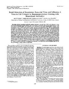
~repared normal human granulocytes by an indirect binding radioimmunoassay using 5I-labeled rabbit antibodies against mouse F(ab')2 (23). Preparation of F(ab')2 and Fab Fragments. F(ab')2 fragments of the four selected purified MAb and control IgG were obtained by incubation for 22 h at 370C with 5% (wt/wt) pepsine, (Worthington, Freehold NJ) in acetate buffer, pH 4 (24), followed by filtration on Sephadex G-150 (Pharmacia, Uppsala, Sweden). Fab fragments of Mab 202 and 35 and of control IgG were obtained by incubation for 6 h at 37°C with 2 or 3% (wt/wt) papain (Sigma Chemical Co., St. Louis, MO) in a 0.075 M phosphate buffer, pH 7, containing 0.075 M NaCI, 0.01 M L-cysteine hydrochloride, and 0.002 M EDTA (25). Fab fragments were separated from partially digested IgG by filtration on Sephadex G150 and from Fc fragments by a DE-52 ion exchange chromatography column equilibrated in 0.01 M phosphate buffer, pH 8. The Fab fragments were eluted with the void volume, whereas the Fc fragments were retained. Purified intact MAb, and F(ab')2 and Fab fragments were analyzed by sodium dodecyl sulfate (SDS)-polyacrylamide gel (8.5%) electrophoresis. They gave single bands with the expected molecular weights of about 150,000, 105,000, and 50,000, respectively (Fig. 1). The affinities of two Fab fragments were measured after labeling with 1251 by the following method. First, increasing amounts of labeled Fab were incubated with a constant amount of unlabeled CEA. The CEA was then precipitated after incubation with a second, noncompetitive, intact Mab by addition of ammonium sulfate. The affinities in 0.02 M Tris buffer were 6.7 × 10⁹ and 9.3 x 10⁹, and in PBS, 4.1 x 10⁹ and 1.1 x 10s M-, for Fab of Mab 192 and 202, respectively. Iodine Labeling of Intact lgG and of Their Fragments. 20 #g of purified MAb or of their fragments were labeled with 1 mCi of ~31I using the Iodobeads method (Pierce Chemicals, Rockford, IL). Control IgG or its fragments were labeled under the same conditions with ~5I. Free iodine was removed from the labeled proteins by dialysis or by Sephadex G-25 chromatography. The specific radioactivity ranged from 20 to 40 ~Ci/~g of MAb for both isotopes. Labeled antibodies or their fragments were tested for binding activity by overnight incubation in 0.15 M PBS at 25 °C with 10 #g CEA bound to cyanogen bromide (CNBr)-activated Sepharose 4B (Pharmacia,). Injection of Nude Mice Bearing Grafts of Human Colon Carcinoma. The human colon carcinoma (Co-112) (7) maintained by serial transplantations into BALB/c nude mice produces CEA as demonstrated using immunoperoxidase (26). Transplanted tumors contain 50-100 #g CEA/g whereas sera from mice bearing tumors of ~ 1 g contain 520 ng of CEA per ml of serum as determined by an enzyme immunoassay using anti-CEA Mab (27). Mice having tumors of 0.1-1.5 g (2-4 wk after transplantation) were chosen for localization experiments. 200 #Ci of 131-1I-labeled Mab or F(ab t )2 fragments and 300 #Ci of labeled Fab fragments, representing 5-10 #g of protein, were injected intravenously together with 80 #Ci of ~25I-iabeled control IgG or its fragment, representing 2-4 #g of protein. The thyroids of the mice were blocked by addition of iodine (Lugol) solution in
FIGURE 1. SDS-polyacrylamidegel (8.5%) electrophoresis of purified MAb 35 and 202 and their fragments. (A) intact 35, (B) F(ab')2 35, (C) Fab 35, (D) Fab 202, (E) F(ab')2 202, (F) intact 202.
416
T U M O R L O C A L I Z A T I O N OF FRAGMENTS OF M O N O C L O N A L ANTIBODIES
the drinking water.
Scanning and Dissection of Injected Animals. Whole-body scannings of mice anesthesized with Tribromoethanol (Merck, Mfinchen, Federal Republic of Germany [FRG]) were obtained by using a thyroid scanning unit (Picker Magnascanner 500) with a 131icollimator. After preliminary scanning studies performed at different times, we selected day 3 after injection for intact MAb and day 2 for both types of fragments as the best times for systematic comparison of scannings. Similarly, days 4-5 for intact MAb, day 3 for F(ab')2, and day 2-3 for Fab were chosen for dissection of the animals and direct measurement of the radioactivity in the different organs. The mice were killed by ether anesthesia and 0.5 ml of blood was taken from the vena cava. The tumor as well as all different organs including head and carcass were dissected, weighed, and counted for both iodine isotopes in a dual channel gamma counter. After differential radioactivity analysis, antibody and normal IgG concentrations were expressed in percentages of the total radioactivity recovered. Antibody and normal IgG (NIgG) uptakes into tumor were calculated by dividing the radioactivity concentration in the tumor by the corresponding concentration in individual normal organs or in the whole mouse (without the tumor). Finally, the specificity index of tumor localization was calculated by dividing the antibody uptake into the tumor by the normal IgG uptake according to the following formula: (MAb in tumor/ MAb in normal tissue)/(NIgG in tumor/NIgG in normal tissue). Autoradiography. The histological distribution of injected, radiolabeled MAb in tumors was analyzed by autoradiography. Mice bearing the tumor Co-112 (7) were injected intravenously with *~SI-labeled MAb or fragments, or with control IgG (160 ~Ci for intact MAb or F(ab')~ fragments and 240 t~Ci for Fab fragments corresponding to 5-10 #g of proteins). The mice were sacrificed the 4th d after injection of intact MAb and the 2nd d after injection of fragments. Half of the resected tumors were fixed in 2.5% glutaraldehyde and embedded in methacrylate, the other half were frozen in isopentane cooled by liquid nitrogen. Sections of 2 t~m for methacrylate-embedded tumors and of 8 ~m for frozen tumors were dipped in Ilford Nuclear Emulsion type L 4 (Ilford, Essex, UK) and developed 2-4 mo later. Tissue sections were stained with nuclear fast red (Merck, Darmstadt, FRG). The CEA present in the same tumor was assessed by immunoperoxidase staining of the frozen section using MAb anti-CEA and the avidin-biotin system (26). These sections were counterstained with Gill's hematoxylin (Polysciences, Warrington, PA). Results
In Vitro Selection and Testing of Intact MAb and Fragments.
Four anti-CEA MAb were selected for the present study on the basis of the following criteria: MAb 35 showed the highest specificity for CEA with no cross-reaction with NCA and no binding to h u m a n granulocytes. MAb 192 was selected because of its high affinity for CEA in physiologic molarity buffer (1.8 x 101° M-). However, its cross-reaction with NCA and h u m a n granulocytes would limit its use in h u m a n immunoscintigraphy. MAb 202 was taken because it appears to recognize the same epitope on CEA as the previously used MAb 23 but had a higher affinity in physiologic molarity buffer. MAb 23 had been used for immunoscintigraphy in patients with encouraging results and therefore served here as reference. MAb 202 and 23 did not bind to purified radiolabeled NCA, but both b o u n d to the surface of granulocytes. T h e percentages of binding of the radiolabeled, intact MAb, and their F(ab')2 and Fab fragments to CEA immobilized on CNBr-activated Sepharose, obtained in PBS are shown in Table I. T h e y ranged between 50 and 78% for MAb and their fragments. Labeled control normal IgG and fragments gave 2.3-6.4% of binding to the same CEA-Sepharose. Tumor Localization of MAb Studied by Paired Labeling Experiments. Groups of 4 -
417
BUCHEGGER ET AL. TABLE I
Percentages of Binding of Radiolabeled MAb and Fragments to CEA Coupled to Sepha rose MAb
MAb form
Control IgG
35
192
202
23
(P3×63Ag8)
Intact
70 (2.1)*
67 (3.6)
78 (4.0)
58 (1.9)
6.4
F(ab')2 Fab
77 (2.6) 65 (5.5)
67 (2.9) ND
76 (3.2) 50 (4.1)
54 (3.2) ND
3.5 2.3
* Percentage of antibody radioactivity bound to CEA-Sepharose is followed by the individual background value (in parenthesis) obtained for each MAb or fragment with Sepharose containing bovine serum albumin. ND, not done. Mab 35 intact
==
I
138
F(ab')2
Fab
u~ 80 ¢L 70 oJ > o ca
60 50
>
o
¢
40 30 20
u
10 0 T
S Li K GI H L u S G C
B
T
S Li K G! H L u S G C
B
T
S Li K GI H Lu SG C B
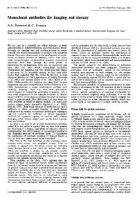
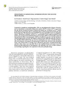
FIGURE 2. Distribution of MAb 35 or its fragments and control IgG or fragments injected simultaneously into nude mice bearing grafts of a human carcinoma. The concentration of antibody (shaded bars) and control IgG (open bars) per gram of tissue is expressed in percentages of total specific radioactivity recovered at various times after injection as indicated in the text. The vertical lines represent the standard deviations calculated from groups of four to seven animals per MAb or fragment. T, tumor; S, spleen; Li, liver; K, kidneys; GI, gastrointestinal tract; H, heart; Lu, lungs; SG, salivary glands; C, carcass and head; B, blood. 7 n u d e mice b e a r i n g grafts o f the h u m a n colon c a r c i n o m a Co-112 were injected simultaneously with ~3~I-labeled M A b or their f r a g m e n t s a n d with their n o r m a l I g G c o u n t e r p a r t labeled with l~SI, a n d were dissected 2 - 5 d later. T h e results o f a n t i b o d y a n d n o r m a l I g G concentrations p e r g r a m o f t u m o r s a n d n o r m a l organs (expressed in p e r c e n t a g e o f total radioactivity r e c o v e r e d for each isotope) are shown in Figs. 2 - 5 . T h e t u m o r uptakes for a n t i b o d y a n d n o r m a l I g G calculated by c o m p a r i s o n with individual n o r m a l organs a n d with the whole m o u s e are shown in Fig. 6. T h e specificity indices o f t u m o r localization c o m p a r e d with the whole animal are shown in T a b l e II. For intact M A b the t u m o r a n t i b o d y uptakes as c o m p a r e d with the whole m o u s e
418
T U M O R LOCALIZATION OF FRAGMENTS OF MONOCLONAL ANTIBODIES
Mab
202
w m
127
F(ab') 2
intact
=. 9O
¢b
Fab
89
80
o
¢L 70 O
>
o o
6O 50
>
40
o o
30
"o
c o o @
20 10
a.
O
.
• T ' S ' L i ' K 'GI' H 'Lu'SG' C ' B '
, , , , , • . . . . T S Li K GI H LuSG C B
' T ' S'Li' K'GI'H'Lu'SG'C'
B'
Distribution of MAb 202 as described in the legendto Fig. 2.
FIGURE 3.
Mab
23 F(ab') 2
intact
o w
90 80
cL
70 60 50
>
40
eJ o
30
U
20 o
10
@
D. 0 T "S ' Li' K 'GI' H 'Lu'SG' C ' B ' FIGURE 4.
• T ' S ' Li' K 'GI" H 'Lu'SG' C ' B '
Distribution of MAb 23 as described in the legend to Fig. 2.
ranged between 7 and 15 and the specificity indices between 3.4 and 6.8. The lowest values were obtained with MAb 35 and the highest with MAb 202. Compared with intact MAb, the F(ab')2 fragments gave markedly increased tumor uptakes with values ranging between 12 and 24, as well as higher specificity indices, ranging between 5.3 and 8.2. The highest results for F(ab')~ fragments were observed with MAb 35 followed by MAb 202. Fab fragments from the two most promising MAb (clone 35 and 202) were
BUCHEGGER Mab
419
AL.
192
intact o o
ET
F(ab') 2
90
80 o.
~' @
7O 6O
~
50
.~
4O
o
30
.
20
o
~
10 0 T S Li K G! H L u S G C
FIGURE 5.
B
T S Li K G! H L u S G C B
D i s t r i b u t i o n o f M A b 1 9 2 as d e s c r i b e d in t h e l e g e n d t o Fig, 2.
prepared and tested. They both gave very high tumor uptakes of 34 and 82, and specificity indices of 12 and 19, for Fab 202 and 35, respectively. It is interesting that the Fab fragment of MAb 35 gave the best results, because MAb 35 is also the most specific anti-CEA MAb, since it does not cross-react with granulocyte glycoprotein(s). The specificity indices calculated for different organs (data not shown) were rather constant for one type of MAb or fragment and consequently very close to the mean values shown in Table II. The stability of these specificity indices is also reflected by the fact that tumor uptakes of antibodies plotted against those of control IgG fall on a straight line, whose slope is the expression of the specificity of tumor localization (Fig. 6). T u m o r uptakes can show large variations in relation to the degree of vascularization of different organs but specificity indices are more constant and thus represent more meaningful values. T h e marked increase of tumor uptakes and specificity indices observed with MAb fragments is in part due to their more rapid elimination, as shown by the decrease of concentration of recovered radioactivity from the whole animal (Table III.) The absolute concentration of fragments in tumors was also decreased (as compared to intact MAb) but to a lesser degree than their concentration in normal tissues (Table III). External Scanning Studies. Intact MAb gave clear tumor detection by scanning only 3 d after injection, for relatively large tumors of 0.5-1 g as shown in Fig. 7A. The failure to detect tumors of smaller size or at earlier times was due mostly to the abundance of labeled intact MAb in the blood and thus in the wellvascularized organs, as determined in a few animals sacrificed and dissected at day 2 and 3 (data not shown), and by the results obtained at day 4-5 (Fig. 2-5). F(ab')~ fragments gave earlier positive tumor detection at day 2 (Fig. 7B) but these fragments did not give very contrasted scannings for tumors smaller than
.c..b3s I .c..b,o,
420 T U M O R LOCALIZATION OF FRAGMENTS OF MONOCLONAL ANTIBODIES Mab
100
]-
UI
< IG. : >a O m m DZ < =J < Z
O
=J O O Z O :t M
Mab
192
-"
oI ~
80
23
-40
70
Om /
60-
Ac -30
3C •
LI AC
50"
-20
40-
c
-c
AC 30-
g
. 20-
)E
~m
K
K
&Li ~B TK
10"
md~ c
/LIB
K
o---'T-1 ~r'~ ~'~--" - o 0 3 6 0 3 6 0 3 6
II Fab
K
II F(ab') 2
Intact
I I
II Fab
-10
/
01 21 01 2 3 01 23 01 2301
I
C
/li.J u,, i .',, t I',, /
10
I
B
II F(ab') 2 intact
II
2
&K
LIJ
1"~2 01 23
II
II
F(ab') 2 intact
F(ab') 2 Intact
I
1 2 5 I C O N T R O L IgG U P T A K E
FIGURE 6. Tumor uptakes of MAb or fragments (ordinate) plotted against those of normal IgG or fragments (abscissa). Tumor uptakes were calculated by comparison with the whole mouse without the tumor (fia), carcass (C), liver (Li), blood (B), and kidneys (K). First panel: Results obtained with MAb 35. Fab fragment (O), F(ab')2 fragment (&), and intact MAb (O). Panels 2-4: Similar analysis of MAb 202, 23, and 192, respectively, and their fragments. The slope of the straight line joining the zero of the two axes with the tumor uptake obtained by comparison with the whole mouse (ill) is the expression of the specificity of tumor localization.
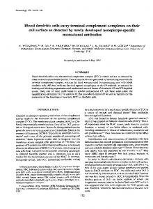
0.5g. The best scanning results were obtained with Fab fragments which allowed the clear detection of tumors weighing only 0.1-0.3 g 2 d after injection (Fig. 7, C and D). It should be noted that all scanning pictures shown represent raw data without any computerized background subtraction. Autoradiographs of Tumor Sections. Autoradiographs demonstrate that injected labeled MAb and fragments localized in tumor nodules and not in the stromai connective tissue of mouse origin, as shown for intact MAb (Fig. 8A). However,
BUCHEGGER ET AL.
421
TABLE II Spec~city Indices of Tumor Localization Obtained with Intact MAb and Fragments MAb form
MAb
35 192 202 23 Intact 3.4 ± 0.7* 3.6 ± 0.3 6.8 ± 2.7 5.0 ± 0.7 F(ab')2 8.2 ± 5.7 7.5 ± 5.0 7.9 ± 3.3 5.3 ± 1.3 Fab 19.0 ± 5.5 ND 11.9 ± 4.3 ND * The specificity indices ± the standard deviation were calculated by comparison with the normal tissue of the whole mouse according to the formula given in Materials and Methods. ND, not done. TABLE III Absolute Concentration of Antibody Radioactivity Recovered in Tumors and in Whole Mice
MAb
MAb form Tumor Mouse normal tissues Intact (day 4-5)* 18.2 _ 6.3' (9.1)e 2.61_+0.46'(1.31) 0 MAb 35 F(ab')2 (day 3) 4.9 _+2.5 (2.5) 0.20 - 0.03 (0.10) Fab (day 2-3) 7.5 _ 1.2 (2.5) 0.09 + 0.02 (0.03) MAb 202 Intact (day 4-5) 9.1 ± 2.5 (4.5) 0.58 ± 0.20 (0.29) F(ab')2 (day 3) 3.4 ± 1.0 (1.7) 0.15 ± 0.06 (0.08) Fab (day 2-3) 2.9 ± 1.3 (1.0) 0.09 ± 0.02 (0.03) * Days of dissection and counting. * Absolute concentration of antibody radioactivity recovered in ~Ci/g of tissues _ standard deviation (corrected for physical half-life of isq). a Concentration of antibody radioactivity recovered per gram of tissue expressed in percent of injected dose (corrected for physical half-life of zslI). the labeled intact M A b were not distributed h o m o g e n e o u s l y in the entire t u m o r tissue. T h e r e were areas o f intense c o n c e n t r a t i o n o f radioactivity in n o n n e c r o t i c t u m o r tissue, whereas o t h e r areas morphologically very similar c o n t a i n e d m u c h less radioactivity. T h e high c o n c e n t r a t i o n o f radioactivity at the p e r i p h e r y o f t u m o r nodules suggests that the vascularization was an i m p o r t a n t factor for this a n t i b o d y distribution. In contrast, necrotic areas o f the t u m o r showed diffuse a n d very low concentrations o f radioactivity. Labeled F(ab')~ a n d Fab f r a g m e n t s a p p e a r e d to p e n e t r a t e d e e p e r into the t u m o r nodules a n d to c o n c e n t r a t e in the p s e u d o l u m e n o f the malignant glands (Fig. 8, B a n d C) that c o n t a i n e d the highest concentration o f CEA, as shown by in vitro i m m u n o p e r o x i d a s e staining on frozen sections f r o m the same t u m o r (Fig. 8D). Mice injected with labeled control n o r m a l I g G or f r a g m e n t showed very low diffuse radioactivity in the t u m o r (data not shown). Discussion W e describe herein the first t h o r o u g h c o m p a r a t i v e study o f t u m o r localization in vivo o f intact M A b a n d their F(ab')~ a n d Fab f r a g m e n t s directed against a tumor-associated antigen. Radiolabeled fragments, in particular Fab, gave m u c h h i g h e r t u m o r uptakes a n d b e t t e r t u m o r detection by external scanning than did intact M A b in the m o d e l o f n u d e mice g r a f t e d with a h u m a n colon carcinoma. T h i s e x p e r i m e n t a l m o d e l was chosen because h u m a n colon carcinomas g r a f t e d
422
TUMOR LOCALIZATION OF FRAGMENTS OF MONOCLONAL ANTIBODIES
FIGURE 7. Whole-bodyscannings of nude mice bearing grafts of a human colon carcinoma obtained after intravenous injection of In'I-labeled MAb or fragments. (A) Mouse injected with intact MAb 202; tumor weighing 1 g, scan obtained at day 3 after injection. (B) MAb 202 F(ab')2 fragment, tumor of 0.7 g~ scan at day 2. (C) MAb 202 Fab fragment, tumor of 0.3 g, scan at day 2. (D) MAb 35 Fab fragment, tumor of 0.1 g, scan at day 2. Scans shown at the right of the corresponding mice represent raw data without any background subtraction.
in nude mice retain the same histological m o r p h o l o g y as the primary h u m a n t u m o r (28), and synthetize and release CEA (7), as observed in patients. T h e specificity o f the results was assessed by using the paired labeling m e t h o d described by Pressman et al. (29) in which a control mouse IgG or its fragments labeled with 1~5I is injected simultaneously with the 13~I-labeled MAb. This internal control allows one to correct for any nonspecific accumulation o f labeled proteins in t u m o r tissues and the calculated specificity indices represent the most relevant evaluation o f the MAb's capacity to bind to antigens present in the tumor. It was o f particular interest to evaluate antibody fragments, since o u r preliminary clinical results using F(ab')2 fragments f r o m o u r first anti-CEA MAb (2) and f r o m a MAb directed against a n o t h e r colon carcinoma antigen (30) suggested that these fragments gave less nonspecific accumulation o f radioactivity in the
FIGURE 8. (A-C) Autoradiographs of methacrylate-embedded, 2-/*m sections from the human colon carcinoma C o l 12 xenografted into nude mice, obtained after injection of l~5I-labeled intact MAb 202 (A), F(ab')z fragments of MAb 23 (B), and Fab fragments of MAb 35 (C). Note that in A, the stromal connective tissue (which is very lightly stained by the nuclear fast red) contains almost no radioactivity. (D) Immunoperoxidase staining demonstrating the
CEA distribution on a frozen section from the same transplanted colon carcinoma. MAb 2~ and the avidin-biotin system (26) were used to reveal CEA. Note that the positive immunoperoxidase staining corresponds with the silver grains of autoradiographs obtained after injection of MAb fragments (B and C). All sections, × 250.
t~ t~
>
©
C3
424
T U M O R L O C A L I Z A T I O N OF F R A G M E N T S OF M O N O C L O N A L ANTIBODIES
reticuloendothelium than intact MAb. However, clinical results are often difficult to interpret since they are derived from the evaluation of photoscanning pictures, and direct measurement of radioactivity can be obtained only from surgically resected malignant and normal tissues. Thus, the present experimental results represent a much stronger argument in favor of using fragments of MAb for the detection of tumor in patients. In particular, the fragments of MAb 35, which gave the highest tumor uptakes and the least cross-reaction with human granulocytes, appear to be the best candidate for localization of colorectai carcinoma by immunoscintigraphy in patients. Our autoradiographic results confirmed the specificity of tumor localization of anti-CEA MAb and fragments at the histological level and differ from those of Lewis et al. (31) who reported that radiolabeled intact goat anti-CEA antibodies localized in the stromal connective tissue of mouse origin present within the grafted human tumor. Despite their specificity, however, our results provide a warning for those who are already considering the use of radiolabeled anti-CEA antibodies for therapy (32), since the autoradiographs show that the labeled MAb localized in areas with high CEA concentration but not on all carcinoma cells. Therefore if radiotherapy with radiolabeled MAb is considered one should select isotopes capable of destroying tumor cells within a radius of 50-100 #m. High energy alpha- or beta-emitting isotopes with these properties can be coupled to antibodies by metal chelates such as diethylenetriamine pentaacetic acid (33, 34). Using this chelate, we have recently coupled l l q n d i u m to MAb 35 and shown that after injection into nude mice, it gave similar tumor uptakes as those obtained with a3q-labeled MAb. Thus, the MAb and the nude mouse model described here should provide a means to evaluate the possibility of destroying human solid tumors by radioimmunotherapy. Summary Four monoclonal antibodies against carcinoembryonic antigen (CEA) have been selected from 32 hybrids that produce antibodies against this antigen, by the criteria of high affinity for CEA and low cross-reactivity with granulocyte glycoprotein(s). T h e specificity of tumor localization in vivo of the four MAb, and their F(ab')2 and Fab fragments was compared in nude mice bearing grafts of a serially transplanted, CEA-producing, human colon carcinoma. T h e distribution of radiolabeled MAb and their fragments after intravenous injection was analyzed by direct measurement of radioactivity in tumor and normal organs, as well as by whole-body scanning and by autoradiography of tumor sections. Paired labeling experiments, in which l saI-labeled antibody or fragments and l~SI-labeled control IgG are injected simultaneously, were undertaken to determine the relative tumor uptakes of each labeled protein. T h e tumor antibody uptake divided by that of control IgG defines the specificity index of localization. T u m o r antibody uptakes (as compared with the whole mouse), ranging between 7 and 15, and specificity indices ranging between 3.4 and 6.8, were obtained with the four intact MAb at day 4-5 after injection. With F(ab')2 fragments of the four MAb, at day 3, the tumor antibody uptakes ranged between 12 and 24 and the specificity indices between 5.3 and 8.2. With the Fab fragments prepared from the two most promising MAb, the antibody uptakes reached values of 34 and 82
BUCHEGGER ET AL.
425
at day 2-3 and the specificity indices were as high as 12 and 19. The scanning results paralleled those obtained by direct measurement of radioactivity. With intact MAb, tumor grafts of 0.5-1 g gave very contrasted positive scans 3 d after injection. Using MAb fragments, tumors of smaller size were detectable earlier. T h e best results were obtained with Fab fragments of MAb 35, which gave clear detections of tumors weighing only 0.1 g as early as 48 h after injection. Autoradiographs of t u m o r sections from mice injected with 125I-labeled MAb demonstrated that the radioactivity was localized in the tumor tissues and not in the stromal connective tissue of mouse origin. T h e highest radioactivity concentration was localized in areas known to contain CEA such as the pseudolumen of glands and the apical side of carcinoma cells. T h e penetration of radioactivity in the central part of t u m o r nodules and the pseudolumen appeared to be increased with the use of MAb fragments. We thank Christine Mettraux and Manuela Rosenberger for excellent technical assistance. We are also grateful to Prof. J.-C. Cerottini and Prof. H. Isliker for advice and suggestions and to Dr. H. Engers for reviewing the manuscript. Receivedfor publication 14 April 1983.
1. 2.
3.
4. 5. 6. 7. 8. 9.
References K6hler, G., and C. Milstein. 1975. Continuous cultures of fused cells secreting antibody of predefined specificity Nature (Lond.). 256:495. Mach, J.-P., F. Buchegger, M. Forni, J. Ritschard, C. Berche, J.-D. I,umbroso, M. Schreyer, C. Girardet, R. S. Accola, and S. Carrel. 1981. Use of radiolabeled monoclonal anti-CEA antibodies for the detection of human carcinomas by external photoscanning and tomoscintigraphy, lmmunol. Today (Amst.) 2:239. Berche, Ch., J.-P. Mach, J.-D. Lumbroso, C. Langlias, F. Aubry, F. Buchegger, S. Carrel, Ph. Rougier, C. Parmentier, and M. Tubiana. 1982. Tomoscintigraphy for detecting gastrointestinal and medullary thyroid cancers: first clinical results using radiolabelled monoclonal antibodies against carcinoembryonic antigen. Brit. Med. J. 285:1447. Gold, P., and S. O. Freedman. 1965. Demonstration of tumor-specific antigens in human colonic carcinomata by immunological tolerance and absorption techniques. J. Exp. Med. 121:439. Accolla, R. S., S. Carrel, and J.-P. Mach. 1980. Monoclonal antibodies specific for carcinoembryonic antigen and produced by two hybrid cell lines. Proc. Natl. Acad. Sci. USA. 77:563. Haskell, C. M., F. Buchegger, M. Schreyer, S. Carrel, and J.,P. Mach. Monoclonal antibodies to carcinoembryonic antigen: ionic strength as a factor in the selection of antibodies for immunoscintigraphy. Cancer Res. In press. Mach, J.-P., S. Carrel, C. Merenda, B. Sordat, and J.-C. Cerottini. 1974. In vivo localization of radiolabeled antibodies to carcinoembryonic antigen in human colon carcinoma grafted into nude mice. Nature (Lond.). 248:704. Goldenberg, D.-M., D.-F. Preston, F.-J. Primus, H.-J. Hansen. 1974. Photoscan localization of GW-39 tumors in hamsters using radiolabeled anti-carcinoembryonic antigen immunoglobulin G. Cancer Res. 34:1. Warenjus, H.-M., G. Galfre, N. M. Bleehan, and C. Milstein. 1981. Attempted targeting of a monoclonal antibody in a human tumor xenograft system. Eur. J. Cancer. 17:1009.
426
TUMOR LOCALIZATION OF FRAGMENTS OF MONOCLONAL ANTIBODIES
10. Moshakis, V., M.J. Bailey, M. G. Ormerod, J. H. Westwood, A. M. Neville. 1981. Localization of human breast-carcinoma, xenografts using antibodies to carcinoembryonic antigen. Br. J. Cancer 43:575. 11. Pimm, M. V., M. J. Embleton, A. C. Perkins, M. R. Price, R. A. Robins, G. R. Robinson, and R. W. Baldwin. 1982. In vivo localization of anti-osteogenic sarcoma 791T monoclonal antibody in osteogenic sarcoma xenografts. Int. J. Cancer 30:75. 12. Hedin, A., B. Wahren, and S. Hammarstr6m. 1982. Tumor localization of CEAcontaining human tumors in nude mice by means of monoclonal anti-CEA antibodies. Int. J. Cancer. 30:547. 13. Ghose, T., S. Ferrone, K. Imai, St.-T. Norveil Jr., St.-J. Luner, R. H. Martin, and A. H. Blair. 1982. Imaging of human melanoma xenografts in nude mice with a radiolabeled monoclonal antibody.J. Natl. Cancer Inst. 69:823 14. Herlyn, D., J. Powe, A. Alavi, J.-A. Mattis, M. Herlyn, C. Ernst, R. Vaum, and H. Koprowski. Radioimmunodetection of human tumor xenografts by monoclonal antibodies. Cancer Res. In press. 15. Colcher, D., M. Zalutsky, W. Kaplan, D. Kufe, F. Austin, and J, Schlom. 1983. Radiolocalization of human mammary tumors in athymic mice by a monoclonal antibody. Cancer Res. 43:736. 16. Fritsche, R.,andJ.-P. Mach. 1977. Isolation and characterization ofcarcinoembryonic antigen (CEA) extracted from normal human colon mucosa. Immunochemistry. 14:119. 17. Stfihli, C., T. Staehelin, V. Miggiano, J. Schmidt, and P. Hfiring. 1980. High frequencies of antigen-specific hybridomas: dependence on immunization parameters and prediction by spleen cell analysis.J. Immunol. Methods, 32:297. 18. K6hler, G., S. C. Howe, and C. Milstein. 1976. Fusion between immunoglobulinsecreting and nonsecreting myeloma cell lines. Eur. J, Immunol. 6:292. 19. Mach, J.-P., and G. Pusztaszeri. 1972. Carcinoembryonic antigen (CEA): demonstration of partial identity between CEA and a normal glycoprotein. Immunochemistry. 9:1031. 20. Von Kleist, S., G. Chavanel, P. Burtin. 1972. Identification of an antigen from normal human tissue that cross-reacts with carcinoembryonic antigen. Proc. Natl. Acad. Sci. USA. 69:2492. 21. Burtin, P., P. C. Quan, and M. C. Sabine. 1975. Nonspecific cross-reacting antigen as a marker for human polymorphs, macrophages and monocytes. Nature (Lond.) 255:714. 22. Buchegger, F., R. S. Accolla, S. Carrel, A. Carmagnola, C. Girardet, J.-P. Mach. 1980. Use of monoclonal anti-CEA antibodies in immunoadsorbent columns and solid-phase radioimmunoassay. Protides Biol. Fluids Proc. Colloq. 28:511. 23. Carrel, S., R.-S. Accolla, A. L. Carmagnola, andJ.-P. Mach. 1980. Common human melanoma-associated antigen(s) detected by monoclonal antibodies. Cancer Res. 40:2523. 24. Nisonoff, A., F. C. Wissler, and D. L. Woernley. 1960. Properties of univalent fragment of rabbit antibody isolated by sPecific adsorption. Arch. Biochem. Biophys. 88:241. 25. Porter, R. R. 1959. The hydrolysis of rabbit gamma globulin and antibodies with crystalline papain. Biochem. J. 73:119. 26. Schreyer, M., C. M. Haskell, F. Buchegger, C. Girardet, S. Carrel, and J.-P. Mach. Localisation of tumor-associated antigens on sections of human colon carcinoma grafted into nude mice with mouse monoclonal antibodies using the avidin-biotinimmunperoxidase reaction. In Proceedings Fourth International Workshop of Immune-deficient Animals in Experimental Research. S. Karger AG, Basel. In press. 27. Buchegger, F., C. Mettraux, R. S. Accolla, S. Carrel, andJ.-P. Mach. 1982. Sandwich
BUCHEGGER ET AL.
28.
29. 30.
31.
32. 33.
34.
427
enzyme immunoassay using three monoclonal antibodies against different epitopes of CEA. Immunol. Lett. 5:85. Povlsen, C. O., and J. Rygaard. 1971. Heterotransplantation of human adenocarcinomas of the colon and rectum to the mouse mutant nude. A study of nine consecutive transplantations. Acta Pathol. Microbiol. Scand. Sect. A Pathol. 79:159. Pressman, D., E. D. Day, and M. Blau. 1975. The use of paired labeling in the determination of tumor-localizing antibodies. Cancer Res. 17:845. Mach, J.-P., J.-F. Chatal, J.-D. Lumbroso, F. Buchegger, M. Forni, J. Ritschard, Ch. Berche, J.-Y. Douillard, S. Carrel, M. Herlyn, Z. Steplewski, and H. Koprowski. Tumor localization in patients by radiolabeled antibodies against colon carcinoma. Cancer Res. In press. Lewis, J. C. M., K. D. Bagshawe, and P. A. Keep. 1982. The distribution of parenterally administered antibody to CEA in colorectal xenografts. Preliminary findings. Oncodev. Biol. Med. 3:161. Order, S. E., J. L. Klein, D. Ettinger, P. Alderson, S. Siegelman, and P. Leichner. 1980. Use of isotopic immunoglublin in therapy. Cancer Res. 40:3001. Khaw, B.,J. T. Fallon, H. W. Strauss, and E. Haber. 1980. Myocardial infarct imaging of antibodies to canine cardiac myosin with indium-111-diethylenetriamine pentaacetic acid. Science (Wash. DC). 209:295. Scheinberg, D. A., M. Strand, and O. A. Gansow. 1982. Tumor imaging with radioactive metal chelates conjugated to monoclonal antibodies. Science (Wash. DC). 215:1511.