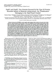
APPLIED AND ENVIRONMENTAL MICROBIOLOGY, Dec. 1998, p. 4918–4923 0099-2240/98/$04.0010 Copyright © 1998, American Society for Microbiology. All Rights Reserved.
Vol. 64, No. 12
Ralstonia solanacearum Pectin Methylesterase Is Required for Growth on Methylated Pectin but Not for Bacterial Wilt Virulence JULIE TANS-KERSTEN, YANFEN GUAN,†
AND
CAITILYN ALLEN*
Department of Plant Pathology, University of Wisconsin—Madison, Madison, Wisconsin 53706 Received 31 July 1998/Accepted 8 October 1998
Ralstonia (Pseudomonas) solanacearum causes bacterial wilt, a serious disease of many crop plants. The pathogen produces several extracellular plant cell wall-degrading enzymes, including polygalacturonases (PGs) and pectin methylesterase (Pme). Pme removes methyl groups from pectin, thereby facilitating subsequent breakdown of this cell wall component by PGs, which are known bacterial wilt virulence factors. R. solanacearum PGs could not degrade 93% methylated pectin unless the substrate was first demethylated by Pme, but as the degree of methylation of the pectin substrate decreased, PG activity increased. Primers derived from a published pme sequence generated an 800-bp DNA probe fragment, which identified Pme-encoding plasmids from a R. solanacearum genomic library. A pme chromosomal mutant had no detectable Pme activity in vitro and no longer grew on 93% methylated pectin as a carbon source. Curiously, the pme mutant, which had no detectable PG activity on highly methylated pectin, was just as virulent as the wild-type strain on tomato, eggplant (aubergine), and tobacco. Since PG activity is required for full virulence, this result suggests that the pectin in these particular hosts may not be highly methylated, or that the breakdown of highly methylated pectin is not a significant factor in the disease process in general. A positive response regulator of PG production called PehR was not required for wild-type Pme production. However, a mutant strain lacking PhcA, which is a global regulator of several virulence genes, produced no detectable Pme activity. Thus, pme expression is directly or indirectly regulated by PhcA but not by PehR. Members of this consortium include an endoglucanase (Egl), which breaks down cellulose (30), and three polygalacturonases (PGs), which hydrolytically degrade the pectic compounds that are a major constituent of the primary plant cell wall and middle lamella (3). Pectins are complex polysaccharides, primarily chains of a-1,4-linked galacturonic acid: they also contain significant amounts of other sugars (12). A variable but substantial fraction of galacturonate residues in plant pectins is methylated (22, 26, 38). In contrast, polygalacturonate is a simple polymer of unmethylated galacturonate. R. solanacearum produces one endo-PG, PehA (also known as PglA); an exo-poly-a-D-galacturonosidase, PehB; and an exo-PG, PehC. PehA cleaves polygalacturonate internally, yielding trimers and larger galacturate oligomers. PehB releases digalacturonic acid, while PehC generates only monogalacturonic acid from the same substrate. Site-directed mutants lacking PehA, PehB, or both are all significantly reduced in virulence, demonstrating that PG activity contributes quantitatively to bacterial wilt disease development (19, 31). However, PGs cannot attack highly methylated pectins, so methyl groups must be removed from pectin substrates before hydrolytic cleavage. R. solanacearum produces one known pectin methylesterase, or Pme (E.C. 3.1.1.11), which cleaves methoxyl groups from methylated pectin, thereby making it susceptible to PG activity. The gene encoding this enzyme was cloned from R. solanacearum 50905 (35), but its role in pectin degradation and pathogenesis has not been studied. Little is known about the biological function of Pme in plant pathogenesis. Erwinia chrysanthemi, which causes soft rot disease, produces two very different Pme’s, PemA and PemB (24, 33). PemA, which is extracellular, is required for wild-type systemic spread of the bacterium in African violets (7). The role in virulence of PemB, which is an outer membrane lipoprotein, has not been determined. To better understand the role of Pme in bacterial wilt disease, we cloned and characterized the pme gene from R. so-
Bacterial wilt is a devastating plant disease that affects economically important hosts, such as potatoes, tomatoes, bananas, and tobacco (14). Ralstonia (formerly Pseudomonas) solanacearum (Smith) Yabuuchi et al., which causes the disease, has an extensive host range, including over 450 plant species in tropical and warm temperate zones worldwide (27). In many parts of the world, this disease is a primary constraint on crop production (10, 14). The bacterium is generally soil borne: it enters host plant roots through wounds or at lateral root emergence points, colonizes the root cortex, and subsequently invades the developing xylem vessels (37, 39). Once established in the xylem, the pathogen spreads rapidly throughout the plant, inducing yellowing, stunting, wilting, necrosis, and death. Bacterial wilt disease appears to be the result of multiple virulence factors working in concert. R. solanacearum produces extracellular polysaccharide (EPS) that is important for disease development, although its precise mechanism of action is uncertain (17, 21, 32). Other known virulence factors belong to an extracellular enzyme consortium that breaks down plant cell walls. These enzymes apparently facilitate bacterial invasion and spread by digesting cortical cell walls, the pectic gels surrounding lateral root emergence points, and the pit membranes that separate adjacent xylem vessels. Enzymatic cell wall degradation probably creates the gels and tyloses typically found in the vessels of wilting plants (6). In addition, these enzymes may also release nutrients that enable rapid bacterial multiplication. * Corresponding author. Mailing address: Department of Plant Pathology, University of Wisconsin—Madison, 1630 Linden Dr., Madison, WI 53706. Phone: (608) 262-9578. Fax: (608) 263-2626. E-mail:
[email protected]. † Present address: Department of Molecular Biology and Microbiology, Sackler School of Graduate Biomedical Sciences, Tufts University, Boston, MA 02111. 4918
R. SOLANACEARUM PECTIN METHYLESTERASE
VOL. 64, 1998
4919
TABLE 1. Strains and plasmids used in this study Strain
E. coli DH5a
R. solanacearum K60 K61-B K60-309 K60-509 K71 K60-phcA K71-phcA Plasmids pBluescript KS1 pT7Blue pLAFR3 pMeB1 pMe71 pMeL1 pMe71DSacII pUM24 pMe7SB
Relevant characteristicsa
Reference or source
F2 endA1 relA f80 lacZDM15 hsdR17 supE44 thi-1 recA1 gyrA96
13
Wild-type race 1, biotype 1 pme::sacB pehA::V pehB::aacC1 Smr Gmrb pehA::V pehB::aacC1 pme::sacB pehR::Tn3-uidA Kmr phcA::V Smr pehR::Tn3-uidA phcA::V Kmr Smr
23 This study 19
Apr Apr Tcr 2.7-kb ClaI fragment of pme in pBluescript 1.8-kb EcoRI fragment of pme in pT7Blue 1.8-kb EcoRI fragment of pme in pLAFR3 Internal 0.55-kb deletion of pme in pMe71 NptI sacB sacR Kmr 3.8-kb SacB-Kan cartridge inserted into SacII deletion of pMe71DSacII
Stratagene Novagen 36 This study
This study 1 1 1 FIG. 1. Subcloning and deletion mutagenesis of pme. A 2.7-kb ClaI fragment of strain K60 DNA encoding Pme activity was cloned into pBluescript to create pMeB1. From this, an internal 1.8-kb EcoRI-ClaI fragment encoding Pme activity was subcloned into pLAFR3 and pT7Blue to create pMeL1 and pMe71, respectively. A 3.8-kb DNA fragment containing an NptI-sacB-SacR cartridge (28) was cloned into the remaining SacII site of SacII deletion construct pMe71 to create pMe7SB.
This study This study This study 28 This study
a Ap, ampicillin; Tc, tetracycline; Km, kanamycin; Gm, gentamicin; Sm, streptomycin. b pehA::V denotes the endo-PG-encoding pehA gene interrupted by the V gene cartridge, which encodes streptomycin resistance. pehB::acc1 denotes the pehB exo-poly-a-D-galacturonosidase gene interrupted by a gentamicin resistance gene cartridge.
lanacearum K60. We then constructed a chromosomal pme mutant strain to determine how the loss of this activity affected bacterial virulence and ability to use highly methylated pectin as a sole carbon source. In addition, we studied the regulation of Pme expression by measuring Pme activity in various regulatory mutant backgrounds. MATERIALS AND METHODS Bacterial strains, plasmids, and culture conditions. The bacterial strains and plasmids used in this research are listed in Table 1. R. solanacearum strains were cultured at 28°C in CPG broth (1 g of Casamino acids/liter, 10 g of peptone/liter, 5 g of glucose/liter) (15) or on TZC plates containing CPG plus 1.8% agar and 0.05% 2,3,5-triphenyltetrazolium chloride (23). Boucher’s minimal medium (BMM) (8) supplemented with 0.2% (wt/vol) citric acid was used to grow strains for the PG reaction product analysis or the quantitative Pme assay. Antibiotics were added at standard concentrations as required. Growth on various carbon sources was assayed in BMM supplemented with 0.2% (wt/vol) carbon source. Pectin or polygalacturonic acid used as a sole carbon source or for reaction product analysis was washed four times in 70% ethanol to remove low-molecular-weight soluble nutrients or contaminants. For carbon source utilization experiments, cultures were inoculated in 250-ml Erlenmeyer flasks containing 25 ml of medium to an initial optical density at 600 nm (OD600) of 0.020 and incubated in a 300-rpm shaker at 28°C; bacterial growth was measured spectrophotometrically as culture OD600. Tobacco extract for plant induction studies was obtained by a method similar to that of Beaulieu and Van Gijsegem (5). Fully expanded tobacco leaves (cv. Bottom Special) were surface sterilized, rinsed with sterile water, cut into 1-cm strips, and shaken overnight in BMM without carbon. The resulting extract was
filter sterilized. Bacterial cultures were then grown in BMM supplemented with 0.2% carbon source plus 0.5% (vol/vol) tobacco extract. Chemicals and enzymes. Growth medium components were purchased from Difco Laboratories (Detroit, Mich.). Highly methylated pectin (P9561; 93% methylated from citrus) was from Sigma Chemical Co. (St. Louis, Mo.); apple pectin (76282; 70 to 75% methylated) and citrus pectin (76280; 63 to 66% methylated) were from Fluka Biochemika (Buchs, Switzerland). DNA restriction and modification enzymes were from Promega Corp. (Madison, Wis.). Radiochemicals were from NEN (Boston, Mass.). Electrophoresis chemicals and nylon transfer membranes were from Bio-Rad Laboratories (Richmond, Calif.). Oligonucleotides were purchased from the University of Wisconsin Biotechnology Center (Madison, Wis.). All other chemicals were from Sigma Chemical Co. DNA manipulations. Recombinant DNA manipulations were performed in accordance with standard procedures (4). R. solanacearum strains were transformed by electroporation as previously described (2). To clone the strain K60 pme gene, an internal DNA fragment of the pme structural gene was amplified by PCR using primers derived from the strain 50905 pme gene (35). The primer sequences were 59GCCGGCACCTACAACGAACT39 and 59GCCCCCGTGTT GTTGTACTC39. The resulting 800-bp fragment was radiolabeled and used to probe a R. solanacearum K60 chromosomal DNA library (2). Construction of pme mutant and PG-pme triple mutant strains. A 0.55-kb SacII fragment was deleted from the pme coding sequence by using the three internal SacII sites in the gene. This deletion generated plasmid pMe71DSacII (Fig. 1). We inserted a 3.8-kb fragment of the pUM24 NptI-sacB-sacR gene cartridge, which encodes resistance to kanamycin (28), into the remaining SacII site of pMe71DSacII to create pMe7SB. This disabled pme::sacB construct was exchanged into the genomes of wild-type strain K60 and K60-309 (pehA pehB double mutant) by gene replacement as previously described (19). Enzyme activity assays. Pme and PG activities were measured qualitatively by using a modification of the pectin overlay method, which allows detection of both activities on a single petri plate (33). Colonies grown 24 to 36 h on CPG were overlaid with two successive layers of substrate containing 0.5% 93%-methylated pectin, 20 mM EDTA (pH 8.0), 50 mM Tris (pH 8.0), 0.2% Triton X-100, and 0.8% agarose. The overlaid plates were incubated overnight at 28°C, stained with 0.5% (wt/vol) ruthenium red with gentle shaking, and destained with distilled water. Pme activity appeared as a dark purplish-red halo surrounding colonies; PG activity was manifested as a clear zone surrounding the colonies. Pme activity was measured quantitatively as production of methanol from methylated pectin (40). Pme activity was calculated as micrograms of methanol produced per minute per CFU for each strain, based on a methanol standard curve. Apoplastic fluids from tobacco leaves infused with 5 3 104 bacteria/cm2 were obtained after 18 h of bacterial growth in planta as previously described (2); Pme activity in apoplastic fluids was measured quantitatively as described above. PG reaction product analysis. Three hundred microliters of cleared supernatant from cultures grown to an OD600 of .1.0 was reacted with 700 ml of 0.4% (wt/vol) pectin or polygalacturonic acid; 25 ml of each reaction was separated by thin-layer chromatography (TLC) and enzyme reaction products were detected with phosphomolybdate spray as previously described (25). Pure mono-, di-, and trigalacturonic acid (Sigma G2125, D4288, T7407) were used as standards.
4920
TANS-KERSTEN ET AL.
APPL. ENVIRON. MICROBIOL. TABLE 2. Pme activity produced by various R. solanacearum strains Strain
Relevant phenotype
% Endo-PG activitya
Pme activityb
K60 K61B K71 K60-phcA K71-phcA
Wild type Pme2 PehR2 PhcA2 PehR2 PhcA2
100 100 5 180 5
3.32 6 0.03 0.06 6 0.05 3.26 6 0.05 0.09 6 0.07 0.13 6 0.08
a Endo-PG activity determined previously (1) expressed as a percentage of that of the wild type. b Pme activity expressed in micrograms of methanol produced per minute per cell 3 10210 6 standard error.
FIG. 2. Overlay activity assay for Pme production. Twenty-four-hour colonies grown on CPG were overlaid with two successive layers of highly methylated pectin substrate and stained with 0.5% (wt/vol) ruthenium red. This assay detected both PG and Pme activity. Clear zones correspond to low-molecularweight galacturonate oligomers degraded by extracellular endo-PG. Dark haloes indicate the formation of polygalacturonic acid from highly methylated pectin by Pme.
Additional cell-associated or plant-inducible Pme activities. K61B (pme) cells grown in the presence of tobacco extract were sonicated in 50 mM MES (morpholineethanesulfonic acid) buffer (pH 5.7) containing 0.1% (wt/vol) PMSF (phenylmethylsulfonyl fluoride) and centrifuged at 10,000 3 g for 10 min. Enzyme reaction product analysis was performed on both the supernatant (cytoplasmic fraction) and the residual pellet (membrane, cell wall, and periplasmenriched fractions), using highly methylated pectin as the substrate. Next, 0.1% Triton X-100 was added to the cell membrane-cell wall-periplasm reaction to dissociate membrane-bound enzymes. Virulence assays. The virulence of R. solanacearum strains was tested on 21-day-old eggplants (aubergines) (cv. Black Beauty), 15-day-old tomatoes (cv. Bonnie Best), or 40-day-old tobacco (cv. Bottom Special). Plants were grown in 80 g (dry weight) of Jiffy Mix per 4-in. pot. Bacteria from freshly streaked plates were grown overnight in BMM supplemented with 0.2% glucose, centrifuged to pellet the cells, and resuspended in sterile water. Eggplant and tobacco roots were wounded by cutting vertically down across each pot 1 cm away from the base of the stem. Tomato roots were not wounded. Fifty milliliters of a water suspension of washed bacteria was poured over the dry soil to yield a final bacterial concentration of 5 3 105 to 1 3 106 CFU/g of soil for eggplants and tobacco or 6.25 3 107 CFU/g of soil for tomatoes. For each assay, inoculum concentrations were determined by dilution plating. Plants were coded and inspected daily for signs of wilting by a rater blind to treatment identity and were rated on a zero-to-four disease index scale as follows: 0, no wilting; 1, 1 to 25% wilting; 2, 26 to 50% wilting; 3, 51 to 75% wilting; and 4, 76 to 100% wilted or dead. Each assay contained 16 plants per treatment, and was repeated three times. Results were analyzed using Systat Version 5.2 (Systat, Inc., Evanston, Ill.).
Characterization of R. solanacearum pme mutants. We used gene replacement to construct a pme mutant strain, called K61B, and a pme pehA pehB triple mutant called K60-509. Both mutants had wild-type colony morphology when grown on TZC plates and a wild-type growth curve when grown in CPG broth (data not shown). Southern blot analysis confirmed that the mutants carried a single interrupted copy of pme (data not shown). Neither mutant displayed Pme activity in the qualitative plate overlay, although Pme was easily detected in the wildtype strain and in E. coli carrying the cloned pme gene (Fig. 2). Similarly, both strains carrying interrupted chromosomal copies of pme produced only background levels of methanol in the quantitative Pme assay (Table 2). pme mutants cannot grow on highly methylated pectin. Wild-type strain K60 grew equally well in minimal medium containing either polygalacturonic acid or 93% methylated pectin as a carbon source. However, although pme mutant K61B grew as well as K60 on polygalacturonic acid, K61B could not utilize highly methylated pectin as a carbon source. In fact, the growth curve of K61B on highly methylated pectin
RESULTS Cloning and analysis of the pme gene. The sequence of an 800-bp DNA fragment amplified from R. solanacearum K60 chromosomal DNA was 96% identical to an internal region of the pme locus from strain 50905 (data not shown). This pme probe hybridized to 10 cosmids in an R. solanacearum K60 genomic library; one cosmid including a 2.7-kb ClaI fragment encoding Pme activity in Escherichia coli was chosen for further subcloning and study (Fig. 1). Plasmid constructs pMe71 and pMeL1 were electroporated into E. coli DH5a and screened for Pme production on activity overlay plates. Both plasmids conferred on E. coli the ability to demethylate pectin; in addition, pMeL1 in trans restored Pme production to chromosomal pme mutant K61B (see below) (Fig. 2).
FIG. 3. Growth of wild-type and Pme mutant strains on various substrates. Wild-type strain K60 (open squares) and K61B (pme) (closed squares) grown in minimal medium plus 93% methylated pectin carbon source, K60 (open triangles) and K61B (closed triangles) grown in minimal medium plus polygalacturonic acid, and K60 (open circles) and K61B (closed circles) grown in minimal medium with no added carbon source. Results shown are the means of three replicated experiments; within each experiment, every strain was replicated four times.
VOL. 64, 1998
R. SOLANACEARUM PECTIN METHYLESTERASE
4921
FIG. 4. PG reaction product analysis. Culture supernatant from either wild-type strain K60 or pme mutant K61B was combined with polygalacturonic acid or with pectin of differing methylations. Twenty-five microliters of each reaction mixture was separated by TLC. Lanes: 1, K60 with 93% methylated pectin; 2, K60 with 70 to 75% methylated pectin; 3, K60 with 63 to 66% methylated pectin; 4, K60 with polygalacturonic acid; 5, monogalacturonic acid standard; 6, digalacturonic standard; 7, trigalacturonic standard; 8, K61B (pme) with 93% methylated pectin; 9, K61B with 70 to 75% methylated pectin; 10, K61B with 63 to 66% methylated pectin; 11, K61B with polygalacturonic acid.
was indistinguishable from that of either strain in medium lacking a carbon source (Fig. 3). Pme is required for PG activity on highly methylated pectin. TLC analysis of PG reaction products indicated that wild-type strain K60 culture supernatant degraded the following substrates equally well: 93% methylated pectin, 70 to 75% methylated apple pectin, 63 to 66% methylated citrus pectin, and polygalacturonic acid (Fig. 4, lanes 1 to 4). In each case, the reaction product profiles were identical, showing mostly digalacturonic acid but also mono- and trigalacturonic acid and small amounts of higher-molecular-weight products, visible as a faint smear below the trimer band. In contrast, supernatants of the pme mutant K61B generated no reaction products from 93% methylated pectin (Fig. 4, lane 8), indicating that without Pme activity, the strain’s PGs could not act on this substrate. K61B could generate some faint mono- and digalacturonic acid and higher-molecular-weight products from 70 to 75% methylated pectin (lane 9); when the substrate was only 63 to 66% methylated, this strain produced noticeably more reaction products (lane 10). On nonmethylated polygalacturonic acid, the reaction profile of K61B was similar to that of the wild-type strain, although the relative concentrations of the reaction products were slightly different (lane 11). Virulence of pme mutant strains is unaffected. Soil inoculation virulence assays of wild-type strain K60, PG mutant strains, and the pme mutant strain were conducted in eggplants (aubergines), tomatoes, and tobacco. Wild-type strain K60 wilted eggplants completely by day 6 postinoculation (Fig. 5). The disease induced by pme mutant K61B was statistically indistinguishable from that caused by the wild-type strain. As previously reported (19), the K60-309 (pehA pehB) double mutant was significantly less virulent than K60, with an average disease index of 3 by the end of the assay. Loss of Pme activity also had no effect on virulence in this background; there was no difference between the disease progress curves of K60-309 and K60-509 (pehA pehB pme). Similar results were obtained when tobacco and tomatoes were inoculated with these strains (data not shown). The absence of a functional pme locus did not reduce virulence in either host. Search for additional Pme genes or activities. We did not detect any other gene with homology to the 800-bp pme probe in the strain K60 genome with a low stringency Southern blot
analysis (data not shown). We also detected no inducible or cell-bound Pme activity in the culture supernatant, cytoplasm, or cell membrane-cell wall-periplasm fractions of the pme mutant when cells were grown in the presence of tobacco leaf extract or highly methylated pectin (data not shown). We compared Pme activity in bacterial culture supernatants and in apoplastic fluids of tobacco leaves in which R. solanacearum cells had grown for 18 h. For wild-type strain K60, Pme levels were substantially higher in planta (around 3 3 1028 U/CFU) than in culture (around 3 3 10210 U/CFU). However, although pme mutant K61B produced only background Pme activity in culture, apoplastic fluids from tobacco plants infused with K61B cells contained the same level of Pme activity as
FIG. 5. Virulence of R. solanacearum mutant strains in eggplants. Roots of 21-day-old eggplants (aubergines) were wounded and then inoculated with 5 3 5 10 bacteria per g of soil. Plants were rated daily on a disease index from 0 (no wilt) to 4 (76 to 100% wilted). Points shown are means of three experiments, each containing 16 plants per treatment. K60 (wild type), open square; K61B (pme), closed square; K60-309 (pehA PehB), open triangle; K60-509 (pme, pehA pehB), closed triangle.
4922
TANS-KERSTEN ET AL.
those from plants infused with the wild-type strain. Apoplastic fluids from water-infused control plants contained about half as much Pme activity as fluids from plants infused with either bacterial strain. It seems probable that most, if not all, in planta Pme activity was of host plant origin. Pme expression is regulated by PhcA. To better understand regulation of Pme production, we measured Pme activity produced by three different regulatory mutants. K60-phcA lacks a LysR-type global regulator of virulence genes called PhcA (9); K71 carries a mutation in pehR, which encodes a positive response regulator controlling expression of PG genes (1), and K71-phcA carries both mutations. Both strains lacking phcA produced only trace Pme activity, similar to that of K61B (Pme2). Conversely, K71 produced wild-type levels of Pme activity (Table 2). These results demonstrate that pme expression in vitro is directly or indirectly regulated by PhcA, but not by PehR, and thus is not coordinately regulated with PGs. DISCUSSION We have isolated a pme gene from the bacterial wilt pathogen, R. solanacearum. The DNA sequence of an 800-bp fragment of the cloned gene was 96% identical to an internal region of a published pme sequence from a different strain of the same species (35), and a plasmid carrying the cloned gene conferred Pme activity on E. coli. Moreover, the plasmid-borne cloned gene restored Pme activity to a pme chromosomal mutant. To better understand the biological role of this gene in R. solanacearum, we constructed a pme chromosomal mutant, K61B, and a pme pehA pehB triple mutant, K60-509. R. solanacearum PGs cannot act on highly methylated pectin unless Pme first removes methoxyl groups from the galacturonic acid polymer. K61B could not break down highly methylated pectin into its component galacturonic acid moieties, even though this strain was able to effectively degrade unmethylated polygalacturonic acid into mono-, di-, and trigalacturonic acid. Further, K61B did not grow on highly methylated pectin as a sole carbon source, although it did grow on polygalacturonic acid. In contrast, wild-type strain K60 grew well on highly methylated pectin media. Thus, functional PGs were present in K61B, but they were unable to depolymerize highly methylated pectin without Pme activity. Loss of Pme activity had no measurable effect on pathogen virulence. Strain K61B, which carries an inactivated pme locus, caused bacterial wilt disease on three different host plants in a manner indistinguishable from the wild-type strain, and a pme pehA pehB triple mutant was just as virulent as a pehA pehB double mutant. This result was not expected, because Pme activity is required for PG activity on highly methylated pectin (Fig. 4), and loss of PG activity significantly reduces virulence (2, 19, 31). While it is possible that our virulence assay is not sufficiently stringent to detect a small but potentially ecologically significant reduction in virulence, we do not believe this is likely because our assay unambiguously discriminates virulence differences among strains lacking various PGs. Another plausible reason for the apparent unimportance of Pme in virulence is that host plant pectic compounds may be sufficiently unmethylated that bacterial PGs can easily degrade them. We found that even in the absence of Pme activity, R. solanacearum PGs retained detectable, although much reduced, activity on 70 to 75% and 63 to 66% methylated pectin in vitro. The degree of methylation of native plant pectins varies, ranging from 13 to approximately 70% (38). Pectin from the potato, a solanaceous plant related to the three hosts we used, is between 53 and 61% methylated (22). It may be that the pectic compounds in eggplants, tomatoes, and tobacco are
APPL. ENVIRON. MICROBIOL.
not methylated to such a degree that Pme is required for breakdown of pectin by the bacterial PGs. Surprisingly, similar levels of Pme activity were present in apoplastic fluid from tobacco leaves infused with wild-type and pme mutant strains. Fluids from water-infused control plants contained only about half as much Pme activity. It seems most likely that this apoplastic Pme was produced by the plant. Plant Pmes are often cell wall-bound in an inactive form (29). They may be released or activated during infection, as a result of enzymatic degradation of plant cell walls by bacteria. If this were the case, these plant Pmes could demethylate the plant’s own pectic compounds enough to make them vulnerable to bacterial PGs. It is also possible that R. solanacearum produces more than one pme, and that a second Pme activity, expressed only when the bacterium grows in planta, can compensate for the absence of the enzyme encoded by the mutated pme locus. Pectic enzymes in other plant-associated bacteria are often highly redundant and in some cases are expressed only after induction by plant signals (20). However, we were unable to detect such an additional Pme gene or activity in R. solanacearum. No additional sequences with homology to the pme locus were identified by Southern blot under very low stringency conditions. Moreover, we did not detect additional Pme activities in vitro, either in the supernatant or in association with the cell, even when bacteria were grown on highly methylated pectin as a sole carbon source or in the presence of potentially inducing crude tobacco leaf extracts. Nevertheless, without in planta studies that can distinguish among specific Pme isozymes, we cannot definitively conclude that R. solanacearum produces no other Pme(s). If bacterial Pme does not contribute to bacterial wilt virulence, why does R. solanacearum produce and secrete it? Perhaps the ability to degrade highly methylated pectin substrates contributes to the organism’s fitness during the saprophytic part of its life cycle, when it often survives in association with latent hosts, decaying plant debris, or plant roots (14). This hypothesis could be tested by comparing root colonization, latent infection, and soil survival by wild-type and Pme2 strains. Expression of virulence factors in R. solanacearum is controlled by a complex hierarchical regulatory network (32). In response to the accumulation of an autoinducer at high bacterial cell densities, a LysR-type global regulator called PhcA positively regulates expression of virulence factors EPS and Egl but represses expression of a two-component type regulator called pehSR, which in turn positively regulates PG production and motility (1, 11, 16). Thus, a phcA mutant strain has about twice the wild-type level of PG activity and is hypermotile, but produces dramatically reduced levels of Egl and EPS and is essentially nonvirulent. The current model proposes that R. solanacearum cells are highly motile and produce PG at low population densities, presumably corresponding to saprophytic life and the early stages of host plant infection and colonization. Then at high cell density, presumably corresponding to ideal conditions for full-blown disease, PhcA represses expression of pehSR, thus reducing motility and PG production. At the same time, PhcA induces expression of EPS and Egl. We found that in culture, production of Pme is positively regulated by PhcA, and is independent of PehR. These results are not consistent with the current model for regulation of R. solanacearum virulence genes. First, Pme, which was not required for virulence in our assays, appears to be coregulated with the known virulence factors Egl and EPS. Moreover, although Pme presumably acts on methylated pectins in concert with or even prior to the PGs, if pme is regulated by PhcA,
R. SOLANACEARUM PECTIN METHYLESTERASE
VOL. 64, 1998
it would be expressed at high population density, while the PGs are expressed earlier, at low population density. Thus, in a sense our regulatory findings are consistent with the lack of a role in virulence for Pme, but leave us without an understanding of Pme’s true biological function. It must be noted that all these results were obtained in culture, and a very different pattern of gene expression may prevail in the host (34). Further, while PhcA may be the ultimate regulator in the hierarchical control of pme expression, it is likely that, as has been shown for EPS (17, 18), pme expression is also affected by a number of intervening regulators responsive to additional signals. The current regulatory model may need to be re-evaluated in light of in planta gene expression studies. ACKNOWLEDGMENTS This research was supported by USDA NRICGP 94-37303-0950, University of Wisconsin Hatch Project 3735, and by the University of Wisconsin College of Agricultural and Life Sciences. We gratefully acknowledge Mark Schell, Qi Huang, and Mark Kainz for helpful discussions. REFERENCES 1. Allen, C., J. Gay, and L. Simon-Buela. 1997. A regulatory locus, pehSR, controls polygalacturonase production and other virulence functions in Ralstonia solanacearum. Mol. Plant-Microbe Interact. 10:1054–1064. 2. Allen, C., Y. Huang, and L. Sequeira. 1991. Cloning of genes affecting polygalacturonase production in Pseudomonas solanacearum. Mol. PlantMicrobe Interact. 4:147–154. 3. Allen, C., L. Simon, M. Atkinson, and S. Sequeira. 1993. Analysis of polygalacturonase as a component of bacterial wilt disease, p. 238–244. In G. L. Hartman and A. C. Hayward (ed.), Bacterial wilt. ACAIR Press, Camberra, Australia. 4. Ausubel, F., R. Brent, R. Kingston, D. Moore, J. Seidman, J. Smith, and K. Struhl. 1987. Current protocols in molecular biology. John Wiley and Sons, New York, N.Y. 5. Beaulieu, C., and F. Van Gijsegem. 1990. Identification of plant-inducible genes in Erwinia chrysanthemi 3937. J. Bacteriol. 172:1569–1575. 6. Beckman, C. H. 1987. The nature of wilt disease of plants. APS Press, St. Paul, Minn. 7. Boccara, M., and V. Chatain. 1989. Regulation and role in pathogenicity of Erwinia chrysanthemi 3937 pectin methylesterase. J. Bacteriol. 171:4085– 4087. 8. Boucher, C., P. Barberis, A. Trigalet, and D. Demery. 1985. Transposon mutagenesis of Pseudomonas solanacearum: isolation of Tn5-induced avirulent mutants. J. Gen. Microbiol. 131:2449–2457. 9. Brumbley, S. M., B. F. Carney, and T. P. Denny. 1993. Phenotype conversion in Pseudomonas solanacearum due to spontaneous inactivation of phcA, a putative lysR transcriptional regulator. J. Bacteriol. 175:5477–5487. 10. Buddenhagan, I., and A. Kelman. 1964. Biological and physiological aspects of bacterial wilt caused by Pseudomonas solanacearum. Annu. Rev. Phytopathol. 2:203–230. 11. Clough, S. J., A. B. Flavier, M. A. Schell, and T. P. Denny. 1997. Differential expression of virulence genes and motility in Ralstonia (Pseudomonas) solanacearum during exponential growth. Appl. Environ. Microbiol. 63:844– 850. 12. Dey, P. M., and J. B. Harborne. 1997. Plant biochemistry. Academic Press, London, United Kingdom. 13. Hanahan, D. 1983. Studies on transformation of Escherichia coli with plasmids. J. Mol. Biol. 166:557–580. 14. Hayward, A. C. 1991. Biology and epidemiology of bacterial wilt caused by Pseudomonas solanacearum. Annu. Rev. Phytopathol. 29:65–87. 15. Hendrick, C., and L. Sequeira. 1984. Lipopolysaccharide-defective mutants of the wilt pathogen Pseudomonas solanacearum. Appl. Environ. Microbiol. 48:94–101. 16. Huang, J., B. F. Carney, T. P. Denny, A. K. Weissinger, and M. A. Schell. 1995. A complex network regulates expression of eps and other virulence
4923
genes in Pseudomonas solanacearum. J. Bacteriol. 177:1259–1267. 17. Huang, J., and M. Schell. 1995. Molecular characterization of the eps gene cluster of Pseudomonas solanacearum and its transcriptional regulation at a single promoter. Mol. Microbiol. 16:977–989. 18. Huang, J., W. Yideeyoungyeon, R. Garg, T. Denny, and M. Schell. 1998. Joint transcriptional control of xpsR, the unusual signal integrator of the Ralstonia solanacearum virulence gene regulatory network, by a response regulator and a LysR-type transcriptional activator. J. Bacteriol. 180:2736–2743. 19. Huang, Q., and C. Allen. 1997. An exo-poly-a-D-galacturonosidase, PehB, is required for wild-type virulence of Ralstonia solanacearum. J. Bacteriol. 179:7369–7378. 20. Hugouvieux-Cotte-Pattat, N., G. Condemine, W. Nasser, and S. Reverchon. 1996. Regulation of pectinolysis in Erwinia chrysanthemi. Annu. Rev. Microbiol. 50:213–257. 21. Husain, A., and A. Kelman. 1958. Relation of slime production to mechanism of wilting and pathogenicity in Pseudomonas solanacearum. Phytopathology 48:155–165. 22. Keijbets, M. J. H., and W. Pilnik. 1974. Some problems in the analysis of pectin in potato tuber tissue. Potato Res. 17:169–177. 23. Kelman, A. 1954. The relationship of pathogenicity of Pseudomonas solanacearum to colony appearance in a tetrazolium medium. Phytopathology 44:693–695. 24. Laurant, F., A. Kotoujansky, G. Labesse, and Y. Bertheau. 1993. Characterization and overexpression of the pem gene encoding pectin methylesterase of Erwinia chrysanthemi strain 3937. Gene 92:17–25. 25. Lojkowska, E., C. Masclaux, M. Boccara, J. Robert-Baudouy, and N. Hugouvieux-Cotte-Pattat. 1995. Characterization of the pelL gene encoding a novel pectate lyase of Erwinia chrysanthemi 3937. Mol. Microbiol. 16:1183– 1195. 26. McFeeters, R. F., and S. A. Armstrong. 1984. Measurement of pectin methylation in plant cell walls. Anal. Biochem. 139:212–217. 27. Prior, P., C. Allen, and J. Elphinstone (ed.). 1998. Bacterial wilt disease: molecular and ecological aspects. Springer Verlag, Berlin, Germany. 28. Reid, J. L., and A. Collmer. 1987. An nptI-sacB-sacR cartridge for constructing directed, unmarked mutations in gram-negative bacteria by marker exchange-eviction mutagenesis. Gene 57:239–246. 29. Ricard, J., and G. Noat. 1986. Electrostatic effects and the dynamics of enzyme reactions at the surface of plant cells: a theory of the ionic control of a complex multi-enzyme system. Eur. J. Biochem. 155:183–190. 30. Roberts, D. P., T. P. Denny, and M. Schell. 1988. Cloning of the egl gene of Pseudomonas solanacearum and analysis of its role in phytopathogenicity. J. Bacteriol. 170:1445–1451. 31. Schell, M., D. P. Roberts, and T. P. Denny. 1988. Analysis of the Pseudomonas solanacearum polygalacturonase encoded by pglA and its involvement in phytopathogenicity. J. Bacteriol. 170:4501–4508. 32. Schell, M. A. 1996. To be or not to be: how Pseudomonas solanacearum decides whether or not to express virulence genes. Eur. J. Plant Pathol. 102:459–469. 33. Shevchik, V., G. Condemine, N. Hugouvieux-Cotte-Pattat, and J. RobertBaudouy. 1996. Characterization of pectinmethylesterase B, an outer membrane lipoprotein of Erwinia chrysanthemi 3937. Mol. Microbiol. 19:455–466. 34. Smith, H. 1998. What happens to bacterial pathogens in vivo? Trends Microbiol. 6:239–243. 35. Spok, A., G. Stubenrauch, K. Schorgendorfer, and H. Schwab. 1991. Molecular cloning and sequencing of a pectinmethylesterase gene from Pseudomonas solanacearum. J. Gen. Microbiol. 137:131–140. 36. Staskawicz, B., D. Dahlbeck, N. Keen, and C. Napoli. 1987. Molecular characterization of cloned avirulence genes from race 0 and race 1 of Pseudomonas syringae pv. glycinea. J. Bacteriol. 169:5789–5794. 37. Vasse, J., P. Frey, and A. Trigalet. 1995. Microscopic studies of intercellular infection and protoxylem invasion of tomato roots by Pseudomonas solanacearum. Mol. Plant-Microbe Interact. 8:241–251. 38. Voragen, A. G. J., H. A. Schols, and W. Pilnik. 1986. Determination of the degree of methylation and acetylation of pectins by HPLC. Food Hydrocolloids 1:65–70. 39. Wallis, F. M., and S. J. Truter. 1978. Histopathology of tomato plants infected with Pseudomonas solanacearum, with emphasis on ultrastructure. Physiol. Plant Pathol. 13:307–317. 40. Wood, P. J., and I. R. Siddiqui. 1971. Determination of methanol and its application to measurement of pectin ester content and pectin methylesterase activity. Anal. Biochem. 39:418–428.