Rapid Publication Proteins Linked to a Protein Transduction Domain Efficiently Transduce Pancreatic Islets Jennifer Embury, Dagmar Klein, Antonello Pileggi, Melina Ribeiro, Sundararajan Jayaraman, R. Damaris Molano, Christopher Fraker, Norma Kenyon, Camillo Ricordi, Luca Inverardi, and Ricardo L. Pastori
The resounding success of a new immunosuppressive regimen known as the Edmonton protocol demonstrates that islet cell transplantation is becoming a therapeutic reality for diabetes. However, under the Edmonton protocol, a single donor does not provide enough islets to attain the insulin independence of a transplant recipient. This limitation is mainly caused by islet apoptosis triggered during isolation. In this study, we describe a highly efficient system of transiently transferring antiapoptotic proteins into pancreatic islets, thus opening an exciting new therapeutic opportunity to improve the viability of transplantable islets. We fused -galactosidase to the 11–amino acid residues that constitute the protein transduction domain (PTD) of the HIV/TAT protein and transduced pancreatic islets ex vivo with this fusion protein in a dose-dependent manner with >80% efficiency. We observed that transduction of the anti-apoptotic proteins Bcl-XL and PEA-15 fused to TAT/PTD prevented apoptosis induced by tumor necrosis factor-␣ in a pancreatic -cell line, indicating that TAT/PTD anti-apoptotic proteins retained their biological activity. Finally, we demonstrated that TAT-fusion proteins did not affect the insulin secretion capability of islets, as determined by glucose static incubation and by reversion of hyperglycemia in diabetic immunodeficient mice. Diabetes 50:1706 –1713, 2001
T
ransplantation of islets of Langerhans has long been considered a potential curative treatment for diabetes (1). Unfortunately, the results of several clinical trials showed that most transplant recipients failed to achieve complete insulin independence. Very recently, however, a new immunosuppressive regimen developed by Shapiro et al. (2) in Edmonton, From the Diabetes Research Institute, University of Miami School of Medicine, Miami, Florida. Address correspondence and reprint requests to R.L. Pastori, Diabetes Research Institute, University of Miami School of Medicine, 1450 NW 10th Ave., Miami, FL 33136. E-mail:
[email protected]. Received for publication 22 December 2000 and accepted in revised form 15 May 2001. Posted on the World Wide Web at www.diabetes.org/diabetes on 21 June 2001. AFC, 7-amino-4-trifluoromethyl coumarin; CHX, cycloheximide; DED, death effector domain; DEVD, Asp-Glu-Val-Asp; DMEM, Dulbecco’s modified Eagle’s medium; ECM, extracellular matrix; FBS, fetal bovine serum; FITC, fluorescein isothyiocyanate; FMK, fluoromethyl ketone; IEQ, islet equivalent; PBS, phosphate-buffered saline; PCR, polymerase chain reaction; PMSF, phenylmethylsulfonyl fluoride; PTD, protein transduction domain; SI, stimulation index; TNF-␣, tumor necrosis factor-␣; VAD, Val-Ala-Asp. 1706
Canada, has resulted in unprecedented success in achieving insulin independence with transplanted islets. The use of a glucocorticoid-free protocol that included the administration of low-dose tacrolimus, sirolimus, and antibody against the interleukin-2 receptor (daclizumab) was associated with sustained insulin independence in 100% of the patients receiving islet transplants. Despite this success, some problems still persist and influence the outcome of islet transplantation. Patients achieved insulin independence only after receiving two or more islet transplants at 4 – 6 weeks after the first transplant. This observation suggests that despite the progress in islet isolation procedures, a single donor transplant may not provide enough functional islets to attain insulin independence. Moreover, islet primary nonfunction or early loss of islets has been reported in syngeneic islet transplant models (3) as well as in models of T-cell inactivation (4 –5). Collectively, these observations indicate that in addition to the recurrence of autoimmunity and the toxicity of immunosuppressive agents, the intrinsic viability of islets plays a critical role in the outcome of islet transplantation. There is substantial evidence linking early graft loss to apoptosis associated with isolation and purification procedures, which expose islets to osmotic, mechanical, and ischemic stresses. When pancreatic islets are harvested for transplantation, a variable fraction of the cells undergo apoptosis due to either enzymatic or mechanical stress during the cell separation process (6 –7). In addition, detachment of the islets from their surrounding extracellular matrix (ECM) may be a significant factor in the loss of viability (8). The importance of cell-matrix interactions for sustaining -cell function is well documented (9 –10). The disruption of the integrin-mediated cell-matrix contact induces apoptosis in the cells that are detached, an event known as “anoikis” (11). It has been shown that virus-mediated transfer of antiapoptotic genes into islets in culture improved the viability of islets significantly (12–15). Proteins can be transferred directly to cells when they are linked to protein transduction domains (PTDs), small peptide domains that can freely cross cell membranes. Several PTDs have been identified that allow a fused protein to efficiently cross cell membranes in a process known as protein transduction (16 –19). In particular, a PTD designated PTD-5, selected from an M13 phage peptide display library, was reported to successfully transduce eGFP into human islets (20). DIABETES, VOL. 50, AUGUST 2001
J. EMBURY AND ASSOCIATES
Proteins fused to the 11–amino acid PTD of the HIV/TAT protein transduce several different tissues, crossing even the hemato-encephalic barrier (21) when administered to mice and rats (22). Transduction with PTD/TAT-fusion proteins is independent of receptors and transporters and is thought to take place across the lipid bi-layer of the cell membrane. This independence from membrane receptors is highly advantageous for ex vivo delivery of proteins to tissues, organs, or cells. In this study, we show that TAT/PTD-fusion proteins have the capability to transduce pancreatic islets with great efficiency ex vivo, preserving the biological function of the transduced protein, without affecting the insulin secretion capability of the islets. These results raise the possibility of protecting the islets from destructive apoptotic signals generated during islet isolation by transducing pancreatic tissue with proteins that are capable of blocking apoptosis. RESEARCH DESIGN AND METHODS Cloning and related techniques. The recombinant TAT/PTD anti-apoptotic protein constructs were generated by inserting the coding region of the Bcl-XL and PEA-15 DNAs in the EcoRI site of the pTAT expression vector in frame with the TAT leader peptide. The pTAT bacterial expression vector and the TAT/PTD -galactosidase expression plasmid were generously provided by Steven Dowdy from Washington University School of Medicine, St Louis, Missouri. The human Bcl-XL DNA inserted into the pTAT vector was generated by polymerase chain reaction (PCR) amplification of the plasmid pBluescript/ Bcl-XL (generously provided by Larry Boise, University of Miami, Miami, Florida). PCR was performed using the oligonucleotides GAATTCGATGTCTCAGAGCAACCGG and GAATTCTCATTTCCG ACTGAAGAG as forward and reverse primers, respectively. The PEA/15 DNA plasmid was generated by PCR amplification of cDNA synthesized with an oligo (dT) primer from total RNA isolated from human islets and subcloned in the vector pCR2.1 (Invitrogen, Carlsbad, CA). The identity of the PEA/15 DNA was confirmed by DNA sequencing. PEA/15 PCR amplification was performed using the oligonucleotides GCACGATATCGCGTCATGGCTGAGTACGGG and GTTTGCGGCCGCTCAGGCCTT CTTCGGTGG as forward and reverse primers, respectively. To generate the PEA/15 DNA for the recombinant TAT/PTD-PEA-15 construct, a PCR was performed using plasmid pPEA/15 as the template and oligonucleotides GAATTCGATGGCTGAGTACGGG and GAATTCTCAG GCCTTCTTCGGT as forward and reverse primers, respectively. The bacterial expression cassette of the TAT/PTD anti-apoptotic fusion proteins was used to generate proteins with the structure Met– 6(x)His-GlyYGRKKRRQRRR-Gly-Bcl-XL or Met– 6(x)His-Gly-YGRKKRRQRRR-Gly-/ PEA-15. Here, the methionine is followed by an NH2-terminal 6(x)His leader and then by the 11–amino acid TAT PTD (in bold) flanked by glycine (Gly) residues (for free bond rotation of the TAT domain). The 6(x)His-affinity tag allows the purification of the fusion protein by affinity chromatography using the Ni/NTA system (Qiagen, Valencia, CA). Protein isolation and purification. The isolation and purification of TAT/ PTD–-galactosidase, -galactosidase lacking the TAT/PTD domain, TAT/ PTD–Bcl-XL, and TAT/PTD–PEA-15 were as described previously (23). A 100-ml LB/Amp overnight culture of BL21(DE3)LysS bacteria expressing the protein of interest was inoculated into 1 l of LB/Amp and grown overnight at 37°C. Cells were centrifuged and washed with 50 ml of phosphate-buffered saline (PBS). Pellets were resuspended and combined in 10 ml of imidazole (20 mmol/l) in cold PBS plus protease inhibitors, aprotonin (1 g/ml), leupeptin (1 g/ml), and phenylmethylsulfonyl fluoride (PMSF; 5 g/ml). The cells were sonicated on ice three times for 15 s and spun at 12,000 rpm for 25 min. The sonicate was applied to a 5-ml Ni-Nitrilotriacetic acid column preequilibrated with 20 mmol/l imidazole. The column was washed with 50 ml of imidazole (20 mmol/l) in PBS, and the protein was eluted with 100 mmol/l imidazole in PBS plus protease inhibitors. Fractions were monitored by colorimetric determinations using a protein assay kit (Bio-Rad). The protein was desalted on a PD-10 column (Amersham Pharmacia Biotech, Upsala, Sweden), and final protein concentrations were determined spectrophotometrically using the Bio-Rad protein assay kit. The purification of TAT/PTD– PEA-15 required sonication of the bacterial pellet in a denaturing agent (such as 8 mol/l urea) to solubilize the denatured TAT-fusion protein present in the bacterial inclusion bodies. After urea solubilization and purification, the DIABETES, VOL. 50, AUGUST 2001
protein had to be renatured in a fast single step. All of the buffers contained 8 mol/l urea, but the PD-10 desalting step was carried out under the same conditions as in the aqueous method. Purity of fusion proteins was assessed by PAGE using the Phasta system (Pharmacia). Proteins were labeled using the Alexa Fluor 488 protein labeling kit (Molecular Probes, Eugene, OR). Isolation of Islet of Langerhans. Islets were isolated from Lewis rats (Charles River Labs) by dissociation of the pancreatic tissue using Liberase RI purified enzyme blend (Roche Molecular Biochemical, Indianapolis, IN) at a concentration of 0.16 mg/ml, following a procedure described earlier (24). This method routinely yielded islets of ⬎95% purity, as assessed by dithizone staining (Sigma, St Lois, MO). The islets were counted and scored for size. Rhesus monkey islet isolation was undertaken by using Liberase (Roche Molecular Biochemicals) at a concentration of 0.47 mg/ml, as described previously (25). Cell line and pancreatic islet culture. TC-3 cells from the Deutsche Sammlung von Mikroorganismen und Zellkulturen (DSMZ) were cultured in 82.5% Dulbecco’s modifed Eagle’s medium (high glucose, 4.5 g/l), 15% horse serum, and 2.5% fetal bovine serum (FBS) at 37°C and 7.5% CO2. Rat islet cells were dispersed into single-cell suspensions by incubation with 0.05% trypsin and 0.53 mmol/l EDTA for 4 min (Gibco-BRL, Gaithersburg, MD) and cultured in CMRL 1600 supplemented with 10% FBS. Transduction of TAT/PTD-fusion protein into pancreatic -cells and islets. TC3 cells (106) were transduced for 30 min with an appropriate amount of TAT-PTD–-galactosidase labeled with Alexa-Fluor 488. To study the persistence of the fusion protein in the insulinoma cell line, TC3 cells were seeded in 25 cm2 tissue culture flasks at a density of ⬃5 ⫻ 106 cells per flask with Delbecco’s modified Eagle’s medium (DMEM) plus serum, and each flask was transduced for 1 h with 800 g of TAT-PTD–-galactosidase/Alexa 488. The cells were washed twice with DMEM and allowed to incubate in a medium free of TAT/PTD-fusion protein for 0, 2.5, 10, 24, and 48 h at 37°C. The cells were collected from each flask and analyzed by flow cytometry. Rat islets (⬃1,000 islets per well) in the CMRL medium were transduced with 1 and 2 g of fusion protein per islet and allowed to incubate for 2 h. After the incubation, the islets were washed in PBS, briefly fixed in 1% paraformaldehyde (10 –15 min) to avoid leakage of the TAT/PTD-fusion protein, and then dissociated with 0.5% trypsin and 0.53 mmol/l EDTA in Hank’s salt solution for 4 min with continuous pipetting. The enzymatic reaction was stopped with 100% FBS, and the cells were washed in PBS. Altogether, 1 million cells of dissociated pancreatic islets were evaluated by flow cytometry. Cells transduced with the TAT/PTD anti-apoptotic proteins were incubated for 3 h before tumor necrosis factor-␣ (TNF-␣)/ cycloheximide (CHX) treatment to allow the internalized fusion protein to refold. For flow cytometry, cell samples were resuspended in PBS, washed twice in PBS, resuspended in 1% paraformaldehyde, and then analyzed for cell size (forward-angle light scatter), density (side-angle light scatter), and fluorescein isothyiocyanate (FITC) intensity using an Epics Coulter (Coulter, Hialeah, FL) or FACSStar (Becton-Dickison, Mountain View, CA) flow cytometer. Analysis of transduction assessed by -galactosidase activity. After the 4-h transduction, pancreatic islets were washed 1⫻ with PBS, resuspended in 500 l of PBS, and cytospined at 860 rpm for 5 min on Histogrip-treated slides. Then, they were dried for 15 min and fixed with freshly diluted fixative following instructions provided in the -Gal staining kit (Stratagen, La Jolla, CA). Staining was developed for 2 h at 37°C or overnight at room temperature. For analysis of -galactosidase activity using the TC-3 cell line, transduced cells were washed 3⫻ with PBS and fixed, and the staining was developed for 15 min at 37°C. Enzymatic detection of caspase-3 activity (DEVDase activity). After the treatment, cells were washed twice with cold PBS, pelleted, and frozen at ⫺80°C until assays were performed. The pellets were resuspended and briefly sonicated in 10 mmol/l HEPES pH 7.4, 2 mmol/l EDTA, 0.1% 3-[(3-cholamidopropyl)dimethylammonio]-1-propanesulfonate, 5 mmol/l dithiothritol, and the protease inhibitors PMSF, pepstatinA, aprotinin, and leupeptin. After centrifugation at 10,000g for 10 min, the supernatant was removed to assay caspase-3 activity and protein content. Caspase-3 activity was measured in the presence or absence of the caspase-3 inhibitor Aectyl (Ac)-Asp-Glu-Val-Asp (DEVD)fluoromethyl ketone (FMK), using the fluorogenic carbobenzoxy-DEVD–7amino-4-trifluoromethyl coumarin (AFC) peptide as substrate, according to the instructions contained in the FluorAce apopain assay kit (Bio-Rad, Hercules, CA). Caspase-3 activity values were adjusted according to the protein content. Assessment of apoptosis in pancreatic -cells by binding of FITC-VADFMK. Detection of pancreatic -cells with activated caspases was based on a flow cytometry method developed by S.J. After the treatment, cells were resuspended in 10 mol/l of CaspACE FITC-Val-Ala-Asp (VAD)-FMK in situ marker (Promega, Madison, WI), incubated at 37°C for 20 min, centrifuged at 300g for 5 min, washed twice in PBS, and fixed in 1% paraformaldehyde. Cells were analyzed on a FACSStar (Becton-Dickinson) flow cytometer. 1707
TRANSDUCTION OF ISLETS WITH FUSION PROTEINS
FIG. 1. Transduction of pancreatic -cells with TAT/ PTD–-galactosidase. A: 106 TC-3 cells were transduced for 3 h with 800 g of TAT/PTD–-galactosidase or -galactosidase (minus TAT/PTD) and analyzed for -galactosidase activity using X-Gal staining. B: TC-3 were transduced for 30 min with 800 g TAT-PTD–galactosidase/Alexa-Flour 488. The cells were washed twice with DMEM and incubated in a medium free of TAT/PTD-fusion protein at 37°C for 0, 2.5, 10, 24, and 48 h. The cells were analyzed by flow cytometry. The mean fluorescence values of all samples are shown. Static glucose challenge of islets of Langerhans. Aliquots of 50 islet equivalents (IEQs) per column were loaded onto Sephadex G-10 columns containing Krebs low-glucose buffer (40 mg/dl) and preincubated at 37°C for 30 min. The preincubation buffer was then replaced with Krebs low-glucose buffer containing protease inhibitors (complete protease inhibitor cocktail tablets; Roche Biochemicals, Indianapolis, IN), and the columns were incubated for 1 h at 37°C. At the end of this first low-glucose incubation period, samples were collected and the buffer was replaced with Krebs high-glucose buffer (400 mg/dl), and columns were incubated for 1 h as before. The high-glucose buffer was collected, replaced again with low-glucose buffer, and then collected after another incubation. Insulin content was determined with an insulin enzyme-linked immunoassay kit (Alpko, Winham, NH). Reversion of hyperglycemia in diabetic immunodeficient mice Animals. Male Lewis rats (150 –170 g body wt) were purchased from Charles River (Wilmington, MA) and used as donors of pancreatic islets. Recipient animals were immunodeficient male CB17-scid mice purchased from Taconic Farms (Germantown, NY). All animals were kept at the University of Miami animal facilities and used in compliance with regulations from the U.S. Department of Agriculture and the National Institutes of Health. All animal manipulations were conducted and monitored under protocols reviewed and approved by the Institutional Animal Care and Use Committee. Islet recipients (7 weeks old) received a single intravenous injection of 90 mg/kg alloxan to induce diabetes 24 – 48 h before transplant. Chemically induced diabetic mice were used as recipients only if they had nonfasting blood glucose ⬎250 mg/ml. Samples were collected from the tail vein, and blood glucose was measured using a strip glucometer (Elite; Bayer, Tarrytown, NY). Islet transplantation. Before transplantation, islets were divided into aliquots of 900 –1,000 IEQs per recipient. Under general anesthesia (Metofane; Shering-Plough Animal Health, Atlanta, GA), a left lombotomy was performed, and the left kidney was exteriorized and exposed. A tear was made in the kidney capsule at the cranial pole using jeweler forceps, and a polyethylene catheter was introduced and advanced to the caudal pole. Islets were slowly infused and spread throughout the caudal kidney pole, the catheter was gently removed, and the breach was cauterized. The kidney was repositioned into the abdominal cavity, and the muscular layer and skin were sutured. Graft function. To determine the achievement of normoglycemia, blood glucose levels were measured daily after transplantation using whole-blood 1708
samples collected from the tail vein. Graft function was defined as nonfasting blood glucose ⬍200 mg/ml. To rule out residual function of the native pancreas, survival nephrectomy was performed after 30 days from diabetes reversal in order to verify that a quick return to hyperglycemia was obtained.
RESULTS
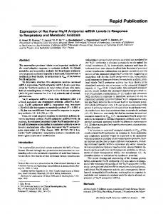
Transduction of TAT/PTD–-galactosidase to pancreatic -cells. To test whether the TAT-fusion protein was capable of transducing pancreatic -cells, insulinoma TC3 cells were incubated for 3 h with 0.8 g of the fusion protein per 1,000 cells in order to allow for the proper refolding of the fusion protein (21), and then they were stained for LacZ activity. As shown in Fig. 1A, the transduction of TAT-PTD–-galactosidase was dependent on the presence of the PTD 11–amino acid domain. Practically 100% of the TC-3 cells were transduced with TAT-PTD– -galactosidase, whereas no activity was detected in cells transduced with -galactosidase missing the TAT/PTD 11–amino acid domain. To study the persistency of TAT/PTD–-galactosidase transduction, cells were transduced with the fusion protein conjugated with the fluorescent marker Alexa-Fluor 488 (similar to FITC). TC-3 cells were transduced for 30 min with the labeled protein (0.8 g per 1,000 cells). Cells were washed and cultured in medium free of TAT/PTDfusion protein, then they were collected at different time points and analyzed by flow cytometry. The labeled protein was detectable for at least 48 h (Fig. 1B). Thus, it appears that TAT/PTD-mediated transduction is a viable DIABETES, VOL. 50, AUGUST 2001
J. EMBURY AND ASSOCIATES
FIG. 2. Transduction of islets with TAT/ PTD--galactosidase. A: Rat islet cells were transduced with 0, 1, and 2 g of the AlexaFluor 488-labeled fusion protein per islet (panels 1, 2, and 3, respectively) and incubated for 2 h. After incubation, islets were dissociated and evaluated by flow cytometry. B: -galactosidase activity in transduced islets assessed by X-Gal staining. Rhesus monkey islets were transduced with 1.25 and 2.5 g of TAT/PTD--galactosidase (panels I and II, respectively) or 2.5 g of -galactosidase (minus TAT/PTD; panel III) per islet. After transduction, islet cells were spread uniformly on the slide using cytospin and then fixed and stained by X-Gal.
option for transferring proteins into pancreatic -cells without affecting their viability. Transduction of TAT/PTD–-galactosidase fusion protein into islets. To investigate the transduction of the TAT/PTD-fusion protein into islets, TAT–-galactosidase fusion protein, either labeled with Alexa-Flour 488 or unlabeled, was incubated with rat and rhesus monkey pancreatic islets, respectively, and the degree of transduction was assessed by flow cytometry and LacZ activity. As shown in Fig. 2 A, the labeled TAT-fusion protein transduction efficiently reached a plateau of 80 and 84% at ratios of 1 and 2 g per rat islet, respectively. Figure 2B shows the transduction of monkey islets with 1.25 (2B-I) and 2.5 g (2B-II) of TAT/PTD–-galactosidase per islet and 2.5 g of -galactosidase without the TAT/PTD domain (2B-III). Islet transduction is a dose-dependent phenomenon as observed by the increasing intensity of LacZ activity in islets transduced with 1.25 or 2.5 g of protein per islet. This experiment demonstrates that the transduced galactosidase protein is functional after TAT/PTD transduction and that the TAT/PTD domain is necessary to transduce protein into islets. Anti-apoptotic proteins expressed as TAT/PTD-fusion proteins blocked TNF-␣–induced caspase activity. To study the effect of internalization on the biological activity of anti-apoptotic proteins after transduction into cells, we generated recombinant TAT/PTD-fusion proteins of Bcl-XL and PEA/15. Both anti-apoptotic proteins exert their antiapoptotic activities via distinct molecular mechanisms. Bcl-XL belongs to the Bcl-2 family, which plays an important role in regulating the response of different cell types (including pancreatic islets) to a wide variety of apoptotic stimuli signaling through the mitochondria (13,26). PEA/15 is a 15-kDa, death effector domain (DED)-containing protein that inhibits apoptosis by binding the DEDs of both DIABETES, VOL. 50, AUGUST 2001
the adapter molecule FADD (Fas-associated death domain) and the effector caspase-8 (27). Both TAT/PTD-Bcl-XL and TAT/PTD-PEA/15 fusion proteins prevented activation of caspase-3, a key enzyme involved in TNF-␣–induced apoptosis (28), in TC-3 insulinoma cells. Inhibition of protein synthesis by CHX is necessary to trigger apoptosis by TNF-␣ in TC-3 cells (D.K. and R.L.P., unpublished data), perhaps because it prevents the de novo synthesis of anti-apoptotic factors triggered by TNF-␣, as previously described in other cell lines (29). Caspase-3 activity was assessed by measuring the release of AFC from the fluorogenic Ac-DEVD-AFC substrate as a consequence of the caspase-3–mediated cleavage. As shown in Fig. 3A, transduction of pancreatic -cells with TAT/PTD–Bcl-XL or TAT/PTD-PEA/15 resulted in a 50 and 48% reduction in TNF-␣–induced caspase-3 activity, respectively, whereas transduction with TAT/PTD–-galactosidase did not prevent TNF-␣–induced apoptosis. Similarly, we observed a general reduction in the activation of caspases in cells treated with the TAT/PTD–Bcl-XL fusion protein, as assessed by the binding of the FITC conjugate of the pan-caspase inhibitor VAD-FMK to activated caspases (Fig. 3B-I). However, as expected, transduction with TAT/ PTD–-galactosidase did not have any preventive effect (Fig. 3B-II). Binding of VAD-FMK/FITC to activated caspases serves as an in situ marker of apoptosis. We did not study inhibition of caspase activation by TAT/PTD-PEA/15 using this method. Furthermore, pancreatic -cells treated with TAT-Bcl-XL or TAT/PTD-PEA/15 fusion proteins had higher viability than control cells after incubation with TNF-␣/CHX (Fig. 4). Using a viability test based on combined acridine orange and ethidium bromide staining (FluoroQuench kit; One Lambda, Canoga Park, CA), we observed that the staining patterns of viable cells (green color) transduced with TAT/PTD–Bcl-XL or TAT/PTD-PEA/ 1709
TRANSDUCTION OF ISLETS WITH FUSION PROTEINS
FIG. 3. Inhibition of TNF-␣–induced caspase activity by anti-apoptotic proteins expressed as TAT/PTD-fusion proteins. A: TC-3 cells were transduced with 30 g of TAT/PTD–Bcl-XL, 25 g of TAT/PTD–PEA-15, or 90 g of TAT/ PTD–-galactosidase. After 3 h, cultures were treated with a combination of TNF-␣ (500 U/ ml) and CHX (10 g/ml) for 18 h. Controls remained untreated during this period. Cells were harvested and lysed. Caspase proteolytic activity was measured using the fluorogenic Ac-DEVD-AFC substrate in the presence or absence of caspase-3 inhibitor, and the activity was then adjusted by protein content. Data represent the means ⴞ SD of five (Bcl-XL–PEA-15) or three (TAT/PTD–-galactosidase) separate experiments. *P < 0.01. B: TC-3 cells nontransduced or transduced with TAT/PTD–BclXL (panel I) or TAT/PTD--galactosidase (panel II) for 3 h were treated with TNF-␣/CHX for 18 h. Controls remained untreated all this time. Pretreatment of pancreatic cells with zVAD-FMK (50 mol/l) completely inhibited TNF-␣–induced apoptosis (marked as control in panel II). Cells were resuspended and incubated in 10 mol/l of FITC-VAD-FMK in situ marker and evaluated by flow cytometry.
15 fusion proteins were comparable to those of the control cells, whereas the morphology of cells treated with TNF␣/CHX in the absence of TAT-fusion protein corresponded to that of an advanced stage of apoptosis (yellow to orange). Similar results were obtained by assessing cell viability using a combination of calcein AM and ethidium homodimer-1 (Molecular Probes, Eugene, OR) (data not shown). The pictures shown in Fig. 4 are representative of three separate experiments. Cells transduced with Bcl-XL– and PEA/15-fusion proteins showed (means ⫾ SD), respectively, 82 ⫾ 8 and 59 ⫾ 13% of the green (viable) cells compared with ⬎93 ⫾ 5% in control cells and only 15 ⫾ 6% in TNF-␣/CHX–treated cells. In addition, the green fluorescence of live cells from control and TAT/PTD-transduced cells was 45 ⫾ 8% more intense than the fluorescence remaining in cells treated only with TNF-␣/CHX, as assessed by the intensity of luminosity of areas of similar pixels using Photoshop 5.5 software. These results indicate that the two anti-apoptotic proteins investigated can be expressed as TAT/PTD-fusion proteins, retaining their biological function and protecting cells from apoptosis. TAT/PTD-fusion protein does not affect insulin secretion capabilities of islets. A key requirement for any molecular manipulation of pancreatic islets is that insulin secretion capability not be affected. We therefore investigated whether the TAT-fusion protein transduction affected islet physiology and insulin secretion; we did this by testing the ability of transduced islets to reverse hyperglycemia in diabetic nude mice. As shown in Fig. 5A, islets transduced with TAT/PTD–Bcl-XL reversed hyperglycemia within the same time frame as control islets. Transduced and nontransduced islets were implanted under the kidney 1710
capsule of immunodeficient CB17-scid mice previously rendered diabetic through chemical induction. All animals returned to normoglycemia after implant of the islet graft in both groups, demonstrating that islet cell performance was not affected by exposure to the TAT/PTD-fusion protein in vitro. After removal on day 29 of the kidney bearing the graft, a prompt rise in blood glucose was observed in all animals, confirming that euglycemia was sustained by insulin produced by the grafted islets. Furthermore, analysis of hematoxylin-eosin–stained sections did not reveal any histological differences between control and TAT-transduced islets, nor was there any difference in the insulin staining between the two (data not shown). To complement these studies, we performed static glucose challenge stimulation of rat islets transduced with TAT/PTD–Bcl-XL. Transduced islets maintained their ability to secrete insulin physiologically in a manner similar to control nontransduced islets (Fig. 5B). The same population of islets were subjected sequentially to low-glucose (2.2 mmol/l), then high-glucose (22 mmol/l), and again low-glucose (2.2 mmol/l) stimulation, thus allowing a more physiological evaluation of glucose-stimulated insulin secretion. Means of stimulation indexes (SIs) from five independent experiments using control and transduced islets (2.5 ⫾ 0.42 [17%] and 2.8 ⫾ 0.6 [22%]), respectively) did not show a significant statistical difference (P ⫽ 0.19). Note that these values represent the SI, which is assessed without the supplementation of insulin secretogues in the high-glucose medium (30). The Lewis rat islets used in these experiments were in good condition, lacking central core necrosis and maintaining intact islet capsule. The validity of the SI assessments is confirmed by their ability DIABETES, VOL. 50, AUGUST 2001
J. EMBURY AND ASSOCIATES
FIG. 4. Increased viability of pancreatic -cells transduced with TAT/PTD anti-apoptotic proteins. TC-3 cells were transduced with 30 g of TAT/PTD–Bcl-XL and 25 g of TAT/PTD–PEA-15 for 3 h and treated with TNF-␣/CHX as described in the legend for Fig. 3. Cells were harvested and stained with FluoroQuench stain (acridine orange plus ethidium bromide) and examined by fluorescence microscopy. Viable cells are stained green, whereas dead or dying cells appear yellow and orange. Pictures are representative from three experiments. Pictures were captured by a charge-coupled device camera (Lei-750; Leica) using VideoVixen software and analyzed with Photoshop version 5.5. The bars below each picture represent the percentage of viable (green) and dead (orange) cells in each experiment.
to return to basal insulin secretion levels when subjected to second low-glucose stimulation. These results indicate that the process of transduction of pancreatic islets with TAT/PTD–Bcl-XL fusion protein did not alter or impair insulin secretion in transduced islets. DISCUSSION
In this study, we used TAT/PTD methodology to deliver proteins to pancreatic islets. Utilizing a TAT/PTD–-galactosidase fusion protein labeled with Alexa 488, we found that TAT-fusion proteins are capable of transducing pancreatic -cells (Fig. 1). The labeled protein was still detectable after 48 h of transduction when cultured in medium free of TAT/PTD-fusion protein (Fig. 1B). Interestingly, all of the cells were fluorescent, suggesting that the TAT/PTD-fusion protein was passed onto newly divided cells as well. Although we have not yet studied the turnover of any TAT/PTD-fusion proteins, we speculate that it depends on the intrinsic turnover of the expressed protein. Similar to that which was observed in transduction mediated by PTD-5 (20), the TAT/PTD domain mediated both cytoplasmic and nuclear localization of -galactosidase in TC-3 cells. Fluorescence microscopy showed that immediately after transduction, the fluoresDIABETES, VOL. 50, AUGUST 2001
cence associated with the TAT/PTD–-galactosidase fusion protein was present throughout the cell, although after 24 h it was found to be more localized in the nuclear compartment (J.E. and R.L.P., unpublished observations). TAT/PTD–-galactosidase also has the ability to transduce pancreatic islets by diffusion in culture with ⬎80% efficiency (Fig. 2). These results indicate that transduction of islets in suspension using the TAT/PTD system is much more efficient than the viral transduction (31). Transduction of pancreatic cells or islets is totally dependent on the presence of the TAT/PTD domain in the protein (Figs. 1A and 2B). Although the mechanism by which cells take up TAT/ PTD fusion proteins is currently unknown, it has been suggested that an unfolding step is involved in its internalization (32) and that in vivo refolding of the protein is subsequently required (16). It was therefore important to investigate whether an anti-apoptotic protein, expressed as a TAT/PTD-fusion protein, retains biological activity after transduction into cells. The experiments shown in Fig. 3 and Fig. 4 indicate that Bcl-XL and PEA/15, two anti-apoptotic proteins that use different molecular mechanisms to block apoptosis, retain their anti-apoptotic properties once transduced to pancreatic cells. 1711
TRANSDUCTION OF ISLETS WITH FUSION PROTEINS
FIG. 5. Transduction of islets with TAT/PTD fusion proteins does not affect insulin secretion capability. A: Reversal of hyperglycemia in diabetic immunodeficient mice with pancreatic islets transduced with TAT/PTD–Bcl-XL. Lewis rat islets were transplanted under the kidney capsule of chemically induced diabetic CB17-scid mice. Animals received islets cultured for 24 h either in the presence (triangles) or absence (circles) of TAT-fusion protein (day ⴚ1). TAT-treated islets restored normoglycemia (blood glucose