Indian J Nephrol 2005;15: 105-107
105
CASE REPORT
Recurrence of oxalosis in isolated renal allograft recipient J. Thanka*, E. Indhumathi**, P. Soundararajan**, S. Rajendiran*. Department of Pathology* and Nephrology** Sri Ramachandra Medical College & R.I (D.U), Chennai
Abstract Renal oxalosis is the final stage of hyperoxaluria when reduction of GFR produces oxalate accumulation. It involves bones, arteries, eyes, heart, nerves and other structures. Liver transplantation is curative for patients with Type I primary hyperoxaluria. For oxalosis patients with minor enzyme deficiencies, renal transplantation may be the therapy of choice although concern exists about recurrent of oxalosis in the transplanted kidney. We are presenting a case of recurrence of oxalosis in a post transplant kidney in an adult male without any evidence of systemic oxalate deposition, with a functioning renal allograft for one year. Key words: Oxalosis, renal transplantation
Introduction Oxalosis can be primary (an inborn error of oxalate metabolism) or secondary due to increased ingestion or absorption (bowel disease) or decreased excretion (Chronic renal failure)1. In the kidney it shows widespread birefringnent tubular and interstitial oxalate crystal deposition. Interstitial oxalate may induce an inflammatory reaction occasionally with granulomas. The consequence is chronic interstitial nephritis leading to end stage renal disease.
Clinical Summary Thirty three year old male, status post live related renal transplantation 6 months, was admitted for a sudden rise in blood urea nitrogen and creatinine on routine follow up. The donor was his own sister. He was on triple immunosuppression (azathioprine, cyclosporin and prednisolone). One year prior to this transplantation patient developed severe renal failure and was evaluated elsewhere that showed normal sized kidneys with severe hypertension. A renal biopsy done at that time showed histological features of End Stage Renal Disease with oxalate crystals in the tubules. The patient was not willing to continue long-term dialysis. The patient was evaluated for oxalate deposition in other tissues. Risk of recurrence of oxalosis following renal transplantation was explained to the patient. In view of the absence of other co-morbid features of oxalosis and Address for Correspondence: Dr. P. Soundararajan Professor and Head of Nephrology 1, Ramachandra Nagar Porur, Chennai – 600 116. e-mail:
[email protected]
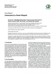
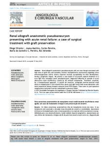
oxalate deposition in other tissues isolated renal transplantation was considered. His sister was evaluated for oxalosis prior to donation and found to have no evidence of oxalosis. Her urinary oxalate values were within normal limits. The patient underwent adequate preoperative dialysis to reduce the oxalate load, and was given oral sodium citrate, magnesium and pyridoxine. Renal transplantation was done. There was immediate diuresis on table and good graft function subsequently. The patient was on triple immunosupression with azathioprine, cyclosporin, prednisolone and patient continued to take oral sodium citrate, magnesium and pyridoxine with high fluid intake. The postoperative period was uneventful and at the time of discharge patient had normal renal function with normal levels of 24 hours urinary oxalate. The patient was on monthly follow-up and 6 months later presented with elevated blood urea nitrogen and creatinine. A renal biopsy was done which showed Banff Grade IA rejection with superimposed pyelonephritis and deposition of birefringent oxalate crystal deposition in the tubules (Fig 1). The interstitium showed scattered granulomas. Urine examination also showed birefringent oxalate crystals (Fig 2). Patient was pulsed with methyl prednisolone and was treated with appropriate antibiotics for urinary tract infection. The renal functions improved and for the past one year he is maintaining serum creatinine of 2 mg%.
Discussion Primary hyperoxaluria (PH) is a heterogenous disease with a variable age of onset and a variable progression to kidney failure2. As a group primary hyperoxaluria comprise three inborn errors of metabolism with recessive Copyright © 2005 by The Indian Society of Nephrology
106
Indian Journal of Nephrology
Indian J Nephrol 2005;15: 105-107
follow a more benign course than in Type I disease. Type III PH is due to increased intestinal absorption of oxalate of unknown pathophysiology3. All the three types of hyperoxaluria have the same clinical features, may be differentiated biochemically on the basis of urinary organic acid excretion4. PH Type I disease excrete an excess of glycolic acid while those with PH Type II excrete an excess of L-glyceric acid PH Type III does not show any elevation of organic acid excretion. 30% of patients are diagnosed only at end stage renal disease. Diagnosis is usually based on history and urinary oxalate excretion. Glycolate and L-glyceric acid excretion and liver biopsy for enzyme defect is confirmatory2. Fig 1: H & E, x 200. Renal biopsy showing intratubular crystalloid structures. Left upper corner : H & E x 100. Partially polarized intratubular oxalate crystals along with background renal structures. Left Lower Corner : Completely polarized oxalate crystals.
autosomal transmission that present similarly with recurrent urinary calculi and hyperoxaluria. Typically plasma oxalate is elevated even when the glomerular filtration is still with in the normal range. The main symptoms at diagnosis are urolithiasis (54%) and nephrocalcinosis (30%). PH Type I is due to the deficiency of hepatic specific peroxisomal enzyme alanine - glycolate amino transferase (AGT) which utilizes pyridoxine (Vit B6) as a coenzyme. Metabolic defect in Type II PH is the deficient activity of D-Glycerate dehydrogenase and often
Historically renal transplantation has yielded very poor results in PH Type I because of recurrent oxalosis of the graft. In the last 10 years, combined hepatorenal transplantation has been successfully applied simultaneously correcting the metabolic lesion in the liver and replacing the damaged kidneys. However there are successful live related selective renal transplantation with excellent renal function for patients with PH Type I are also reported. Especially there are isolated successful renal transplantation for patients without significant systemic oxalosis and have evidence of residual AGT activity5. Studies also have shown that it is possible to perform successful renal transplantation in small children and adults with primary oxalosis and completely prevent the deposition of oxalate in renal allograft. A specialized strategy for medical management included intensive pretransplant hemodialysis, post transplant long-term diuresis, administration of neutral phosphate, magnesium and pyridoxine6. Renal transplantation with a strict medical protocol would appear to be the initial treatment of choice for renal failure due to primary oxalosis in centres where liver transplantation facilities are not well developed. Overall renal transplant is better for patient survival than no transplant7.
Conclusion
Fig 2: Urine Sediment, unstained x 400, showing typical oxalate crystals.
Renal transplantation experience for oxalosis is limited. Even though combined liver and kidney transplantation is ideal still isolated kidney transplant improves survival as in our case.
References 1.
2.
Hill G.S.Calcium and the Kidney, hydronephrosis, in Heptinstall’s Pathology of the Kidney, ed Jennette JC, Olson JL, Schwartz MM, Silva FG, New York. Lippincott Raven, 1988 P 891 - 936. Hoppe B, Langman C B. A United States survey on diagnosis, treatment and outcome of primary hyperoxaluria. Pediatr Nephrol 2003; 18(10): 986-91.
Copyright © 2005 by The Indian Society of Nephrology
3. 4.
Toussaint C, De Pauw L. Primary hyperoxaluria Nephrologie 1995; 16(6): 339 - 406. Stephen H. Morgan. The Primary hyperoxalurias, in Oxford Textbook of Clinical Nephrology, ed Alex M Davidson, J.Stewart Cameron, Jean - Pierre Grunfeld, David N.S.Kerr, Eberhard Ritz and Christopher G Winearls, New York. Oxford Medical Publications, 1998 P 2464 - 2468.
Indian J Nephrol 2005;15: 105-107
5.
6.
Allen AR, Thompson EM, Williams G, Watts RW, Pusey CD. Selective renal transplantation in primary hyperoxaluria type I. Am J Kidney Dis 1996; 27(6): 891-5. Scheinman JI, Najarian JS, Mauer SM. Successful strategies for renal transplantation in primary oxalosis. Kidney Int.1984; 25(5): 804 - 11.
Recurrence of oxalosis in renal allograft recipient
7.
107
Jon I. Scheinman. Recent data on results of isolated kidney or combined kidney / liver transplantation in the U.S.A. for primary hyperoxaluria.J of Nephrol 1998; 2:4245.
Copyright © 2005 by The Indian Society of Nephrology