Dec 4, 1989 - DdrasG gene expression during the early development of Dictyostelium discoideum has been examined in detail. The amount of ...
MOLECULAR AND CELLULAR BIOLOGY, Mar. 1990, p. 918-922 0270-7306/90/030918-05$02.00/0 Copyright C) 1990, American Society for Microbiology
Vol. 10, No. 3
Regulation of DdrasG Gene Expression during Dictyostelium Development MEENAL KHOSLA,' STEPHEN M. ROBBINS,' GEORGE B. SPIEGELMAN,12 AND GERALD WEEKS' 2 Departments of Microbiology' and Medical Genetics,2 University of British Columbia, Vancouver, British Columbia V6T I W2, Canada Received 29 August 1989/Accepted 4 December 1989
DdrasG gene expression during the early development of Dictyostelium discoideum has been examined in detail. The amount of DdrasG-specific mRNA increased approximately twofold during the first 2 to 3 h of development and then declined rapidly, reaching negligible levels by the aggregation stage. The increase in mRNA levels that occurred during the first 2 to 3 h of development also occurred during differentiation in cell suspensions and was enhanced when cells were shaken rapidly. This initial increase was unaffected by cell density. When cells were set up to differentiate on filters, the addition of a glucose-amino acid mixture slightly delayed differentiation and had a similar effect on the expression of the gene. The decline in DdrasG expression during development did not occur when cells were treated with cycloheximide, suggesting that the expression of a developmentally regulated gene product is essential for the reduction of DdrasG gene mRNA. There was no decrease in DdrasG mRNA level during differentiation in shake suspension, but the decrease did occur upon application of pulses of cyclic AMP to shaking cultures. The application of a continuously high level of cyclic AMP delayed the increase in expression of the gene and did not result in the subsequent decline. These results suggest that the induction of a functional cyclic AMP relay system is important in reducing DdrasG gene mRNA levels.
Dictyostelium discoideum development is initiated when amoeboid cells are deprived of their food source, and within a few hours the hitherto solitary amoebae aggregate in response to pulses of cyclic AMP. The aggregate then elongates to form a pseudoplasmodium within which distinct populations of prestalk and prespore cells are discernable. Eventually the cell mass culminates to produce a fruiting body composed of a rounded mass of spore cells supported off the substratum by a slender, linear array of stalk cells. This simple differentiation of amoeboid cells into either stalk or spore cells, along with the regulated proportioning and specific positioning of the cell populations, makes D. discoideum an attractive organism for studies on the mechanism of cellular differentiation (13). In addition to being the chemoattractant during aggregation, cyclic AMP regulates the expression of a number of genes during early development (8). In addition, it is involved in the induction of both prespore- and prestalkspecific genes during the postaggregative period. Some genes respond to a high extracellular concentration of cyclic AMP, while others respond to pulses of low concentrations (8). These roles for cyclic AMP have been deduced from a variety of in vitro differentiation systems, including studies with shaken cell suspensions. Ras genes are found in a variety of eucaryotic organisms, ranging from yeasts to humans, and most organisms possess two or more distinct genes (1). Two ras genes, Ddras and DdrasG, have been identified in D. discoideum, and the two genes are expressed at different developmental stages (21, 22). Ddras is expressed maximally at the pseudoplasmodial stage of development, and the mRNA is enriched in the prestalk cell population (21). In contrast, DdrasG is expressed maximally during growth and early development *
(22). The combined expression of the two genes can account for the changes in the rates of ras-protein synthesis that are observed during the differentiation process (20), although this does not preclude the possibility that there are additional ras genes that are yet to be discovered. Ddras expression occurs precociously in response to the addition of cyclic AMP during in vitro differentiation in shake suspension, and in this regard the expression is similar to that of a number of other prestalk-enriched genes (21). During normal development, these genes are probably induced when the concentration of cyclic AMP reaches a sufficient level within the developing cell mass. In this report we examine the factors involved in regulating DdrasG gene expression during early development. MATERIALS AND METHODS Organisms and growth and differentiation conditions. D. discoideum V12-M2 was grown on nutrient agar plates in association with Enterobacter aerogenes (25). Upon visible clearing of the bacterial lawn, the vegetative amoebae were separated from residual bacteria by four low-speed centrifugations (700 x g for 2 min). To initiate normal differentiation on a substratum, 108 washed cells were plated onto 4.0-cm membrane filters (Millipore Corp.) resting on support pads saturated with 20 mM potassium phosphate buffer, pH 6.0. The filters were incubated at 22°C for the times indicated in the text. To initiate in vitro differentiation in suspension, washed cells were routinely suspended at 5 x 10⁶ cells per ml in 20 mM potassium phosphate buffer, pH 6.0, in an Erlenmeyer flask and subjected to gyratory shaking at the speeds indicated in the figure legends. In one experiment the cell density was varied, as indicated in the legend to Fig. 5. RNA extraction and Northern (RNA) blot analysis. Total cytoplasmic RNA from the various stages of development was isolated by a previously described procedure (2). The
Corresponding author. 918
REGULATION OF DdrasG GENE EXPRESSION IN D. DISCOIDEUM
VOL. 10, 1990
0
1
2
3
4
5
-0 .M._.f_. ..1
2 3 4
0 1
6
919
5 6 7
S.
.. ...
B
4*90
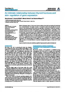
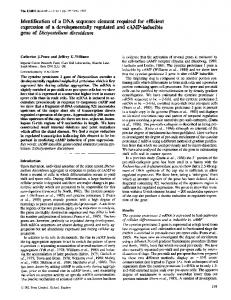
B FIG. 1. The effect of cycloheximide on DdrasG expression. Cells were plated for development on filters saturated with 20 mM potassium phosphate, pH 6.0, in the absence (A) or the presence (B) of cycloheximide at 400 p.g/ml. RNA was extracted at the times (hours) indicated above each lane, processed as described in Materials and Methods, and hybridized with the Ddras-c3 clone.
RNA samples (20 ,ug) were adjusted to 50% formamide-40 mM 3-(N-morpholino)propanesulfonic acid, pH 7.0-10 mM sodium acetate-1 mM EDTA-6% formaldehyde. The samples were heat denatured for 10 min at 65°C and then size fractionated on 1.25% formaldehyde-agarose gels (12). The RNA was transferred to nitrocellulose, and the filters were prehybridized at 37°C in 30% formamide-Denhardt solution (0.02% Ficoll, 0.02% bovine serum albumin, 0.02% polyvinylpyrrolidone)-5x SSC (lx SSC is 0.15 M NaCl plus 0.015 M sodium citrate)-20 mM sodium phosphate buffer, pH 6.5-sonicated salmon sperm DNA (100 ,ug/ml)-0.5% sodium dodecyl sulfate-polyadenylic acid (30 ,ug/ml) and then hybridized at 37°C for 16 h following the addition of DdrasG that had been labeled by the random oligonucleotide method (5). The filters were washed twice (30 min per wash) in 0.1 x SSC-0. 1% sodium dodecyl sulfate at 65°C and then exposed to X-ray film. Levels of DdrasG mRNA were assessed relative to IG7 mRNA as an internal control. The IG7 gene has been shown to be expressed constitutively during differentiation (4). RESULTS Expression of DdrasG during development. In a previous report, we showed that DdrasG gene expression was maximal in vegetative and early developing cells but was negligible by 8 h of development (22). A more detailed analysis of the developmental expression of the DdrasG gene revealed a slight but consistent increase in the level of DdrasG mRNA during the first 2 to 3 h of development of strain V12-M2, followed by a rapid decline to low levels by 6 h (Fig. 1 and 2). These results are consistent with previous findings on the rates of ras-protein synthesis during early development of this strain (20). Effect of cycloheximide on the developmental expression of the DdrasG gene. Cycloheximide is a potent inhibitor of protein synthesis in D. discoideum (24). Recently, Singleton and co-workers showed that a set of vegetatively expressed genes which are normally repressed during early differentiation can be divided into two groups. One group is induced in response to cycloheximide, whereas the second group is unaffected by the antibiotic (24). Furthermore, Firtel has shown that the gene F9 that is normally induced during early
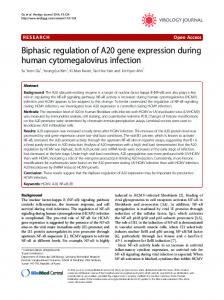
FIG. 2. The expression of DdrasG mRNA in the presence of glucose and an amino acid mixture. Cells were plated for development on filters saturated with 20 mM potassium phosphate buffer, pH 6.0, containing no addition (A) or 0.5 mg of each amino acid (except cysteine and tyrosine) per ml and 0.5 mg of glucose per ml (B). RNA was extracted at the times (hours) indicated above each lane, processed as described in Materials and Methods, and then hybridized with the DdrasG-c3 clone.
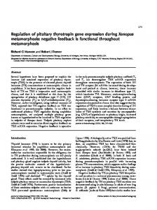
development is superinduced in response to cycloheximide (16). We found that the initial increase in DdrasG expression was unaffected by cycloheximide but that the subsequent decline in mRNA level did not occur in the presence of the drug (Fig. 1). This result suggests that the initial increase in expression does not require the production of a developmentally regulated gene product but that the subsequent decrease in expression does. Effects of amino acids on DdrasG gene expression during development. Amino acids prevent starvation and inhibit the initiation of differentiation in the axenic strain A-3 (18, 19). In order to determine whether amino acid starvation could be a stimulus for the initial increase in DdrasG gene expression, a mixture of amino acids and glucose was added to cells of strain V12-M2 at the onset of differentiation. In this strain, the combination of the amino acid mixture and glucose produced only a slight delay in the initiation of differentiation (data not shown) and there was a similar delay in the increase and subsequent decline of DdrasG mRNA (Fig. 2). This result suggests that the increase in expression of DdrasG requires the amino acid starvation signal for the initiation of development. Expression of DdrasG during incubation in shake suspension. In order to further investigate the factors involved in the regulation of DdrasG gene expression during development, cells were shaken in suspension in the absence of nutrients. In slow-shake conditions that allow cell contact, the initial elevation of DdrasG gene expression was considerably lower and the subsequent decline did not occur (Fig. 3). The presence of a continuous, high extracellular concentration of cyclic AMP delayed the increase even more (Fig. 3). When cells were rapidly shaken and cell contact was prevented, the initial increase in the level of DdrasG mRNA was rapid and considerably greater than that observed when differentiation occurred on a substratum or in slowly shaken cell suspensions, and again the subsequent decline was not observed (Fig. 3). The application of pulses of cyclic AMP to rapidly shaken cells had no effect on the initial increase, which remained rapid and enhanced, but the subsequent decrease in mRNA level occurred (Fig. 4). This result suggests that the reduction of DdrasG mRNA level that occurs during the onset of aggregation is due to a response of the cyclic AMP relay system to pulses of cyclic AMP.
920
KHOSLA ET AL.
0 1
A -W a
MOL. CELL. BIOL.
2
3 4
5
6 7
a b
c
d e
f g
0
4# %# li
U ~~~~~~~~~~~~~~~~~~~~~~~~~~~~~
kUU~~~~~~~~~~~~~~~~~~~~~~~~~~~
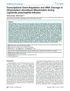
FIG. 5. The effect of cell density on DdrasG expression. Washed cells were suspended in 20 mM potassium phosphate buffer, pH 6.0, and shaken at 250 rpm at the following densities: 1 x 101 cells per ml (lane b), 3 x 10' cells per ml (lane c), 1 x 10⁶ cells per ml (lane d), 3 x 10⁶ cells per ml (lane e), 1 x 10⁷ cells per ml (lane f), and 3 x 10⁷ cells per ml (lane g). RNA was extracted after 2.5 h. RNA extracted at the time of the initial cell suspension was run in lane a. RNA samples were processed as described in Materials and Methods and were hybridized with DdrasG-c3. FIG. 3. The accumulation of DdrasG mRNA in shaken cell suspension. Washed cells were suspended in 20 mM potassium phosphate buffer, pH 6.0, at a density of 5 x 10⁶ cells per ml. Cell suspensions were shaken at 70 rpm (A), shaken at 70 rpm in the presence of 500 p.M cyclic AMP with additional cyclic AMP added to a concentration of 100 ,uM every hour (B), or rapidly shaken at 250 rpm (C). RNA was isolated at the times (hours) indicated above each lane, processed, and hybridized with DdrasG-c3.
Consistent with this view is the finding that the decrease in DdrasG expression did not occur when cyclic AMP pulses were applied in the presence of caffeine (Fig. 4). In D. discoideum, caffeine prevents the activation of adenylate cyclase and, hence, inhibits part of the cyclic AMP signal relay response (3, 26). 0
A
2
4
6
8
Careful examination of the gels in Fig. 1A and 4B shows that the RNA hybridizing to the DdrasG-c3 lane appears to migrate more slowly as the overall signal declined. One possibility is that there is more than one form of RNA encoded by DdrasG. If so, the data presented here represent the sum of the two forms. The general conclusions on the effects of the growth conditions on the transcription activity of the DdrasG do not change, although there may be a level of posttranscriptional regulation overlaid on our analysis. The effect of cell density on DdrasG gene expression. The initial increase in DdrasG mRNA level in rapidly shaken cells was greater than the increase that occurred when cells were slowly shaken (Fig. 3), suggesting the possibility that the induction of expression was cell density dependent. Cells were therefore rapidly shaken at varying densities, and the amount of DdrasG mRNA was determined after 2.5 h. The results indicated that cell density had no effect on DdrasG gene expression (Fig. 5).
DISCUSSION Although most organisms possess more than one ras gene, D. discoideum is thus far unique in that its two ras genes, Ddras and DdrasG, are expressed at different times (21, 22). Ddras mRNA is absent during growth and early development and does not appear until the pseudoplasmodial stage of development, when it is enriched in prestalk cells (21). Induction occurs precociously under in vitro conditions in response to the addition of cyclic AMP, a pattern that is similar to that for a number of other prestalk-enriched genes (21). In contrast, DdrasG gene expression occurs during growth and early development (22). We have shown here that DdrasG mRNA levels increased approximately twofold during the first 2 to 3 h of development and then declined to low levels by the aggregation stage (Fig. 1 and 2). Figures 1 C and 2 show an apparent decline in DdrasG mRNA at 1 h ' .~~~~~~~~~~~~~~~~~~~~~~~~~~~~~~~ relative to the level in vegetative cells. A possible interpretation of this decline is that there is a vegetatively expressed FIG. 4. The effects of cyclic AMP and caffeine on DdrasG expression. Washed cells were suspended at 5 x 10⁶ cells per ml in ras-related gene whose synthesis is halted before the in20 mM potassium phosphate buffer, pH 6.0. The sample was divided crease in DdrasG expression. However, in other experiinto three; one sample was shaken at 250 rpm (A), a second was ments (not shown) the decline at 1 h was not observed, shaken at 250 rpm and pulsed with cyclic AMP to a concentration of although the later pattern of DdrasG mRNA was unchanged. 25 nM every 5 min (B), and a third was shaken at 250 rpm in the we do not consider the apparent 1-h decline in Fig. 1 Thus, presence of 2 mM caffeine, with additional caffeine added to a and 2 indicative of additional ras-gene expression. concentration of 1 mM every 2 h and cyclic AMP added to a In rapidly shaken cell suspensions, there is negligible cell concentration of 25 nM every 5 min (C). RNA was extracted at the contact and the cyclic AMP signal relay system does not times (hours) indicated above each lane, processed, and hybridized function (11). Under these conditions, DdrasG was exwith DdrasG-c3.
B
VOL. 10, 1990
REGULATION OF DdrasG GENE EXPRESSION IN D. DISCOIDEUM
pressed at higher levels than during normal differentiation on a substratum and the subsequent decrease in expression did not occur. When rapidly shaken cells were treated with pulses of cyclic AMP, DdrasG expression was reduced at around the same time as during development on filters. The response of D. discoideum to pulses of cyclic AMP involves at least two distinct pathways (6-8). In the first, the binding of cyclic AMP to a cell surface receptor activates adenylate cyclase, which generates a transient rise in intracellular cyclic AMP and in turn a relay of the signal to other cells. In the second pathway, the binding of cyclic AMP to a cell surface receptor leads to the activation of guanylate cyclase and movement of cells towards the initial signal. It has been proposed that the first pathway is involved in the cyclic AMP-mediated regulation of some genes, while the second pathway is involved in the regulation of others (6). The cyclic AMP-mediated repression of DdrasG expression did not occur in the presence of caffeine. Since caffeine inhibits adenylate cyclase activation (3, 26), this result indicates that the repression of DdrasG expression requires the transient increase in intracellular cyclic AMP concentration or some other component involved in this branch of the cyclic AMP signal relay system. The expression of a number of developmentally regulated genes in slowly shaken cell suspensions more closely resembles that observed during differentiation on a substratum than that observed under fast-shake conditions (10, 23, 27). In most of our experiments, the initial increase in expression of DdrasG occurred in slowly shaken suspensions but the subsequent decline did not. However, in one experiment with slowly shaken cells, DdrasG expression did decline (data not shown) and we attribute this variability to the extreme sensitivity of the cyclic AMP relay system to slight perturbations in the shake suspension conditions. The response of DdrasG to cyclic AMP is similar to that of the previously characterized M4-1 gene (10, 11). M4-1 is expressed at moderate levels in vegetative cells, and its expression is reduced at around 5 h of development, with kinetics that are similar to those for DdrasG. Like DdrasG, M4-1 expression does not decline during starvation of a rapidly shaken cell suspension if a high continuous level of cyclic AMP is applied to the culture. However, in rapidly shaken cells, the application of pulses of cyclic AMP leads to a reduction of M4-1 mRNA and the addition of caffeine prevents the response to the cyclic AMP pulses. The delay between the application of cyclic AMP pulses to rapidly shaken cultures and the decrease in M4-1 and DdrasG mRNA levels mimicked the time course of these mRNA levels observed in cells undergoing development on filters. The consistency of the lag between the onset of the cyclic AMP pulses and the decline in the mRNA levels suggests that some cellular response system has to be established before the modulation of the mRNA levels occurs. Consistent with this idea is our finding that the addition of cycloheximide early in development prevented the reduction of DdrasG mRNA levels, since this latter result suggested that a developmentally regulated gene product(s) was essential for the decrease to occur. Thus, it would appear that both the reduction of DdrasG and M4-1 gene transcription might be regulated by similar mechanisms. However, M4-1 expression differed from that of DdrasG in that it did not appear to display the transient increase in expression during very early development. We should emphasize that the Northern data represent steady-state mRNA levels. Thus, it is possible that the reduction of DdrasG mRNA (and presumably M4-1 mRNA as well) is due to a specific developmentally regu-
921
lated nuclease. This scheme would be consistent with the cycloheximide data, although we have no direct evidence for such a nuclease. K5 gene expression is also reduced during early development by pulses but not by continuous high levels of cyclic AMP, and the effect does not occur in the presence of caffeine (15, 17). However, the expression of the K5 gene differs somewhat from that of M4-1 and DdrasG. K5 is expressed at negligible levels in vegetative cells and then increases dramatically during the first 2 to 3 h of development, prior to its rapid decline in expression. Furthermore, the reduction of KS occurs immediately after the application of the cyclic AMP pulse and if cells are pulsed at the onset of differentiation, the initial induction of gene expression does not occur (5, 17). The expression of the phosphodiesterase inhibitor is also different from that exhibited by M4-1 and DdrasG in that it is inhibited during early development both by pulses of cyclic AMP and by continuous high levels (9, 28). The transient increase in DdrasG gene expression occurred immediately upon the onset of differentiation. The signals involved in mediating the transition between growth and differentiation are poorly understood, although it is recognized that cells need to be both nutrient starved and at a sufficiently high density for differentiation to be initiated (14, 18, 19). The transient increase in DdrasG expression was not dependent upon cell density but was slightly delayed by the presence of a mixture of glucose and amino acids, a condition that in V12-M2 slows the onset of the differentiation process, presumably by preventing starvation. Thus, amino acid starvation may be the direct stimulus for the transient increase in DdrasG gene expression. The physiological function of the two ras gene products in D. discoideum is not yet known, but their differential expression suggests that DdrasG may function during vegetative growth and early development, whereas Ddras may function during multicellular development. Continued expression of DdrasG might have deleterious effects on late development, since the DdrasG gene product might compete with the very similar Ddras gene product. In this regard, it is interesting that the signal that leads to lower DdrasG mRNA levels (Fig. 4) stimulates the expression of Ddras (21), ensuring that the two genes are not expressed at the same time during development. We are presently attempting to determine whether the expression of DdrasG during late development is deleterious by examining transformants that aberrantly express DdrasG. ACKNOWLEDGMENTS We thank J. G. Williams for supplying the IG7 cDNA clone. We thank the Medical Research Council and the British Columbia Health Care Research Foundation for grants that supported this work. LITERATURE CITED 1. Barbacid, M. 1987. ras genes. Annu. Rev. Biochem. 56:779827. 2. Birnboim, H. C. 1988. Rapid extraction of high molecular weight RNA from cultured cells and granulocytes for Northern analysis. Nucleic Acids Res. 161:1487-1497. 3. Brenner, M., and S. D. Thoms. 1984. Caffeine blocks activation of cyclic AMP synthesis in Dictyostelium discoideum. Dev. Biol. 101:136-146. 4. Early, A. E., and J. G. Williams. 1988. A Dictyostelium prespore-specific gene is transcriptionally repressed by DIF in vitro. Development 103:519-524. 5. Feinberg, A. P., and B. Vogelstein. 1983. A technique for
922
6.
7.
8. 9.
10. 11.
12.
13. 14. 15.
16.
17.
KHOSLA ET AL.
radiolabelling DNA restriction endonuclease fragments to high specific activity. Anal. Biochem. 132:6-13. Firtel, R. A., P. J. M. van Haastert, A. R. Kimmel, and P. N. Devreotes. 1989. G protein linked signal transduction pathways in development: Dictyostelium as an experimental system. Cell 58:235-239. Janssens, P. M. W., and P. J. M. van Haastert. 1987. Molecular basis of transmembrane signal transduction in Dictyostelium discoideum. Microbiol. Rev. 51:396-418. Kessin, R. H. 1988. Genetics of early Dictyostelium discoideum development. Microbiol. Rev. 52:29-49. Kessin, R. H., S. J. Orlow, R. I. Shapiro, and J. Franke. 1979. Binding of inhibitor alters kinetic and physical properties of extracellular cyclic AMP phosphodiesterase from Dictyostelium discoideum. Proc. Natl. Acad. Sci. USA 76:5450-5454. Kimmel, A. R. 1987. Different molecular mechanisms of cAMP regulation of gene expression during Dictyostelium development. Dev. Biol. 122:163-171. Kimmel, A. R., and B. Carlisle. 1986. A gene expressed in undifferentiated vegetative Dictyostelium is repressed by developmental pulses of cyclic AMP and reinduced during dedifferentiation. Proc. Natl. Acad. Sci. USA 83:2506-2510. Lehrach, H., D. Diamond, J. M. Wozney, and H. Boedtker. 1977. RNA molecular weight determinations by gel electrophoresis under denaturing conditions, a critical examination. Biochemistry 16:4743-4751. Loomis, W. F. 1975. Dictyostelium discoideum. A developmental system. Academic Press, Inc., New York. Loomis, W. F. 1987. Regulation of cell-type-specific in Dictyostelium discoideum. Cold Spring Harbor Symp. Quant. Biol. 50:769-777. Mann, S. K. O., and R. A. Firtel. 1987. Cyclic AMP regulation of early gene expression in Dictyostelium discoideum: mediation via the cell surface cyclic AMP receptor. Mol. Cell. Biol. 7:458-469. Mann, S. K. O., and R. A. Firtel. 1989. Two phase regulatory pathway controls cyclic AMP mediated expression of early genes in Dictyostelium. Proc. Natl. Acad. Sci. USA 86:192419288. Mann, S. K. O., C. Pinko, and R. A. Firtel. 1988. cAMP regulation of early gene expression in signal transduction mu-
MOL. CELL. BIOL.
tants of Dictyostelium. Dev. Biol. 130:294-303. 18. Marin, F. T. 1976. Regulation of development of Dictyostelium discoideum. 1. Initiation of growth to development transition by amino acid starvation. Dev. Biol. 48:110-117. 19. Marin, F. T. 1977. Regulation of development of Dictyostelium discoideum. 2. Regulation of early cell differentiation by amino acid starvation. Dev. Biol. 60:389-395. 20. Pawson, T., T. Amiel, E. Hinze, N. Auersperg, N. Neave, A. Sobolewski, and G. Weeks. 1985. Regulation of a ras-related protein during development of Dictyostelium discoideum. Mol. Cell. Biol. 5:33-39. 21. Reymond, C. D., R. H. Gomer, M. Mehdy, and R. A. Firtel. 1984. Developmental regulation of a Dictyostelium gene encoding a protein homologous to mammalian ras protein. Cell 39:141-148. 22. Robbins, S. M., J. G. Williams, K. A. Jermyn, G. B. Spiegelman, and G. Weeks. 1989. Growing and developing Dictyostelium cells express different ras genes. Proc. Natl. Acad. Sci. USA 86:938-942. 23. Singleton, C. K., P. A. Gregoli, S. S. Manning, and S. J. Northington. 1988. Characterization of genes which are transiently expressed during the preaggregative phase of development of Dictyostelium discoideum. Dev. Biol. 129:140-146. 24. Singleton, C. K., S. S. Manning, and Y. Feng. 1988. Effect of protein synthesis inhibitors on gene expression during the early development of Dictyostelium discoideum. Mol. Cell. Biol. 8:10-16. 25. Sobolewski, A., N. Neave, and G. Weeks. 1983. The induction of stalk cell differentiation in submerged monolayers of Dictyostelium discoideum. Characterization of the temporal sequence for the molecular requirements. Differentiation 25:93-100. 26. Thiebert, A., and P. N. Devreotes. 1984. Adenosine and its derivatives inhibit cAMP signalling response in Dictyostelium discoideum. Dev. Biol. 106:166-173. 27. Town, C., and J. Gross. 1978. The role of cyclic nucleotides and cell agglomeration in post-aggregative enzyme synthesis in Dictyostelium discoideum. Dev. Biol. 63:412420. 28. Yeh, R. P., F. K. Chan, and M. B. Coukell. 1978. Independent regulation of the extracellular cyclic AMP phosphodiesteraseinhibitor system and membrane differentiation by exogenous cyclic AMP in Dictyostelium discoideum. Dev. Biol. 66:361374.