66,499-560. Dono hue, M 'Ernst, H., Wentworth, B., Nadal-Ginard, B., and Rosenthal,. Slater ... &ne Tr&scription (Cohen, P., and Foulkes, J. G., eds) pp. 101-116,.
Vol. 267, No. 22, Issue of August 5, PP. 15673-15680.1992 Printed in U.S. A.
THEJOURNAL OF BIOLOGICAL CHEMISTRY
0 1992 by The American Society for Biochemistry and Molecular Biology, Inc.
Regulation of Gene Expression of Rat Skeletal Muscle/Liver 6-Phosphofructo-2-kinase/Fructose-2,6-bisphosphatase ISOLATION AND CHARACTERIZATION OFA GLUCOCORTICOID RESPONSE ELEMENT IN THE FIRST INTRON OF THE GENE* (Received for publication, January 24, 1992)
Alex J. Lange, Carme Espinet, Robert Hall, M. Raafat El-Maghrabi, Albert0 M. Vargas, Richard J. MiksicekS, Daryl K. Grannert, andSimon J. Pilkis From the Department of Physiology and Biophysics and the $Departmentof Pharmacology, Health Science Center, State Uniuersitv of New York. Stonv Brook. New York 11 794-8661 and the §Departmentof Molecular Physiology and Biophysics, Vanderbiit University, Nashville, Tennessee 37232
At least two genes encode isoenzymesof rat 6-phos- notransferase (TAT)’ is induced by dexamethasone (11, and phofructo-2-kinase/fructose-2,6-bisphosphatase.Al- investigations of the regulation of this and other genes by ternative splicing of one of these genes generates a glucocorticoids led to the identification of DNA response skeletal muscle-specific transcript from an upstream elements (GREs) which bind ina site-specific manner to promoter and a liver-specific transcript from a down- glucocorticoid hormone-receptor complexes (2,3). The recepGREsite presumably stream promoter. A potent glucocorticoid response ele- tor-DNA complex formed atthe ment was identified in the first intron of the gene, i.e. enhances gene transcription by placing the transactivating between liver exon I and exon 11. The element is ap- domain of the receptor in a position where it can interact proximately 3.5 kilobase pairs (kb) downstream of the with one or more components of the transcription initiation liver isoenzyme transcription start site and13 kb up- complex (4,s). The consensus sequence for these GREs is an stream of exon I1 of the gene and confers dexametha- imperfect palindrome (T/G)GTACAnnnTGTTCT (6). Tsai sone-sensitive expression of chloramphenicol acetyl- et al. (7) demonstrated that multiple GREs enhance binding transferase (CAT) activity from a heterologous thy- and increase function, especially when located at great dismidine kinase promoter and from both homologous 5’- tances from the transcription start site. The liver PEPCK flanking regions of the gene. This glucocorticoid re- gene isalso positively regulated by glucocorticoids,but in this sponse element also exhibits androgen- but not estro- case the response element is more complex. ThePEPCK gen-sensitive expression of CAT activity inHeLa cells GRU is comprised of two glucocorticoid receptor binding sites, cotransfected with the appropriate receptor expression which have little homology to the GRE consensus sequence, and binding of other protein factors is required to elicit the vector. DNase footprintand sequence analysisrevealed that theelement is comprised minimally of two hormone response (8). In addition, Strahle et al. (9) have adjacent 15-mer glucocorticoid receptor dimer binding shown that transcription factors such as NF-1, Oct-1, and sites situated in opposite orientations. Glucocorticoid Spl can greatly enhance the potency of single GREs. These regulation of 6-phosphofructo-2-kinase/fructose-2,6-findings reflect an increasing weight of evidence that GREs are very complex and quite diverse in nature (10). bisphosphatase gene expression in liver and skeletal muscle is mediated by a single complex glucocorticoid Rat liver 6-PF-2-K/Fru-2,6-P2ase is encoded by a gene response element located in the first intron of the skel- whose expression is subject to a complex pattern of hormonal etal muscle/liver gene. regulation, which includes regulation by glucocorticoids (10). This bifunctional enzyme catalyzes the synthesis and degradation of Fru-2,6-P2, a potent positive effector of 6-phosphofructo-1-kinase and negative effector of fructose-l,6-bisphosphatase. 6-PF-2-K/Fru-2,6-P2ase thusprovides a mechanism An important physiological action of glucocorticoids is the for the liver cell to switch between being a net consumer or a modulation of the expression of hepatic genes encoding regregulation of this ulatory enzymes of intermediary metabolism. Dexamethasone net producer of glucose (11).The short term enzyme in liver is modulated by CAMP-dependent phosinduction of specific proteins has been determined by twophorylation (12). The long term regulation of both the liver dimensional high resolution electrophoresis in two hepatoma and skeletal muscle 6-PF-2-K/Fru-2,6-P2ase in different encell lines (1).It hasbeen demonstrated that theexpression of docrine and nutritional statesis via changes in mRNA levels. a small subset (approximately 1%)of the total liver proteins The amountof this bifunctional enzyme in liver is decreased is affected by the hormone. Gene expression of tyrosine ami- during starvation andin diabetesand is restored by refeeding * This work was supported by National Institutesof Health Grants DK-38354 (to S. J. P.) and CA-43384 (to R. J. M.). The costs of publication of this article were defrayed in part by the payment of page charges. This article must therefore be hereby marked “advertisement” in accordance with18 U.S.C. Section 1734 solely to indicate this fact. The nucleotide sequence(s) reported in this paperhas been submitted t o the GenBankTM/EMBLDataBankwith accession number(s) M89910 (see Fig. 5).
The abbreviationsusedare: TAT, tyrosineaminotransferase; PEPCK, phosphoenolpyruvate carboxykinase; 6-PF-2-K/Fru-2,6P2ase, 6-phosphofructo-2-kinase/fructose-2,6-bisphosphatase; GRE, glucocorticoid response element; GRU, glucocorticoid response unit; Fru-2,6-P*, fructose-2,6-bisphosphatase; CAT, chloramphenicol acetyltransferase; TK, thymidine kinase; DMEM, Dulbecco’s modified Eagle’s medium; bp, base pair(s); kb, kilobase pair(s); SDS, sodium dodecyl sulfate; GRBD, glucocorticoid receptor binding domain; IRE, insulin response element.
15673
15674
6-Phosphofructo-2-kinase/Fructose-2,6-P2ase Regulation Gene
a high carbohydrate dietor by insulin administration, respectively (13). The increase in mRNA that occurs with refeeding or insulin administration correlates with the increase in enzyme protein. Liver and skeletal muscle 6-PF-2-K/Fru-2,6Pnase are generated by alternative splicing of first exons of a single gene, and glucocorticoids regulate the gene expression of both isoenzymes. The amount of 6-PF-2-K/Fru-2,6-P2ase and its cognate mRNA is reduced in livers of adrenalectomized rats (14). Levels of liver 6-PF-2-K/Fru-2,6-P2ase mRNA from adrenalectomized rats increase 90-fold upon administration of the synthetic glucocorticoid, triamcinolone, and the skeletal muscle isoenzyme mRNA also increases, albeit to a lesser degree (14). Dexamethasone treatment increased the rateof 6-PF-2-K/Fru-2,6-P2ase gene transcription in vivo as well as in rat hepatoma cells (FTO-2B and FAO) (15).' Addition of glucocorticoids during plating preventsthe loss of 6-PF-2-K/Fru-2,6-P2ase mRNA when hepatocytes are placed into primary culture, which suggests that glucocorticoids are the most important hormonal factor in maintaining bifunctional enzyme gene expression. Also, glucocorticoid addition toprimary culturesof hepatocytes in serum-free media results inan approximately 100-foldinduction of bifunctional enzyme mRNA (17, 18). In order to study this pronounced effect of glucocorticoids on the regulation of gene expression of 6-PF-2-K/Fru-2,6Pnase, we isolated the gene for rat skeletal muscle/liver 6-PF2-K/Fru-2,6-P2ase andits 5"flanking region. Restriction fragments of the gene wereexamined in a reporter gene system for theirability to confer glucocorticoid-responsive expression of CAT activity intransfected cells. We report here the identification and characterization of a DNA element in the gene of rat skeletal muscle/liver 6-PF-2-K/Fru-2,6-P2ase which confers hormone-sensitive expression on a CAT gene reporter system via botha heterologous thymidinekinase (TK) promoter as well as the two cognate promoters of the gene. EXPERIMENTALPROCEDURES
Materials-Genomic X libraries in Charon 4A and EMBL3 were from Clontech (Palo Alto, CA.) and Dr. Gunter Schutz (Heidelberg, Germany), respectively. [a-32P]dATPwas from ICN (Irvine, CA). a"'S-dATP andThreo ['4C]chloramphenicol, and Hybond+ nylon membranes were from Amersham Corp. Acetyl-coA was from Pharmacia (Uppsula, Sweden). Sequenase and sequencing reagents were fromUnited States Biochemical. pBluescript/SK+ was purchased from Stratagene (San Diego, CA). Restrictionendonucleases were from either NEB (Beverly, MA) or Boehringer Mannheim. Luciferin was from Analytical Luminescence Laboratories (San Diego,CA). Dexamethasone and DNase I was from Sigma. RPMI media, DME media, fetal calf serum, and bovine calf serum were from GIBCO. All other reagents were from Fisher. GenomicCloning-Rat genomic libraries in X Charon 4A and EMBL3 were screened.Phage DNA was transferred to Hybond+ nylon membranes, denatured in 0.5 M NaOH and 1.5 M NaC1, and neutralized in 0.5 M Tris-C1, pH 7.0, and 1.5 M NaCI. The probes used for hybridization were random primed 3ZP-labeled(19) fragments of the ratliver 6-PF-2-K/Fru-2,6-P2asecDNA (500 and 1400 bp)(13). Hybridization was carried out at 42 "C in 50% formamide, 1 X SSPE (150 mM NaCl, 10 mM NaP04,and 1 mM EDTA, pH 7.41, 5% Denhardt's, 0.1% SDS, and 0.1 mg/ml salmon sperm DNA. Filters were washed three times a t 50 "C with 0.1 X SSC and 1.0% SDS. DNA Sequencing and Analysis-All sequencing was done by the Sanger dideoxy method (20) using the Sequenasesequencing kit, United States Biochemical. Charon 4A and EMBL3 phage clones were restriction digested and subcloned into the pBSplasmid. Exon placement was done by restriction mapping and sequencing. Templates were primed with either M13 primers or by exonic primers based on cDNA sequence. A t least 100 bp of intronic sequence was C. Espinet, A. M. Vargas, M. R. El-Maghrabi, A. J. Lange, and S. J. Pilkis, submitted for publication.
determined at exon/intron borders. Sequence analysis was carried out using the GCG software package, Madison, WI ( X ) , and consensus transcription factor binding sequences were identified with the program SIGNALSCAN (22). SI Nuclease Protection-A 430-bp single-stranded probe labeled with [a-32P]dATPwas synthesized with Escherichia coli DNA polymerase (Klenow fragment) from asingle-strandedtemplate which contained the liver exon I and approximately 1 kb of the 5"flanking region (18,231. The probe was primed with the reverse oligonucleotide (5'-TTCTGCTCCGAGATGTACCAG-3') complementary to the 5' end of the published liver 6-PF-2-K/Fru-2,6-P2ase cDNA (13) and trimmed to 430 bp by restriction digestion with AccI. The probe was then electrophoresed on a 8 M urea, 6% acrylamide gel, visualized by autoradiography, and extracted with Maxam-Gilbert elution buffer (500 mM NH40Ac, 10 mM MgOAc, 1 mM EDTA, 0.1% SDS, and 10 pg/ml tRNA). This single-stranded probe was hybridized with 20 pg of total liver RNA and then incubated with SI nuclease for 1 h at 42 "C. The protected RNA/DNA duplex was then electrophoresed on a 8 M urea, 6% acrylamide gel with a sequencing ladder for reference. CAT Reporter Plasmid Constructions-All CAT constructs were made from either pBLCAT2 or pBLCAT3 (24) which were supplied by the laboratory of Gunter Schutz (Heidelberg, Germany). pBLCAT3 is a promoterless CAT gene plasmid construct into which genomic fragments were cloned andtested for promoter activity. pBLCAT2, which contains the TK promoter upstream of the CAT gene, was used to test enhanceractivity of genomic fragments. Fragments were cloned into the vector a t the small polylinker region upstream of the TK promoter either by using the existing restriction sites or by ligation into the blunt-ended Sal1 site. The Sal1 site was filled in with dNTPs using Klenow fragment. The 5"flanking region of the muscle form of the 6-PF-Z-K/Fru-2,6-P2ase gene, a 1.7 kb HindIIIIBspMI(bluntedas described above) fragmentending80 bases 3' of the muscle transcription start site (23), was cloned into the vector between the HindIII and the blunted Sal1 sites. The 5'flanking region of the liver form of the 6-PF-2-K/Fru-2,6-P2asegene, a800-bpblunted (as described above) Hinfl fragmentending 133 bases 3' of the liver transcription startsite, was cloned into thevector at theblunted Sal1 site of the polylinker. The SV40 enhancer element was the 72-bp repeat located in SV40 at positions 49 and 190 (25) linked with BamHI sites a t both ends. This 72-bp repeat was bluntended with DNA polymerase (Klenow fragment) and subsequently cloned into the similarly blunt-ended HindIII site. Transient Transfections of Cultured Cell Lines-Four cultured cell lines were used for transient transfections. They were: 1) FTOBB cells (26, 27), a rat hepatoma cell line maintained on DMEM,5% calf serum, 2 mM glutamine; 2) FA0 cells (26, 27), a highly differentiated rat hepatoma cell line maintained on RPMI, 10% fetal calf serum, 2 mM glutamine; 3) DDT cells (28), a Syrian hamster smooth muscle cell line derived from ductus deferens maintained on DMEM, 5% fetal calf serum, 2 mM glutamine and 1%nonessential amino acid; 4) HeLa cells (29), a de-differentiated human cervical carcinoma cell maintained on DMEM, 10% calf serum, 2 mM glutamine. Transfections were done by electroporation using a Bethesda Research Laboratories Cell-porator set at 400 V, 1185 microFarads for the liver cell lines and 300 V, 1185 microFarads for the smooth muscle cell line. Cells were grown to near confluence, harvested by trypsinization or EDTA treatment, washed in serum-free media, and electroporated in 1-ml volumes at a concentration of 1 X lo6 cells/ml in the presence of20 pg of reporter plasmid. HeLa cells were transfected by the standard Capo4 precipitation protocol (30). Cells were also cotransfected with the pRSVGR vector (31) to provide ample glucocorticoid receptor or with p6R-AR to provide androgen receptor (32) where indicated. An SV40 enhanced luciferase reporter plasmid (33) was transfected to control for transfection efficiency. Cells were then replated into serum containing media and treated with hormone, where appropriate. After incubation for 12 h at 37 "C, the media were replaced with fresh media containing the appropriatehormone. After 8 h, cells were harvested and extracts were prepared by repeated freeze-thawing in 0.25 M Tris-CI, pH 8.0. CAT and Luciferase Assays-CAT activity determinations were carried out essentially as described by Gorman et al. (34). Cell-free extracts (100 pg of protein) were incubated with 0.4 mM acetyl-coA and 25 PC, of ['JC]chloramphenicol in 250 mM Tris-C1, pH 8.0, for 1 h a t 37 "C. The reactionmixture was then extracted with ethyl acetate, concentrated, and spotted on silica gel thin layer chromatographyplates. Theplates were the chromatographed with chloroform:methanol (955) and exposed for autoradiography. Luciferase activity was assayed as described by De Wet et al. (35).
CH4A-109
6-Pho.~phofructo-2-kina.se/Fructose-2,6-P,a.~~ Gene Hqulation CH4A-124 I
I
EMBL3-22
CH4A-22
P
EMEL3-2.1
S
R
l
1
R 1
5
R I
RR I I
I
RRRR R
R
R R
S
I
I
\ /
i
\I//
I
’
(M 2
0
RR 1 1
R I
EMBL3-7
:E ‘L
I
HfI ! I ! 3456 7
8
9
40
1;;: 11 12f314
1Ohb
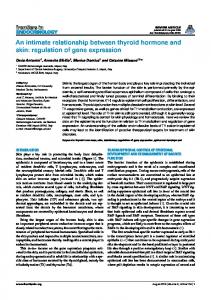
FIG. 1. Gene structure of skeletal muscle/liver 6-PF-2-K/ Fru-2,6-P2ase gene.lipprr horizontal linrs indicate the X genomic clones from two lihraries (see “Experimental Procedures”). The restriction map shows the location of EroRI ( I ? ) sites in the gene. The position and size of the 15 exons of the gene (see “Experimental I’rocedures”) are shown as orrticd [incs on the louvr horizontal linr. T h e position of the glucocorticoid response element is indicated hv the lramrd hox hetween exon I,, and exon 11.
DNase Footprinting-The glucocorticoid receptor binding domain (GRRD) used in this study was a gift from K. R. Yamamoto. It was 11 purified 85-amino acid expressed peptide which hound to the TAT (;RE with high affinity.:’ A 292“bp Hinfl-Sau.?AI fragment of intron I (hetween exon 11, and exon 11) of the skeletal muscle/liver 6-PF-2K/Fru“L,G-P,ase gene was cloned in the forward orientation into the was hlnnted Sal1 site of the pRLCAT2 polylinker. The fragments excised hy HamHI and PstI digestion and end-laheled at the RamHI site hy a fill-in reaction using Sequenase (United StatesBiochemical, version 1) modified T’i DNA polymerase in the presence of [ d T ] dATP and cold 0.4 mM dNTPs. Also, a 185-hp MsrI-MnrI fragment isolated from this intronic region of the gene and cloned into the pH1,CAT vector was excised and laheled as descrihed ahove for the Hinfl-SauXAI fragment. As a positive control for DNase protection by the GRRD, a 229-hp I;coRI-Hg[I fragment containing the consensus TAT (;RE was excised from the pGRETKCAT vector (36) was rllso end-laheled at the I h R I site as descrihed ahove. Approximately 2 ng of laheled prohe was used for each DNase digestion. Laheled fragments were incuhatedwithandwithout purified GRRD in a binding buffer (25 mM Tris-CI, pH 7.9. 6.25 mM MgCI,, 0.5 mM EDTA, 50 mM KCI, 0.5 mM dithiothreitol, and 10% glycerol) at room temperature for 5 min. Protein-DNA mixtures were then treated with 6 ng of DNase I for 1 min a t room temperature. DNase reaction was terminated hy addition of 100 PI of S T O P solution (1% SDS, 20 mM ISDTA. 200 mM NaCI, and 100 pg/ml tRNA) and heated to 90 “C for 6 min. Samples were then extracted with pheno1:chloroform (1:l) and then ch1oroform:isoamyl alcohol ( 2 5 1). Ethanol-precipitated samples weresuhjected to electrophoresis on 6rE polyacrylamide, 8 M urea gels and the hands visualized hy autoradiography.Protectedsites were located hv comparison with either HpnII-pHRS22-laheled standnrd or hy Maxam-Gilhert sequencing ( 3 7 ) . Protrin I~elrrminntion-Proteinconcentration was determined with the BCA Protein Assay Reagent from Pierce Chemical Co. using the enhanced protocol and hovine serum alhumin as a standard.
1.567.5
generates two first exons, Iv and I[,, in the skeletal muscle and liver forms of the enzyme, respectively (13, 17, 23). The remaining 1.7exons are common to both forms of the enzvme. The restriction map of Darville et a/. (2.7) had an additional EcoRI restriction site between exon6 and exon 7 which is not present in our gene map. Our genomic h clones also overlap in the region betweenexon 8 and 9 which was previously undefined (23). Transcription Start Site of Liwr 6-PF-2- K/Fru-2.6-P,aw mRNA-The transcription start site of the liver 6-PF-P-K/ Fru-2,6-P2ase mRNA was determined in order to engineer reporter gene (CAT) constructs to analyze the .i’-flanking region of the gene for promoteractivity.Theseconstructs depend on the inclusion of the native transcription start site for production of CAT message andprotein. SI nuclease mapping was done using a single-stranded probe (see “Experimental Procedures”) which extended 430 hp upstream from base 27 of the published cDNA sequence (13). Theprotected fragments were from 169 to 171 bases in length, which indicates that transcription initiates at the three adjacentnucleotides, GCG. Thelengthandflanking sequence of these fragments are shown in Fig. 2. Although there is no exact consensus sequence for a TATA box upstream of this transcription start site, thereis a TATT sequence 27 bp upstream and a CAATT sequence 107 bp upstream of this transcription start site. There is some controversv (17, 23) regarding the location of the cap site of the liver 6-PF-2-K/Fru-2,6-I’~ase gene. We report here a transcription start site approximately 79 bp further upstream than the site reported by Darville P ! al. (23). Also, no signal at a distance corresponding to the site reported by Darville Pt al. (23) was seen (data not shown). Regardless of these unresolved discrepencies,the 3’ Hinfl boundary usedin making all the liver promoter constructs (see below) was downstream of either of the reported transcription start sites. Promoter Activity of the 5‘-F/mking Rcyionn of thP 6-PF6 0
RESULTS
Isolation and Structure of Rat Liver 6-PF-2-K/Fru-2,6Prase Gene-In order to identify the glucocorticoid response element of the skeletal muscle/liver 6-PF-2-K/Fru-2,6-P2ase gene it was necessary to isolate the gene and characterize its genomic organization. Six overlapping genomic phage clones that spanned the entire 6-PF-2-K/Fru-2,6-P,ase gene were isolated from two genomic libraries:a Charon 4A phage library and an EMBL3 phage library. A schematic representation of the gene isolated from these clones is shown in Fig. 1, includingthe exon locationsand EcoRI restrictionsites.These findings extend our original report on the organizationof the gene (17) and are also in good agreement with the genomic organizationreported by Darvilleet al. (23). The gene is approximately 55 kb in length and is comprised of 15 exons. There is a tissue-specific use of two promoters which thereby K. R. Yamamoto, personal communication.
FIG. 2. Transcription start site for the liver form of 6-PF2-K/Fru-2,6-P2ase. The triplet hand Idenotcd with l t n c . ~t o hate.; (;CG, indicating the start site) is shown i n Inn