THE JOURNAL OF BIOLOGICAL CHEMISTRY © 2004 by The American Society for Biochemistry and Molecular Biology, Inc.
Vol. 279, No. 39, Issue of September 24, pp. 40545–40559, 2004 Printed in U.S.A.
Regulation of Histone Acetylation during Memory Formation in the Hippocampus* Received for publication, February 27, 2004, and in revised form, July 20, 2004 Published, JBC Papers in Press, July 23, 2004, DOI 10.1074/jbc.M402229200
Jonathan M. Levenson, Kenneth J. O’Riordan, Karen D. Brown, Mimi A. Trinh, David L. Molfese, and J. David Sweatt‡ From the Baylor College of Medicine, Department of Neuroscience, Houston, Texas 77030
Formation of long term memory requires several steps that end with regulation of gene expression. First, high frequency patterns of synaptic activity lead to activation of NMDA1 re-
ceptors (NMDA-Rs) and an influx of Ca2⫹ into the cytoplasm (1–3). The increase in intracellular Ca2⫹ concentration results in the activation of a variety of signaling pathways that ultimately converge to activate ERK (4). Once activated, ERK then regulates the expression of a variety of genes by either directly or indirectly regulating the activities of several transcription factors (5–7). These transcription factors effect a highly coordinated pattern of transcription that results in the formation and stabilization of long term memory. In the nucleus, DNA is tightly packaged into chromatin, a DNA-protein complex (8). In its native state, chromatin is highly inhibitory to transcription. The major protein component of chromatin consists of a group of highly basic proteins known as histones (8). Positively charged lysine residues in the N-terminal tail of histones mediate most of the DNA-histone interaction. Unmodified, native histones tightly bind to DNA, preventing other protein interactions, including the RNA polymerase II enzyme interaction required for transcription (8). The native structure of chromatin must be disrupted for transcription to occur. Acetylation of the ⑀-amino group of lysine residues by histone acetyltransferases (HATs) neutralizes the positive charge of histones, disrupting the interaction between histone and DNA (8). Some HATs have been shown to preferentially acetylate histones on specific lysine residues. For example, the HATs HPA2 and Gcn5 appear to preferentially acetylate histone H3 on Lys-14, a residue that has been implicated in regulation of transcription (9, 10). Histone acetylation appears to increase transcription initiation by facilitating the binding of transcription factors and the RNA polymerase II enzyme holoenzyme to DNA (11, 12). It should also be noted that acetylation of histones can in some cases be self-perpetuating, which in development may serve as a long term cellular memory, creating a functionally stable chromatin state and thus chronic changes in the rates of specific gene expression (13–15). Several studies have suggested that regulation of histones, and thus chromatin structure, is an important step in modulating transcription and facilitating long term changes in neuronal physiology. Gross changes in chromatin structure have been demonstrated in the suprachiasmatic nucleus when ani-
* This work was supported by National Institute of Mental Health Grant MH57014 and by NICHD, National Institutes of Health Grant HD24064. The costs of publication of this article were defrayed in part by the payment of page charges. This article must therefore be hereby marked “advertisement” in accordance with 18 U.S.C. Section 1734 solely to indicate this fact. ‡ To whom correspondence should be addressed: Baylor College of Medicine, Dept. of Neuroscience, 1 Baylor Plaza, Houston, TX 77030. Tel.: 713-798-3107; Fax: 713-798-3946; E-mail:
[email protected]. edu. 1 The abbreviations used are: NMDA, N-methyl-D-aspartic acid; NMDA-R, NMDA receptor; ERK, extracellular signal-regulated kinase;
HAT, histone acetyltransferase; PKC, protein kinase C; PKA, protein kinase A; LTP, long term potentiation; CS, cutting saline; ACSF, artificial cerebrospinal fluid; fEPSP, field excitatory postsynaptic potential; TBS, Tris-buffered saline; MAP, mitogen-activated protein; ANOVA, analysis of variance; PPF, paired-pulse facilitation; P-ERK2, phosphoERK2; MEK, MAP kinase/ERK kinase; PDA, phorbol 12,13-diacetate; FSK, forskolin; dFSK, 1,9-dideoxyforskolin; 4␣P, 4-␣-phorbol 12,13-didecanoate; TSA, trichostatin A; DRB, 5,6-dichlorobenzimidazole riboside; ActD, actinomycin D; NaB, sodium butyrate; HDAC, histone deacetylase; REST, repressor element 1 silencing transcription factor; AMPA, ␣-amino-3-hydroxy-5-methyl-4-isoxazoleproprionic acid; AMPA-R, AMPA receptor.
Formation of long term memory begins with the activation of many disparate signaling pathways that ultimately impinge on the cellular mechanisms regulating gene expression. We investigated whether mechanisms regulating chromatin structure were activated during the early stages of long term memory formation in the hippocampus. Specifically, we investigated hippocampal histone acetylation during the initial stages of consolidation of long term association memories in a contextual fear conditioning paradigm. Acetylation of histone H3 in area CA1 of the hippocampus was regulated in contextual fear conditioning, an effect dependent on activation of N-methyl-D-aspartic acid (NMDA) receptors and ERK, and blocked using a behavioral latent inhibition paradigm. Activation of NMDA receptors in area CA1 in vitro increased acetylation of histone H3, and this effect was blocked by inhibition of ERK signaling. Moreover, activation of ERK in area CA1 in vitro through either the protein kinase C or protein kinase A pathways, biochemical events known to be involved in long term memory formation, also increased histone H3 acetylation. Furthermore, we observed that elevating levels of histone acetylation through the use of the histone deacetylase inhibitors trichostatin A or sodium butyrate enhanced induction of long term potentiation at Schaffer-collateral synapses in area CA1 of the hippocampus, a candidate mechanism contributing to long term memory formation in vivo. In concert with our findings in vitro, injection of animals with sodium butyrate prior to contextual fear conditioning enhanced formation of long term memory. These results indicate that histone-associated heterochromatin undergoes changes in structure during the formation of long term memory. Mimicking memory-associated changes in heterochromatin enhances a cellular process thought to underlie long term memory formation, hippocampal long term potentiation, and memory formation itself.
This paper is available on line at http://www.jbc.org
40545
40546
Histone Acetylation in Long Term Memory Formation
mals are exposed to phase-resetting light pulses, and chromatin structure is regulated in hippocampal neurons in response to activation of a variety of neurotransmitter pathways (16, 17). Ischemic insults increase the expression of REST, a gene silencer known to recruit chromatin remodeling enzymes to DNA, within areas CA1 and CA3 of the hippocampus (18). Some studies have also examined the structure of chromatin within specific gene-associated promoters. Status epilepticus induces changes in the structure of chromatin associated with the GluR2 and BDNF genes in area CA3 (19). Global ischemia alters chromatin structure associated with GluR2 in areas CA1 and CA3 (18). Other studies have found regulation of chromatin structure around the ApC/EBP gene in Aplysia using a pharmacologic paradigm that triggers long term facilitation of neurotransmitter release at the sensory-motor neuron synapse in this species (20, 21). All of these studies indicate that chromatin structure is highly dynamic within the nervous system and suggest the possibility that chromatin structure itself might be recruited as a target of plasticity-associated signal transduction mechanisms. In the present study, we examined the structure of chromatin, as assessed by histone acetylation, during induction of long term memory formation in area CA1 of the hippocampus. Contextual fear conditioning increased acetylation of histone H3, but not H4. Furthermore, stimulation of a known memoryassociated signaling pathway, ERK, through activation of either the protein kinase C (PKC) or protein kinase A (PKA) pathways also increased acetylation of histone H3 without affecting H4. Artificially elevating levels of histone acetylation in vitro enhances the induction of a lasting form of synaptic strengthening, long term potentiation (LTP) in hippocampal area CA1. Finally, treatment of animals in vivo with a histone deacetylase inhibitor enhanced formation of long term memory. Taken together these observations implicate alterations in histone acetylation and, by extension, modulation of chromatin structure, in mammalian associative learning and long term memory formation. EXPERIMENTAL PROCEDURES
Animals—Young adult, male Sprague-Dawley rats (150 –200 g) were used for all experiments. Animals were housed under light/dark 12 h/12 h and allowed access to rodent chow and water ad libitum. Animals were allowed to acclimate to laboratory conditions at least 3 days prior to use in experiments. All procedures were performed with the approval of the Baylor College of Medicine Institutional Animal Care and Use Committee and according to national guidelines and policies. Fear Conditioning—Animals were transported to the laboratory at least 30 min prior to fear conditioning. Animals were placed into the training chamber and allowed to explore for 2 min, after which they received an electric shock (1 s, 0.5 mA). The 2 min/1 s shock paradigm was repeated for a total of three shocks. After the last shock, animals were allowed to explore the context for an additional 1 min prior to removal from the training chamber. For experiments investigating the effect of sodium butyrate on long term memory, animals were exposed to only 1 shock. Latent inhibition training was performed by placing the animals into the training chamber 2 h prior to administering the standard fear conditioning paradigm outlined above. In experiments where animals received an injection of either saline (1.25 l/kg) or MK-801 (100 or 300 g/kg), injections were performed 1 h prior to fear conditioning. In experiments where animals received an injection of either Me2SO (2.9 ml/kg) or SL327 (100 mg/kg), injections were performed immediately after the training session. For behavioral experiments, freezing behavior was measured during and either 1 or 24 h after fear conditioning. Freezing behavior was measured by observing the animals for 2 s every 10 s. Age-matched animals that were handled by the experimenter but did not receive any experimental manipulations (naı¨ve) were used as controls in all fear conditioning experiments. Hippocampus Slice Preparation—Animals were sacrificed using a rodent guillotine. The brain was immersed in ice-cold cutting saline (CS (in mM): 110 sucrose, 60 NaCl, 3 KCl, 1.25 NaH2PO4, 28 NaHCO3, 0.5 CaCl2, 7 MgCl2, 5 glucose, 0.6 ascorbate) prior to isolation of the caudal portion containing the hippocampus and entorhinal cortex. Transverse
slices (400 m) were prepared with a Vibratome (The Vibratome Company, St. Louis, MO). During isolation, slices were stored in ice-cold CS. After isolation, cortical tissue was removed and hippocampal slices were equilibrated in a mixture of 50% CS and 50% artificial cerebrospinal fluid (ACSF (in mM): 125 NaCl, 2.5 KCl, 1.25 NaH2PO4, 25 NaHCO3, 2 CaCl2, 1 MgCl2, 25 glucose) at room temperature. Slices were further equilibrated in 100% ACSF for 45 min at room temperature, followed by a final incubation in 100% ACSF at 32 °C for 1 h. All solutions were saturated with 95%/5% O2/CO2. Slices were treated with the appropriate drugs or vehicle after the last equilibration at 32 °C. For experiments investigating the regulation of histone acetylation in vitro, slices from four animals were pooled, randomized, and divided into two treatment groups: vehicle control and drug-treated. For electrophysiology experiments, slices from one animal were recorded at a time. Therefore, each electrophysiology experiment had matched vehicle and drug-treated slices from the same animal. Isolation of Area CA1—For isolation of area CA1 from whole brain, the brain was immersed in oxygenated (95%/5% O2/CO2) ice-cold CS immediately after removal from the animal. For isolation of area CA1 from slices of hippocampus, slices were immersed in ice-cold CS immediately after the treatment period. For whole brain and slice dissections, area CA1 was dissected away from other hippocampal subfields under a dissecting microscope; tissue from two animals was pooled for each treatment group. Once isolated, Area CA1 was placed in 4 ml of ice-cold homogenization buffer (in mM: 250 sucrose, 50 Tris, pH 7.5, 25 KCl, 0.5 phenylmethylsulfonyl fluoride, 1% protease inhibitor mixture (Sigma), 0.9 Na⫹-butyrate) and homogenized for 12 strokes at 4,000 RPM with a Teflon-on-glass grinder (VWR). Slice Electrophysiology—Electrophysiology was performed in an interface chamber (Fine Science Tools, Foster City, CA). Oxygenated ACSF (95%/5% O2/CO2) was perfused into the recording chamber at a rate of 1 ml/min. Electrophysiological traces were digitized and stored using Digidata (models 1200 and 1320A) and Clampex software (Axon Instruments, Union City, CA). Extracellular stimuli were administered on the border of areas CA3 and CA1 along the Schaffer-collaterals using Teflon-coated, bipolar platinum electrodes. fEPSPs were recorded in stratum radiatum with an ACSF-filled glass recording electrode (1–3 M⍀). The relationship between fiber volley and fEPSP slopes over various stimulus intensities was used to assess baseline synaptic transmission. All subsequent experimental stimuli were set to an intensity that evoked a fEPSP that had a slope of 50% of the maximum fEPSP slope. Paired-pulse facilitation was measured at various interstimulus intervals (20, 50, 100, 200, and 300 ms). Long term potentiation (LTP) was induced with 2,100 Hz tetani (1 s), with an interval of 20 s between tetani. Synaptic efficacy was monitored 0.5 h prior to and 3 h following induction of LTP by recording fEPSPs every 20 s (traces were averaged for every 2-min interval). Drug or vehicle was introduced 20 min prior to LTP induction. Histone Extraction—All procedures were performed on ice, and all solutions were chilled to 4 °C prior to use unless otherwise indicated. All centrifugation steps were performed at 4 °C. Tissue homogenates were centrifuged at 7,700 ⫻ g for 1 min. The supernatant (cytoplasmic fraction) was aspirated and stored at ⫺80 °C. The pellet (nuclear fraction) was resuspended in 1 ml of 0.4 N H2SO4. Histones were acidextracted from the nuclear fraction for 30 min. Acid extracts were centrifuged at 14,000 ⫻ g for 10 min. The supernatant was transferred to a fresh tube, and proteins were precipitated with the addition of 250 l of 100% trichloroacetic acid containing 4 mg/ml deoxycholic acid (Na⫹ salt, Sigma) for 30 min. Precipitated proteins were collected by centrifugation at 14,000 ⫻ g for 30 min. The supernatant was discarded, and the protein pellet was washed with 1 ml of acidified acetone (0.1% HCl) followed by 1 ml of acetone for 5 min each. Protein precipitates were collected between washes by centrifugation (14,000 ⫻ g, 5 min). The resulting purified proteins were resuspended in 10 mM Tris (pH 8) and stored at ⫺80 °C. Western Blotting—Loading buffer was added (final concentration: 6.25 mM Tris, pH 6.8, 2% SDS, 10% glycerol, 1.25% 2-mercaptoethanol, 0.1% bromphenol blue), and samples were incubated at room temperature for 20 min prior to SDS-PAGE. Samples were run on a discontinuous polyacrylamide gel consisting of a 20% acrylamide resolving and a 4% acrylamide stacking gel, after which proteins were transferred to polyvinylidene difluoride membranes for immunoblotting. Membranes were blocked in 3% bovine serum albumin in TTBS (in mM: 150 NaCl, 20 Tris, pH 7.5, 0.05% Tween 20) for 45 min at room temperature. Membranes were incubated in primary antibodies overnight at 4 °C and in horseradish peroxidase-conjugated secondary antibodies for 2.5 h at room temperature. Immunolabeling of membranes was detected via chemiluminescence (ECL, Amersham Biosciences; SuperSignal,
Histone Acetylation in Long Term Memory Formation Pierce). Luminescence was recorded with BioMax MR film (Kodak Scientific Imaging Systems, Rochester, NY) and digitized (Epson Perfection 1240U), and integrated densities of each band were quantified with ImageJ (National Institutes of Health, Bethesda, MD). Several exposures were captured for each immunoblot to ensure that all densitometry was performed on images taken in the linear exposure range. Antibodies—The primary antibodies used, and their dilutions were as follows: anti-MAP kinase 1/2 (1:1000, Upstate Biotechnology Inc.), anti-Phospho-p44/42 MAP kinase (Thr-202/Tyr-204, 1:1000, Cell Signaling), anti-Histone H3 (mouse monoclonal, 1:500, Upstate), antiacetyl Histone H3 (Lys-14, 1:1000, Upstate), anti-Histone H4 (1:500, Upstate), and anti-acetyl Histone H4 (Lys-5/Lys-8/Lys-12/Lys-16, 1:1000, Upstate). The host for all primary antibodies was rabbit unless otherwise specified. The secondary antibodies were horseradish peroxidaseconjugated goat Anti-IgG heavy and light chain (Jackson ImmunoResearch, West Grove, PA). Statistical Analysis—All Western blotting data was analyzed using a Kruskal-Wallis ANOVA followed by a Dunn’s multiple comparison test. Analysis of freezing behavior between the contextual fear conditioning and latent inhibition training paradigms and between saline and sodium butyrate injections were performed with Student’s t-test. The effect of MK-801 on freezing behavior was analyzed with a one-way ANOVA, followed by a Newman-Keuls multiple comparison test. Input/ output relationships were analyzed using either a single exponential function (Y ⫽ slopemax ⫻ 1 ⫺ e⫺KxX) or a linear regression where appropriate. Parameters of the equations used to fit input/output relationships were compared using an F test. Paired-pulse facilitation (PPF) and LTP were analyzed via two-way ANOVA with repeated measures. For analysis of LTP, data acquired before and after tetani were analyzed separately. Post-hoc comparisons after two-way ANOVA were made using the method of Bonferroni. Significance for all tests was set at p ⬍ 0.05. RESULTS
Regulation of Histone Acetylation in Vivo by Contextual Fear Conditioning—Contextual fear conditioning requires ERK activity and leads to an increase in the activity of ERK2 in area CA1 of the hippocampus of rats 1 h after training (22). We confirmed that the behavioral methods employed in the current studies also led to increases in ERK2 activity in area CA1 of the hippocampus. Contextual fear conditioning increased levels of phospho-ERK2 (P-ERK2; Fig. 1A) without affecting levels of P-ERK1 (data not shown) when measured 1 h after fear conditioning. The increase in P-ERK2 was not apparent 24 h after training (Fig. 1A), indicating that the regulation of ERK2 was transient. Injection of animals with the NMDA receptor (NMDA-R) antagonist MK-801 1 h prior to fear conditioning blocked the increase in P-ERK2 normally seen 1 h after fear conditioning (Fig. 1A) and the formation of contextual fear memory (Fig. 2, C and D). Moreover, injection of animals with the MEK inhibitor SL327 immediately after fear conditioning, a treatment that prevents formation of long term contextual fear memory (22), also blocked the increase in P-ERK2 normally seen 1 h after fear conditioning (Fig. 1A). These results indicate that our methods are comparable to previously published reports of biochemical changes associated with fear conditioning in the hippocampus. Furthermore, our results demonstrate that the increase in P-ERK2 seen after fear conditioning requires the activation of NMDA-Rs and the upstream kinase in the ERK cascade, MEK. The above results suggest that ERK2 phosphorylation was induced by formation of a long term memory where an animal associates a novel context with a noxious stimulus (footshock). It was possible, however, that the regulation of ERK2 was due to either formation of new spatial memories upon exposure to the novel context, or was a stress response to the footshock alone in the absence of associative memory formation. A latent inhibition training paradigm was used to determine whether either alternative possibility contributed to the regulation of ERK2 1 h after fear conditioning. Latent inhibition occurs when an animal is pre-exposed to a
40547
novel context prior to receiving an unconditioned stimulus (electric shock) in that context. Latent inhibition is unique in that the animal does not form an association between the noxious stimulus and the novel context, yet the actual latent inhibition is context-specific (23, 24) (Fig. 2, A and B). Therefore, in latent inhibition training the animal has formed a spatial memory that blocks the formation of an associative contextual fear memory. Animals were placed into the novel training context 2 h prior to receiving the three-shock context fear conditioning training paradigm. The latent inhibition training procedure inhibited formation of long term contextual fear memory (Fig. 2, A and B). Exposure of animals to the latent inhibition paradigm blocked the increase in hippocampal P-ERK2 that normally occurs 1 h after fear conditioning (Fig. 1A). Taken together with previously published results (22), these results indicate that the regulation of ERK2 after contextual fear conditioning was specific for the formation of associative contextual fear memories. The lack of regulation of ERK2 after latent inhibition training suggests that regulation of ERK2 in area CA1 of the hippocampus is not involved in the formation of latent inhibition memory and is consistent with the hypothesis that latent inhibition is independent of ERK2. Formation of long term contextual fear memory requires transcription in area CA1 of the hippocampus, and a wide variety of transcripts are regulated with contextual fear conditioning, especially 1 h after conditioning (25). We investigated whether there might be changes in chromatin structure 1 h after fear conditioning, which is a critical period when transcription occurs during long term memory formation in the hippocampus (26). We observed significant increases in the acetylation of histone H3 on Lys-14 in Area CA1 of the hippocampus 1 h after contextual fear conditioning (Fig. 1, B and C). The increase in H3 acetylation was not apparent 24 h after conditioning (Fig. 1C), indicating that the regulation of H3 was transient, and restricted to periods where critical phases of memory-associated transcription occur (26). Similar to behavioral fear conditioning, the increase in acetylation of histone H3 required the activation of NMDA-Rs and MEK, because injection of animals with either MK-801 or SL327 inhibited the increase in H3 acetylation (Fig. 1C). In addition, the increase in acetylation of histone H3 was specific for the formation of associative contextual fear memory; latent inhibition training blocked the increase in acetylation of histone H3 normally seen with fear conditioning (Fig. 1B, p ⬍ 0.05). These results indicate that the increase in acetylation of histone H3 on Lys-14, 1 h after fear conditioning, is specific for formation of NMDA-Rand ERK-dependent associative contextual fear memory. To assess whether regulation of histone acetylation was specific to histone H3, or might be a general phenomenon, the acetylation of histone H4 was also investigated. The antibody we used for these experiments detects any combination of Lys acetylation among the four different residues in the N terminus of histone H4. Acetylation of histone H4 was not significantly affected by contextual fear conditioning when measured either 1 or 24 h after, or by blockade of NMDA-Rs or MEK (Fig. 1, B and D). Interestingly, latent inhibition training resulted in significant increases in acetylation of histone H4 (Fig. 1D), suggesting the intriguing possibility that the molecular processes involved in the storage of hippocampus-dependent latent inhibition memory occur through selective regulation of transcription associated with histone H4. Overall, these results demonstrate differential regulation of hippocampal histone acetylation depending on the specific type of memory formed. Regulation of Histone Acetylation in Vitro by NMDA-Rs— ERK is known to be involved in hippocampus-dependent mem-
40548
Histone Acetylation in Long Term Memory Formation
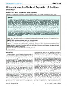
FIG. 1. Contextual fear conditioning increases phosphorylation of ERK2 and acetylation of histone H3. Animals were fear conditioned and area CA1 of the hippocampus was isolated 1 or 24 h after training. For this and subsequent figures illustrating data collected via Western blotting, representative Western blots are shown above graphs of summary quantification. For each representative immunoblot, a matched pair is shown where the control sample is on the left and the experimental sample is on the right. A, significant regulation of ERK2 phosphorylation was seen after contextual fear conditioning (H[5,31] ⫽ 25, p ⬍ 0.0001). Contextual fear conditioning led to an increase in P-ERK2
Histone Acetylation in Long Term Memory Formation
40549
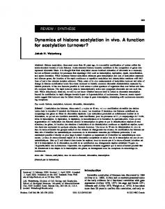
FIG. 2. Contextual fear memory requires proper timing and NMDA-R function. Animals were exposed to the contextual fear conditioning paradigm, and freezing behavior was measured either during training (A and C) or 24 h after training (B and D) in the training chamber. A, animals exposed to the standard contextual fear conditioning paradigm (Context ⫹ Shock, n ⫽ 12) or the latent inhibition training paradigm (2-h pre-exposure to training chamber, n ⫽ 12) exhibited similar amounts of freezing during the training session. B, animals exposed to the contextual fear conditioning paradigm had significantly more freezing behavior than animals exposed to the latent inhibition training paradigm (t ⫽ 4.7, df ⫽ 20, p ⬍ 0.0001). C, animals injected with either saline (n ⫽ 5) or MK-801 (100 g/kg: n ⫽ 3; 300 g/kg: n ⫽ 4) 1 h prior to contextual fear conditioning showed similar amounts of freezing behavior during training (F[2,11] ⫽ 2.4, p ⬍ 0.2). D, animals injected with 300 g/kg MK-801 showed less freezing behavior than animals injected with saline 24 h after contextual fear conditioning (F[2,11] ⫽ 8.5, p ⬍ 0.01). Error bars are S.E.; asterisks indicate significant differences (p ⬍ 0.05).
ory formation, including contextual fear conditioning (22, 27). Moreover, our results suggest that the ERK signaling cascade may be important in the regulation of chromatin structure. Therefore, we tested whether activation of ERK, like behavioral conditioning itself, might trigger regulation of histone acetylation in area CA1. To ensure that our slice cultures were closely modeling the biochemical changes that occur in vivo, we first investigated whether ERK and/or histone acetylation was regulated by activation of NMDA-Rs. Treatment of hippocampal slices with NMDA for 10 min induced significant increases in P-ERK2 (Fig. 3A). The increases in P-ERK2 were blocked when slices were pretreated with either U0126 or PD98059 (Fig. 3A), which are inhibitors of MEK. Moreover, treatment of slices with NMDA increased acetylation of histone H3, which was blocked by pretreatment with either U0126 or PD98059 (Fig. 3B). No change in the acetylation of histone H4 was observed by any of the above treatments (Fig. 3C). These results indicate that in area CA1 of the hippocampus, activation of NMDA-Rs leads to increases in P-ERK2 and acetylation of histone H3, and that these events require the upstream kinase in the ERK cascade, MEK. Regulation of Histone Acetylation in Vitro by ERK Activation—We next investigated whether activation of either the protein kinase C (PKC) or protein kinase A (PKA) pathways in vitro led to activation of ERK, to determine whether we could use these pathways to study ERK-dependent changes in histone acetylation. Activation of PKC with phorbol 12,13-diacetate (PDA) significantly increased levels of P-ERK1 and P-
ERK2 in area CA1 (Fig. 4, A and B; P-ERK1 data not shown). Similarly, activation of PKA with forskolin (FSK) significantly increased levels of P-ERK2 (Fig. 4, A and B), but not P-ERK1 (data not shown). The effects of both PDA and FSK were specific to the active properties of the drugs, because the respective inactive analogs 4-␣-phorbol 12,13-didecanoate (4␣P) and 1,9-dideoxyforskolin (dFSK) had no affect on levels of PERK1 or P-ERK2 (Fig. 4, A and B, and data not shown). The effects of both PDA and FSK on P-ERK were blocked by the MEK inhibitors U0126 and PD98059 (Fig. 4, A and B), confirming that effects of PDA and FSK on ERK were through the activation of the upstream kinase in the ERK cascade, MEK. We next investigated whether activation of ERK via PKC or PKA signaling led to regulation of histone acetylation in hippocampal area CA1. Treatment of slices with either PDA or FSK significantly increased acetylation of histone H3 (Fig. 5, A and B). The increases in acetylation of histone H3 were specific for the active properties of the drugs used, because treatment with either 4␣P or dFSK had no affect on the acetylation of histone H3 (Fig. 5, A and B). Moreover, pretreatment of slices with the MEK inhibitors U0126 or PD98059 blocked the ability of either PDA or FSK to increase histone H3 acetylation, further confirming that the effects of these drugs were due to their actions on the ERK signaling cascade (Fig. 5, A and B). As a positive control, H3 acetylation was measured after exposure of slices to the histone deacetylase inhibitor trichostatin A (TSA). TSA significantly increased acetylation of H3 (Fig. 5, A and B, without affecting levels of P-ERK2 (Fig. 4, A and B). Thus,
1 h after fear conditioning (n ⫽ 10), but not 24 h after (n ⫽ 4). No regulation of ERK2 was seen after latent inhibition training (n ⫽ 11). Injection of animals 1 h prior to training with the NMDA-R antagonist MK-801 (300 g/kg, n ⫽ 6) or immediately after training with the MEK inhibitor SL327 (100 mg/kg, n ⫽ 6) inhibited the increase in P-ERK2. B, acetylation of histones H3 and H4 was measured in area CA1 from naı¨ve (N) and fear conditioned (FC) animals 1 h after contextual fear conditioning. Shown are representative Western blots depicting levels of acetylated histones and total histone protein from acid extracts of nuclear preparations. Below the immunoblots is a diagram illustrating the various behavioral manipulations performed in our studies. C, significant regulation of histone H3 acetylation was observed after contextual fear conditioning (H[5,31] ⫽ 15, p ⬍ 0.01). Contextual fear conditioning led to an increase in the acetylation of histone H3 1 h after training (n ⫽ 11), but not 24 h after (n ⫽ 6). No change in H3 acetylation was observed after latent inhibition training (n ⫽ 10), or injection with either MK-801 (n ⫽ 9) or SL327 (n ⫽ 6). D, no significant regulation of histone H4 acetylation was observed 1 h or 24 h after fear conditioning (n ⫽ 11). Injection of MK-801 (n ⫽ 9) or SL327 (n ⫽ 3) also had no effect on histone H4 acetylation. However, latent inhibition training significantly increased acetylation of histone H4 (n ⫽ 11, H[5,30] ⫽ 10, p ⬍ 0.05). Error bars indicate ⫾ S.E.; asterisks indicate significant differences (p ⬍ 0.05) as determined by post-hoc Dunn’s multiple comparison test.
40550
Histone Acetylation in Long Term Memory Formation
FIG. 3. Activation of NMDA-Rs increases phosphorylation of ERK2 and acetylation of histone H3. Acute hippocampal slices were treated with 100 M NMDA in Mg2⫹-free ACSF for 10 min. After treatment, area CA1 was isolated and processed for Western blotting. A, phosphorylation of ERK2 was significantly increased in area CA1 after treatment with NMDA (n ⫽ 5, H[3,10] ⫽ 9, p ⬍ 0.05). The MEK inhibitors U0126 (20 M, n ⫽ 4) and PD98059 (50 M, n ⫽ 5) inhibited the affect of NMDA on P-ERK2. B, acetylation of histone H3 was significantly increased in area CA1 after treatment of slices with NMDA (n ⫽ 4, H[3,10] ⫽ 7, p ⬍ 0.05). Both U0126 (n ⫽ 4) and PD98059 (n ⫽ 5) inhibited the affect of NMDA on acetylation of histone H3. C, NMDA had no affect on acetylation of histone H4 (H[3,10] ⫽ 1, p ⬍ 0.7). Error bars are ⫾ S.E.; asterisks indicate significant differences (p ⬍ 0.05) as determined by post-hoc Dunn’s multiple comparisons test.
activation of ERK in hippocampal area CA1, either through the PKC or PKA signaling pathways, led to increased acetylation of histone H3 in area CA1 of the hippocampus. To assess whether activation of ERK in vitro led to a general increase in histone acetylation, levels of histone H4 acetylation were also measured. Activation of either PKC or PKA did not significantly affect the acetylation of histone H4 (Fig. 6B), suggesting that activation of ERK does not regulate acetylation of histone H4. Incubation of slices in the presence of TSA increased the acetylation of histone H4 (Fig. 6, A and B), indicating that the antiserum used to measure acetylation of histone H4 was capable of detecting changes in acetylation
state. These results support the hypothesis that the overall acetylation state of histone H4 in area CA1 of the hippocampus does not appear to be affected by activation of the ERK signaling cascade. Increasing Basal Histone Acetylation Enhances Induction of LTP—Our results indicated that acetylation of histone H3 was increased in area CA1 during formation of long term memory in vivo and with activation of ERK in vitro. This suggested that ERK acts to couple the activity of intracellular signaling cascades to the acetylation state of histones in the nucleus as a mechanism for regulating transcription to induce long term changes in neuronal function. Induction of long-lasting LTP at
Histone Acetylation in Long Term Memory Formation
40551
FIG. 4. Regulation of ERK phosphorylation. ERK phosphorylation was monitored in acute hippocampal slices in response to treatments with various drugs for 1 h. Significant regulation of P-ERK2 was observed (H[10,49] ⫽ 37, p ⬍ 0.0001). A, representative immunoblots illustrating the effect of various treatments on levels of P-ERK1/2. Blots for phosphorylated-ERK are shown at the top, and blots of total ERK are shown at the bottom. In each blot, control samples are on the left and experimental samples are on the right. B, summary quantification of the regulation of P-ERK2. Treatment of slices with either PDA (3 M, n ⫽ 12) or FSK (50 M with 100 M Ro20-1724, n ⫽ 9) led to significant increases in levels of P-ERK2. Pretreatment with either U0126 (U0⬘, 20 M) or PD98059 (PD9⬘, 50 M) blocked the effect of PDA (U0: n ⫽ 5; PD9: n ⫽ 3) and FSK (U0: n ⫽ 4; PD9: n ⫽ 3). Levels of P-ERK2 were diminished by treatment with either U0126 (n ⫽ 6) or PD98059 (n ⫽ 3) alone. The inactive analogs of PDA and FSK, 4␣P (3 M, n ⫽ 3), and dFSK (50 M with 100 M Ro20-1724, n ⫽ 3) had no effect on P-ERK2. The HDAC inhibitor TSA (n ⫽ 9) had no effect on P-ERK2. Error bars indicate ⫾ S.E.; asterisks indicate significant differences as determined by post-hoc Dunn’s multiple comparison test. TSA data were analyzed via Wilcoxon signed rank test.
FIG. 5. Regulation of histone H3 acetylation by activation of ERK. Acetylation of histone H3 was monitored in acute hippocampal slices in response to treatments with various drugs for 1 h. Significant regulation of H3 acetylation was observed (H[10,43] ⫽ 38, p ⬍ 0.0001). A, representative immunoblots illustrating the regulation of histone H3 acetylation. Blots for acetylated histone H3 (Lys14) are shown at the top, while blots for total histone H3 are shown at the bottom. B, summary quantification of histone H3 acetylation. Treatment of slices with either PDA (3 M, n ⫽ 11) or FSK (50 M with 100 M Ro20-1724, n ⫽ 11) increased histone H3 acetylation. The effects of PDA and FSK were inhibited by pretreatment with either U0126 (20 M, PDA: n ⫽ 6; FSK: n ⫽ 4) or PD98059 (50 M, PDA: n ⫽ 3; FSK: n ⫽ 3). The inactive analogs of PDA and FSK, 4␣P (3 M, n ⫽ 3) and dFSK (50 M with 100 M Ro20-1724, n ⫽ 3) had no effect on H3 acetylation. Inhibition of MEK using either U0126 (20 M, n ⫽ 4) or PD98059 (50 M, n ⫽ 3) had no effect on basal levels of acetylation of histone H3. The HDAC inhibitor TSA increased acetylation of histone H3 (1.65 M, n ⫽ 7). Error bars indicate ⫾ S.E.; asterisks indicate significant differences (p ⬍ 0.05) as assessed by Dunn’s multiple comparison test. TSA data were analyzed via Wilcoxon signed rank test.
40552
Histone Acetylation in Long Term Memory Formation
FIG. 6. Acetylation of histone H4 is not regulated by activation of ERK. Acetylation of histone H4 was monitored in acute hippocampal slices in response to treatments with various drugs for 1 h. No significant regulation of H4 acetylation was observed (H[10,37] ⫽ 7, p ⬍ 0.7). A, representative immunoblots illustrating the regulation of histone H4 acetylation. Blots for acetylated histone H4 (Lys5/Lys-8/Lys-12/Lys-16) are shown at the top, whereas blots for total histone H4 protein are at the bottom. B, summary quantification of histone H4 acetylation. TSA (1.65 M, n ⫽ 7) was the only treatment that had a significant effect on H4 acetylation. Error bars are ⫾ S.E.; the asterisk indicates significantly different (p ⬍ 0.01) from 100% as assessed by Wilcoxon Signed Rank Test.
Schaffer-collateral synapses in area CA1 of the hippocampus, a candidate mechanism that may contribute to long term memory formation in vivo, requires activation of ERK in addition to translation and transcription (28 –33). Therefore we hypothesized that elevating levels of histone acetylation might facilitate induction of LTP through augmentation of gene transcription. To test this hypothesis, we investigated the effect of the histone deacetylase inhibitor TSA on induction of Schaffercollateral LTP in vitro. Acute hippocampal slices were maintained in an interface chamber into which either TSA or vehicle was perfused. Extracellular field excitatory postsynaptic potentials (fEPSPs) were recorded in stratum radiatum in area CA1 for 10 min prior to perfusion with drug or vehicle. After the start of perfusion, fEPSPs were measured for another 20 min to ensure that the treatments had no acute effect on baseline synaptic efficacy. After the initial perfusion with either TSA or vehicle, slices were exposed to 2,100 Hz tetani (1 s), which resulted in the induction of LTP that lasted at least 3 h (Fig. 7A). Slices treated with vehicle displayed normal LTP, whereas slices treated with TSA exhibited an enhancement of LTP (Fig. 7A). The TSAmediated enhancement of LTP occurred during both the early (0 –120 min) and late (⬎120 min) phases of LTP (Fig. 7A). These results suggest that an inhibitor of histone deacetylase, which significantly increased acetylation of histones H3 and H4 (Figs. 5 and 6), can enhance the amount of potentiation triggered by high frequency stimulation of Schaffer-collateral synapses in area CA1 of the hippocampus. No studies of the short or long term effects of TSA on synaptic properties exist. Therefore, it was possible that the enhancement of LTP by TSA was due to an indirect effect on synaptic properties, and not through modulation of histone acetylation. To determine whether TSA was indirectly affecting
synaptic transmission, we performed a number of control experiments. In the first experiment, slices were exposed to TSA and synaptic transmission was monitored for the same time period as in the LTP experiments. TSA had no effect on synaptic transmission in the absence of high frequency stimulation (Fig. 7A), suggesting that TSA was not affecting basal levels of synaptic efficacy or allowing potentiation due to synaptic stimulation at baseline testing frequencies. To further explore potential effects of TSA on synaptic transmission, slices were exposed to TSA for either 20 min or 3 h, and input/output relationships and paired-pulse facilitation (PPF) were measured. Treating slices for either 20 min or 3 h with TSA had no significant effect on input/output relationships (Fig. 7, B and D, or PPF (Fig. 7, C and E) relative to control slices treated with the vehicle, suggesting that synaptic connectivity and transmitter release were unaffected by TSA. Taken together, the physiologic results presented thus far indicate that the enhancement of LTP by TSA was not due to indirect effects on basal synaptic transmission. In another series of experiments, we investigated the possibility that TSA was affecting properties of NMDA-Rs. To directly measure any long term affects of TSA on NMDA-Rs, field potentials were recorded in the presence of the AMPA-R antagonist 6-cyano-7-nitroquinoxaline-2,3-dione to isolate the NMDA-R-dependent component of synaptic transmission. NMDA-R-mediated synaptic transmission was unaffected by chronic exposure to TSA for over 3 h (Fig. 8A). Furthermore, LTP induced in the presence of TSA required NMDA-R activation, because the NMDA-R antagonist AP5 blocked induction of LTP in the presence of TSA (Fig. 8B). To further explore the potential affects of TSA on synaptic transmission, we measured the depolarization that occurs during the 100-Hz tetanus used to induce LTP. No significant difference was observed in the
Histone Acetylation in Long Term Memory Formation
40553
FIG. 7. Trichostatin A enhanced induction of LTP. The effect of the HDAC inhibitor TSA was assessed on LTP and basal synaptic transmission of Schaffer-collateral synapses. A, induction of L-LTP by 2100-Hz (1 s, 20-s interstimulus interval) tetani was significantly enhanced in slices treated with TSA (1.65 M, open diamonds, n ⫽ 24) relative to slices exposed to the vehicle (0.05% EtOH, closed circles, n ⫽ 18; F[1,2,783] ⫽ 558, p ⬍ 0.0001). Continuous treatment of slices with TSA where LTP was not induced had no effect on baseline synaptic efficacy for up to 3 h after the start of treatment (crosses, n ⫽ 10; H[103,991] ⫽ 50.34, p ⫽ 1.0). Representative traces 4 min before (dashed line) and 180 min after (solid line) induction of LTP are shown to the right of the summary plot. The calibration bar indicates 5 ms and 1 mV. B, input/output relationships for slices treated with vehicle or TSA for 20 min. No significant differences were seen (F[2,10] ⫽ 4, p ⬍ 0.1). C, paired-pulse facilitation was unaffected by treatment of slices with TSA for 20 min (F[1,129] ⫽ 0.01, p ⬍ 1.0). D, input/output relationships for slices treated with vehicle or TSA for 3 h. No significant differences were seen (F[1,13] ⫽ 0.1, p ⬍ 0.8). E, paired-pulse facilitation was unaffected by treatment of slices with TSA for 3 h (F[1,168] ⫽ 0.8, p ⬍ 0.4). These results suggest that the enhancement of LTP by TSA was not due to direct effects on synaptic transmission. In all panels, Error bars are ⫾ S.E.
40554
Histone Acetylation in Long Term Memory Formation
FIG. 8. TSA does not affect NMDA-Rs, the NMDA-R dependence of LTP induction, or tetanus-induced synaptic depolarization. A, slices were exposed to the AMPA-R antagonist CNQX (20 M) and perfused with an ACSF containing 4 mM CaCl2 and no MgCl2. The resulting field potentials represent NMDA-R-mediated synaptic transmission (32). TSA (1.65 M) had no short or long term effects on NMDA-R-mediated synaptic transmission (n ⫽ 6, F[99,472] ⫽ 0.5, p ⫽ 1). Representative traces immediately before (dashed) and 186 min after (solid) introduction of TSA are shown. The calibration bar indicates 5 ms and 1 mV. B, slices were exposed to both TSA (1.65 M) and the NMDA-R antagonist AP5 (50 M) 20 min prior to LTP induction. After induction of LTP, the AP5 was washed out, but slices were still exposed to TSA. Induction of LTP in the presence of TSA was blocked by AP5 (n ⫽ 8, F[89,612] ⫽ 0.5, p ⬍ 1). Representative traces are shown 4 min before (dashed) and 180 min after (solid) LTP induction. The calibration bar indicates 1 mV and 5 ms. C, depolarization was measured during a 100-Hz tetanus in slices treated with either vehicle (0.05% EtOH, n ⫽ 10) or TSA (1.65 M, n ⫽ 10) for 20 min. No significant difference in area under the curve was seen between slices treated with TSA or vehicle (t ⫽ 0.6, df ⫽ 18, p ⬍ 0.6). Representative depolarizations are shown to the right of the graph. The calibration bar indicates 250 ms and 1 mV. In all panels, Error bars are ⫾ S.E.
area under the curve of depolarizations induced in the presence or absence of TSA (Fig. 8C). Taken together, our data indicate that TSA has no detectable affect on many different aspects of
synaptic transmission that are relevant in the induction of LTP. Therefore, we conclude that the enhancement of LTP by TSA must occur through an extrasynaptic mechanism.
Histone Acetylation in Long Term Memory Formation
40555
FIG. 9. Enhancement of LTP by TSA is dependent on transcription. A, slices were exposed to either DRB (60 M, n ⫽ 12) or DRB plus TSA (60 M ⫹ 1.65 M, n ⫽ 15) beginning 20 min prior to induction of LTP (arrow) and continuing for 3 h afterward. Slices exposed to DRB show normal early-phase LTP, which decays to basal levels of synaptic efficacy 2 h after high frequency stimulation. Treatment of slices with DRB (60 M) completely inhibits the enhancement of LTP normally seen with TSA (1.65 M). Potentiation of synaptic transmission in the presence of both DRB and TSA decays to unpotentiated levels seen in the DRBtreated slices. B, slices were exposed to either actinomycin D (25 M, n ⫽ 7) or actinomycin D plus TSA (25 M ⫹ 1.65 M, n ⫽ 8) beginning 20 min prior to induction of LTP (arrow) and continuing for 3 h afterward. Slices exposed to actinomycin D show normal early-phase LTP, which decays to basal levels of synaptic efficacy 2 h after high frequency stimulation. Treatment of slices with actinomycin D completely inhibits the enhancement of LTP normally seen with TSA. Potentiation of synaptic transmission in the presence of both actinomycin D and TSA decays to unpotentiated levels seen in slices treated with actinomycin-alone. In all panels, representative traces shown are 4 min before (dashed line) and 180 min after (solid line) induction of LTP. The calibration bars in both panels indicate 5 ms and 1 mV.
The Enhancement of LTP by TSA Is Dependent on Transcription—Previous studies investigating the mechanisms underlying LTP in acute slices of hippocampus suggested that transcription is not necessary for potentiation occurring prior to 2 h (31). However, we observed an effect of TSA immediately after induction of LTP (Fig. 7A). We therefore sought to determine if the effects of TSA on activity-dependent synaptic potentiation immediately after tetanus were in fact dependent on altered transcription. To this end, we tested the effect of a reversible transcription inhibitor, 5,6-dichlorobenzimidazole riboside (DRB) on the induction of LTP in the presence and absence of TSA. Treatment of slices with DRB had no effect on the expression of LTP up to 2 h after induction (Fig. 9A). However, after 2 h, slices treated with DRB exhibited a decremental LTP that decayed to unpotentiated levels by 3 h after induction (Fig. 9A). Therefore, the LTP induction paradigm used in our study induces a form of LTP whose latter phases are dependent upon transcription for either induction or expression. Having verified that LTP induced by our procedures is dependent upon transcription, we next determined if the effects of TSA were also dependent upon transcription. Slices were perfused with both TSA and DRB 20 min prior to the delivery of high frequency stimulation. When both drugs were present
prior to LTP induction, the augmenting effects of TSA on synaptic potentiation were completely blocked at all times (Fig. 9A). Previous studies have indicated that DRB has no effect on basal synaptic transmission (31). Moreover, detailed analysis of input/output relationships and PPF at 20 min and 3 h after application revealed no significant differences in basal synaptic transmission between slices treated with DRB and slices treated with DRB and TSA (data not shown). Therefore, these results suggest that both the early and late enhancement of LTP by TSA were dependent upon transcriptional modulation. To further verify that the effect of TSA on LTP was transcription-dependent, we investigated the effect of another inhibitor of transcription, actinomycin D (ActD), on the TSA-induced enhancement of LTP. Previous studies have thoroughly documented that ActD has no effect on synaptic transmission (31). Treatment of slices with ActD for 20 min prior and continuing for 3 h after induction of LTP had a qualitatively similar effect on LTP as compared with DRB (Fig. 9B). In the presence of ActD, LTP appeared normal for the first 2 h after induction but decayed to basal levels after 2 h (Fig. 9B). Moreover, ActD blocked the effect of TSA on LTP at all times (Fig. 9B), further indicating that the effect of TSA on LTP was due to an effect on transcription. Together with the data obtained using DRB, these results imply
40556
Histone Acetylation in Long Term Memory Formation
FIG. 10. Sodium butyrate enhanced induction of LTP. The effect of the HDAC inhibitor sodium butyrate was assessed on LTP induced by 2100-Hz (1 s, 20-s interstimulus interval) tetani. A, LTP was significantly enhanced (F[1,2019] ⫽ 7.8, p ⬍ 0.0001) in slices treated with sodium butyrate (300 M, n ⫽ 15) for 40 min (beginning 20 min before LTP induction) relative to slices treated with vehicle (ACSF, n ⫽ 6). Representative traces 4 min before (dashed line) and 180 min after (solid line) LTP induction are shown next to the graph. The calibration bar indicates 5 ms and 1 mV. B, input/output relationships for slices treated with vehicle or sodium butyrate for 20 min. No significant differences were seen (F[2,13] ⫽ 3, p ⬍ 0.1). C, paired-pulse facilitation was unaffected by treatment of slices with sodium butyrate for 20 min (F[1,208] ⫽ 0.3, p ⬍ 0.6). In all panels, Error bars are ⫾ S.E.
that application of TSA rapidly induces (i.e. within 20 min) the expression of one or more genes that facilitate either the induction or expression of LTP but has no effect on baseline synaptic transmission. Alternatively, TSA may act to prime the normal transcriptional response to high frequency synaptic activity, which facilitates the induction or expression of LTP. Overall, our studies demonstrate that an agent that modifies histone acetylation, TSA, selectively augments activity-dependent synaptic potentiation via a transcription-dependent mechanism. Enhancement of LTP by a Second Histone Deacetylase Inhibitor—To determine whether the enhancement of LTP by TSA was due to the inhibition of histone deacetylases, we used a structurally dissimilar inhibitor of histone deacetylases, sodium butyrate (NaB). Exposure of slices to NaB for 40 min elevated acetylation of histones H3 and H4 (data not shown) and enhanced induction of LTP (Fig. 10A). The NaB-dependent
enhancement of LTP was apparent at all times assayed (Fig. 10A), similar to what was observed for TSA (Fig. 7A). In addition, NaB had no significant affect on input/output relationships (Fig. 10B) or PPF (Fig. 10C), indicating that at the concentration used, NaB did not affect synaptic transmission. These results further indicate that inhibition of histone deacetylases can enhance the induction of LTP. An Inhibitor of Histone Deacetylase Enhances Formation of Long Term Memory—Synaptic plasticity is believed to be a mechanism involved in the formation of memory in vivo. We observed that two distinct inhibitors of HDAC activity enhanced induction of LTP in vitro (Figs. 7 and 10). Therefore, we postulated that inhibition of HDAC activity in vivo may enhance the formation of long term contextual fear memory. To investigate this, rats were injected with NaB 1 h prior to one-shock contextual fear conditioning. Injections of NaB had
Histone Acetylation in Long Term Memory Formation
40557
general malaise that was associated with the training context in the absence of the footshock. In a final series of experiments, we investigated the effect of sodium butyrate on short term memory formation. Animals were injected with either saline or NaB and fear conditioned as above, but freezing behavior was tested 1 h after the end of the training session. As before (Fig. 11A), no significant differences were seen in freezing behavior between animals injected with either saline or NaB during training (Fig. 11E), indicating that injection of NaB had no affect on the animal’s ability to perceive or respond to the training stimuli. However, injection of NaB had no effect on freezing behavior exhibited when animals were re-exposed to the training context 1 h after training (Fig. 11F). These results indicate that, when administered in vivo, NaB has no effect on short term memory formation. Therefore, our results suggest that inhibition of HDAC activity in vivo specifically enhanced the formation of long term contextual fear memory. DISCUSSION
FIG. 11. An inhibitor of HDAC activity enhances formation of long term memory. Animals were injected with either sodium butyrate (1.2 g/kg) or saline (1.2 ml/kg) 1 h prior to one-shock contextual fear conditioning. A, freezing behavior during training was unaffected by injection of sodium butyrate (t ⫽ 0.4, df ⫽ 29, p ⬍ 0.8). B, animals injected with sodium butyrate displayed significantly more freezing behavior than animals injected with saline when re-exposed to the training chamber 24 h later (t ⫽ 2, df ⫽ 29, p ⬍ 0.05). C, animals were injected with either sodium butyrate or saline, and then exposed to the training chamber for 3 min. No shock was administered. Spontaneous freezing behavior was unaffected by sodium butyrate (t ⫽ 1, df ⫽ 14, p ⬍ 0.3). D, animals were re-exposed to the training chamber 24 h after their initial exposure shown in panel C. Sodium butyrate had no affect on spontaneous freezing behavior (t ⫽ 1.2, df ⫽ 19, p ⬍ 0.3). E, freezing behavior during training was unaffected by injection of sodium butyrate (t ⫽ 0.6, df ⫽ 14, p ⬍ 0.6). F, animals injected with sodium butyrate displayed similar levels of freezing behavior to animals injected with saline when re-exposed to the training chamber 1 h after training (t ⫽ 0.2, df ⫽ 14, p ⬍ 0.8). Error bars are ⫾ S.E.; asterisks indicate significant differences as assessed via Student’s t-test.
no affect on freezing behavior observed during training, suggesting that NaB does not affect an animal’s ability to perceive and respond to the footshock (Fig. 11A). However, when freezing behavior was assessed 24 h after training, animals injected with NaB displayed significantly more freezing behavior than animals injected with saline (Fig. 11B). To investigate whether the injection of NaB itself was either influencing freezing behavior or inducing general malaise that was associated with presentation of the novel context, animals were exposed to the same paradigm outlined above, but no footshock was administering during the mock training session. No difference in freezing behavior was observed when animals were exposed to the training cage during the mock training session (Fig. 11C), indicating that NaB had no acute effects on freezing behavior. Animals exhibited no increase in freezing behavior upon re-exposure to the training context 24 h after the mock training trial (Fig. 11D), indicating that the footshock was required for the formation of contextual fear. Moreover, there were no significant differences in the amount of freezing behavior between saline- and NaB-injected animals, indicating that injection of NaB did not induce a fearful response or
Formation of long term memory requires the coordinated action of numerous signaling pathways to ultimately effect long term changes in gene expression. Memory-associated signaling begins at the plasma membrane, where activation of NMDA receptors leads to influx of Ca2⫹ and engagement of a variety of signaling pathways that ultimately converge on the ERK signaling cascade (4). Upon activation, ERK performs several functions relevant for establishing short and long term memory. In the case of long term memory, ERK regulates the activity of a number of transcription factors, including CREB and Elk-1 (5, 6). These ERK-modulated transcription factors engage a complex pattern of transcriptional regulation that ultimately results in the establishment and stabilization of long term memory. Several recent studies suggest that chromatin structure must be altered to allow for robust and lasting changes in gene expression, especially in the nervous system (14, 19 –21, 34). The most efficient targets for altering the structure of chromatin are histones, the protein backbone of chromatin. Neutralizing the basic charge of histones through acetylation can lead to changes in chromatin structure, and we hypothesized that formation of long term memory would result in significant changes in chromatin structure. In addressing this hypothesis, we observed a large change in the acetylation of histone H3 in area CA1 of the hippocampus 1 h after contextual fear conditioning. Like formation of memory itself, the change in acetylation of histone H3 required activation of NMDA-Rs, required engagement of the ERK signaling cascade, and was restricted to times where critical phases of memory-associated transcription occur. These results are remarkable for several reasons. First, they imply that changes in hippocampal chromatin structure during the early periods of long term memory formation are widespread and dramatic in this learning paradigm. Second, these results are consistent with the emerging model that the transcription of many genes may be regulated during the early stages of long term memory formation (25). This is not surprising, because several transcription factors have been implicated in the early stages of long term memory formation, including CREB, C/EBP- and ␦, c-fos, erg1/zif268, Elk-1, and NFB (5, 6, 25, 35–39). Finally, at its most basic level these observations indicate that regulation of chromatin structure is one of the fundamental molecular mechanisms contributing to long term memory formation. Our results suggest that the ERK signaling cascade couples the activity of NMDA-Rs and two cytoplasmic signaling pathways, PKA and PKC, known to be involved in long term memory formation (40 – 42) with the functional state of chromatin in
40558
Histone Acetylation in Long Term Memory Formation
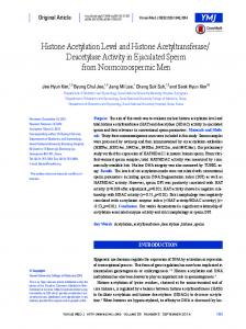
FIG. 12. Model for role of histone acetylation in long term memory formation. Formation of long term memory begins with activation of NMDA-Rs at the plasma membrane. Influx of Ca2⫹ ions activates several different signaling pathways that all converge to activate the MEK/ERK signaling cascade. Upon activation, ERK modulates the activities of several different transcription factors and co-activators. The action of numerous transcription factors and co-activators are integrated by the structure of chromatin and are apparent as an increase in acetylation of histone H3. The changes in chromatin structure ultimately lead to changes in transcription of genes relevant for memory formation. Additionally, latent inhibition training results in changes in chromatin structure apparent as acetylation of histone H4. The modulation of chromatin structure by latent inhibition is not accompanied by ERK activation and does not lead to acetylation of histone H3. MNT-R, modulatory neurotransmitter receptor; VGCC, voltage-gated Ca2⫹ channel; AC, adenylate cyclase.
the nucleus (Fig. 12). Our model shows that ERK accomplishes this through a multicomponent mechanism encompassing: the direct regulation of transcription factors, such as Elk-1 (5, 6); indirectly modulating the activity of transcriptional co-regulators, such as CBP (7); and through recruiting other histonemodifying enzymes to the transcriptional complex such as histone acetyltransferases, methyltransferases, and kinases (Fig. 12). Further work will be required to determine the precise molecular mechanisms through which ERK regulates histone acetylation in the hippocampus. In addition to ERK, other signaling cascades are also likely involved in regulating chromatin structure. For example, we observed that latent inhibition training resulted in a significant increase in acetylation of histone H4, which was independent of an increase in P-ERK2 (Fig. 1D). Moreover, activation of NMDA-Rs, PKA, or PKC had no effect on acetylation of histone H4 in vitro (Fig. 6), suggesting that the signaling pathways governing acetylation of histone H4 are very different from the signaling pathways regulating acetylation of histone H3. Given that latent inhibition training prevents formation of contextual fear memory, it is possible that the resulting changes in transcription mediated by histone H4 might act to antagonize histone H3-mediated changes in transcription. Some evidence suggests that signaling via p38 MAP kinase is independent of and acts to antagonize ERK signaling and vice versa (21, 43, 44). Therefore, it is possible that the regulation of histone H4 by latent inhibition is due to p38 MAP kinase signaling. We observed that inhibition of histone deacetylases, enzymes that promote the formation of transcriptionally silent heterochromatin, augmented the formation of LTP in vitro and long term memory in vivo. How could an inhibitor of histone deacetylase enhance synaptic plasticity and long term memory formation without having any affect on basal properties of synaptic transmission or animal behavior? Previous studies indicate that inhibition of histone deacetylase only affects the expression of a restricted set of genes (45). Moreover, a contem-
porary model of long term memory formation posits that, under normal conditions, transcriptional repressors of memory formation dominate transcriptional activators, so that under normal conditions long term synaptic enhancement and memory formation are prevented (46). Transcriptional repressors recruit histone deacetylases to DNA, resulting in the formation of transcriptionally silent heterochromatin, whereas activators of transcription recruit histone acetyltransferases, which increase histone acetylation and result in the formation of transcriptionally competent euchromatin. Therefore, histone deacetylase inhibitors may be acting to decrease the efficacy of memory repressors, essentially priming the transcriptional machinery. This hypothesis is supported by the observation that inhibiting transcription blocks the effect of HDAC inhibitors on LTP. The transcriptional priming results in enhanced induction of LTP and long term memory formation. Augmentation of long term forms of synaptic plasticity and long term memory formation has also been seen by disrupting the inhibitory forms of CREB, a critical transcription factor in the formation of long term memory (47–50). Therefore, it is possible that HDAC inhibitors might represent a viable route for the treatment of cognitive impairment or for enhancing memory in people with normal cognitive ability. Modification of histones, either through acetylation, phosphorylation, or methylation, is a biochemical memory that can persist beyond the initial signaling event in many cell types (51). To date, these sorts of changes have largely been identified as a mechanism for triggering cellular differentiation that persists indefinitely (52, 53). An interesting current hypothesis in the field of developmental biology is that changes in the acetylation of histones persist as a cellular form of memory of previous cytoplasmic signaling events (51, 54). For example, the transcription factor REST is responsible for inhibiting the expression of a set of genes that would impart a neuronal phenotype to a cell. Once induced to differentiate into a non-neuronal cell, REST recruits a number of proteins, including co-repressors and his-
Histone Acetylation in Long Term Memory Formation tone deacetylases, to effect a permanent change in chromatin structure around neuronal genes (14, 15). Therefore, a brief signaling event that induced differentiation resulted in a lasting change in gene expression effected through changes in chromatin structure. In fact, epigenetic change in chromatin structure is one of the most pervasive forms of cellular memory: “remembered” long term changes in gene expression occur in cells as dissimilar as bacteria and neurons (55). Thus, changes in acetylation of histones might not only represent a mechanism for acute changes in transcription of genes relevant for memory formation in neurons, but rather be a conserved mechanism for cellular memory in general. Our studies have revealed a new molecular correlate of long term memory formation: alterations in chromatin structure. We observed changes in chromatin structure that accompanied long term memory formation and activation of second messenger signaling pathways that are known to mediate induction of long term synaptic potentiation. Interestingly, by circumventing the normal signaling pathways involved in the formation of memory and mimicking these changes in chromatin structure by pharmacologic means, we actually enhanced the capacity for Schaffer-collateral synapses to express LTP in vitro and enhanced the formation of long term memory in vivo. These results suggest the interesting possibility that chromatin structure itself may represent a “memory,” allowing for temporal integration of spaced signals or metaplasticity of synapses. We anticipate that future studies will begin to elucidate the role chromatin structure plays in memory formation and hopefully provide new insights into human cognition and into new treatments for diseases thereof. REFERENCES 1. Dingledine, R. (1983) Fed. Proc. 42, 2881–2885 2. Harris, E. W., Ganong, A. H., and Cotman, C. W. (1984) Brain Res. 323, 132–137 3. Fanselow, M. S., Kim, J. J., Yipp, J., and De Oca, B. (1994) Behav. Neurosci. 108, 235–240 4. Adams, J. P., and Sweatt, J. D. (2002) Annu. Rev. Pharmacol. Toxicol. 42, 135–163 5. Davis, S., Vanhoutte, P., Pages, C., Caboche, J., and Laroche, S. (2000) J. Neurosci. 20, 4563– 4572 6. Sananbenesi, F., Fischer, A., Schrick, C., Spiess, J., and Radulovic, J. (2002) Mol. Cell. Neurosci. 21, 463– 476 7. Merienne, K., Pannetier, S., Harel-Bellan, A., and Sassone-Corsi, P. (2001) Mol. Cell. Biol. 21, 7089 –7096 8. Varga-Weisz, P., and Becker, P. (1998) Curr. Opin. Cell Biol. 10, 346 –353 9. Angus-Hill, M. L., Dutnall, R. N., Tafrov, S. T., Sternglanz, R., and Ramakrishnan, V. (1999) J. Mol. Biol. 294, 1311–1325 10. Cheung, P., Tanner, K. G., Cheung, W. L., Sassone-Corsi, P., Denu, J. M., and Allis, C. D. (2000) Mol. Cell 5, 905–915 11. Freedman, L. (1999) Cell 97, 5– 8 12. Orphanides, G., and Reinberg, D. (2000) Nature 407, 471– 475 13. Turner, B. M. (2002) Cell 111, 285–291 14. Battaglioli, E., Andres, M. E., Rose, D. W., Chenoweth, J. G., Rosenfeld, M. G., Anderson, M. E., and Mandel, G. (2002) J. Biol. Chem. 277, 41038 – 41045 15. Lunyak, V. V., Burgess, R., Prefontaine, G. G., Nelson, C., Sze, S. H., Chenoweth, J., Schwartz, P., Pevzner, P. A., Glass, C., Mandel, G., and Rosenfeld, M. G. (2002) Science 298, 1747–1752
40559
16. Crosio, C., Cermakian, N., Allis, C. D., and Sassone-Corsi, P. (2000) Nat. Neurosci. 3, 1241–1247 17. Crosio, C., Heitz, E., Allis, C. D., Borrelli, E., and Sassone-Corsi, P. (2003) J. Cell Sci. 116, 4905– 4914 18. Calderone, A., Jover, T., Noh, K. M., Tanaka, H., Yokota, H., Lin, Y., Grooms, S. Y., Regis, R., Bennett, M. V., and Zukin, R. S. (2003) J. Neurosci. 23, 2112–2121 19. Huang, Y., Doherty, J. J., and Dingledine, R. (2002) J. Neurosci. 22, 8422– 8428 20. Guan, Z., Giustetto, M., Lomvardas, S., Kim, J. H., Miniaci, M. C., Schwartz, J. H., Thanos, D., and Kandel, E. R. (2002) Cell 111, 483– 493 21. Guan, Z., Kim, J. H., Lomvardas, S., Holick, K., Xu, S., Kandel, E. R., and Schwartz, J. H. (2003) J. Neurosci. 23, 7317–7325 22. Atkins, C. M., Selcher, J. C., Petraitis, J. J., Trzaskos, J. M., and Sweatt, J. D. (1998) Nat. Neurosci. 1, 602– 609 23. Lubow, R. E. (1973) Psychol. Bull. 79, 398 – 407 24. Impey, S., Smith, D. M., Obrietan, K., Donahue, R., Wade, C., and Storm, D. R. (1998) Nat. Neurosci. 1, 595– 601 25. Levenson, J. M., Choi, S., Lee, S. Y., Cao, Y. A., Ahn, H. J., Worley, K. C., Pizzi, M., Liou, H. C., and Sweatt, J. D. (2004) J. Neurosci. 24, 3933–3943 26. Igaz, L. M., Vianna, M. R., Medina, J. H., and Izquierdo, I. (2002) J. Neurosci. 22, 6781– 6789 27. Selcher, J. C., Atkins, C. M., Trzaskos, J. M., Paylor, R., and Sweatt, J. D. (1999) Learn. Mem. 6, 478 – 490 28. Stanton, P. K., and Sarvey, J. M. (1984) J. Neurosci. 4, 3080 –3088 29. Frey, U., Krug, M., Reymann, K. G., and Matthies, H. (1988) Brain Res. 452, 57– 65 30. Frey, U., Krug, M., Brodemann, R., Reymann, K., and Matthies, H. (1989) Neurosci. Lett. 97, 135–139 31. Nguyen, P. V., Abel, T., and Kandel, E. R. (1994) Science 265, 1104 –1107 32. English, J. D., and Sweatt, J. D. (1997) J. Biol. Chem. 272, 19103–19106 33. Frey, U., Frey, S., Schollmeier, F., and Krug, M. (1996) J. Physiol. 490, 703–711 34. Huang, Y., Myers, S. J., and Dingledine, R. (1999) Nat. Neurosci. 2, 867– 872 35. Bourtchuladze, R., Frenguelli, B., Blendy, J., Cioffi, D., Schutz, G., and Silva, A. J. (1994) Cell 79, 59 – 68 36. Jones, M. W., Errington, M. L., French, P. J., Fine, A., Bliss, T. V., Garel, S., Charnay, P., Bozon, B., Laroche, S., and Davis, S. (2001) Nat. Neurosci. 4, 289 –296 37. Taubenfeld, S. M., Wiig, K. A., Monti, B., Dolan, B., Pollonini, G., and Alberini, C. M. (2001) J. Neurosci. 21, 84 –91 38. Fleischmann, A., Hvalby, O., Jensen, V., Strekalova, T., Zacher, C., Layer, L. E., Kvello, A., Reschke, M., Spanagel, R., Sprengel, R., Wagner, E. F., and Gass, P. (2003) J. Neurosci. 23, 9116 –9122 39. Meffert, M. K., Chang, J. M., Wiltgen, B. J., Fanselow, M. S., and Baltimore, D. (2003) Nat. Neurosci. 6, 1072–1078 40. Abeliovich, A., Paylor, R., Chen, C., Kim, J. J., Wehner, J. M., and Tonegawa, S. (1993) Cell 75, 1263–1271 41. Abel, T., Nguyen, P. V., Barad, M., Deuel, T. A., Kandel, E. R., and Bourtchouladze, R. (1997) Cell 88, 615– 626 42. Weeber, E. J., Atkins, C. M., Selcher, J. C., Varga, A. W., Mirnikjoo, B., Paylor, R., Leitges, M., and Sweatt, J. D. (2000) J. Neurosci. 20, 5906 –5914 43. Blum, S., Moore, A. N., Adams, F., and Dash, P. K. (1999) J. Neurosci. 19, 3535–3544 44. Murray, H. J., and O’Connor, J. J. (2003) Neuropharmacology 44, 374 –380 45. Van Lint, C., Emiliani, S., and Verdin, E. (1996) Gene Expr. 5, 245–253 46. Abel, T., and Kandel, E. (1998) Brain Res. Brain Res. Rev. 26, 360 –378 47. Yin, J. C., Del Vecchio, M., Zhou, H., and Tully, T. (1995) Cell 81, 107–115 48. Yin, J. C., Wallach, J. S., Del Vecchio, M., Wilder, E. L., Zhou, H., Quinn, W. G., and Tully, T. (1994) Cell 79, 49 –58 49. Bartsch, D., Casadio, A., Karl, K. A., Serodio, P., and Kandel, E. R. (1998) Cell 95, 211–223 50. Bartsch, D., Ghirardi, M., Skehel, P. A., Karl, K. A., Herder, S. P., Chen, M., Bailey, C. H., and Kandel, E. R. (1995) Cell 83, 979 –992 51. Paro, R. (1995) Trends Genet. 11, 295–297 52. Orlando, V. (2003) Cell 112, 599 – 606 53. Muller, C., and Leutz, A. (2001) Curr. Opin. Genet. Dev. 11, 167–174 54. Jenuwein, T., and Allis, C. D. (2001) Science 293, 1074 –1080 55. Casadesus, J., and D’Ari, R. (2002) BioEssays 24, 512–518