05. Spano
18-02-2002
13:54
Pagina 255
ORIGINAL PAPER
Eur. J. Histochem. 44: 255-267, 2000 © Luigi Ponzio e figlio - Editori in Pavia
Relationship between actin microfilaments and plasma membrane changes during apoptosis of neoplastic cell lines in different culture conditions A. Spano, L. Sciola, G. Monaco, and S. Barni1 Dipartimento di Scienze Fisiologiche Biochimiche e Cellulari - Via Muroni, 25 - 07100 Sassari - Italy and 1Dipartimento di Biologia Animale e Centro di Studio per l’Istochimica, CNR - Piazza Botta, 10 - 27100 Pavia - Italy Accepted: 12/6/00 Key words: apoptosis, actin, PS exposure, blebbing, mitochondria
SUMMARY In this study we investigated the relationship between the reorganisation of actin cytoskeleton and the changes at cell surface level (i.e. PS exposure and blebbing) in two neoplastic cell lines during apoptosis: Chang liver cells (adherent culture) and promyelocytic HL-60 cells (suspension culture), treated with the podophyllotoxin derivative VP16. The morphological analysis, performed by means of conventional fluorescence microscopy and confocal laser scanning microscopy, on Chang cells showed that onset and progress of the two processes are synchronised. The initial disassembly of stress fibers was associated with the early PS exposure on the cell surface. Moreover, the accumulation of actin at cortical level appeared strongly associated with an intense labelling for Annexin V and, in some cases, especially in the areas of membrane blebbing. The double staining for actin and PS exposure, quantitatively analysed by flow cytometry in HL-60 cells after different treatment times, demonstrated that the decrease of Annexin V binding in the late stages of apoptosis is associated with the strong reduction of actin labelling probably also due to a proteolytic cleavage. These events were also partially related to variations of Correspondence to: L. Sciola E-mail:
[email protected]
the functional state of mitochondria, by analysing cytofluorometrically the dissipation of the inner membrane potential (∆Ψm). INTRODUCTION Apoptosis, or programmed cell death, is a fundamental process of cell loss in the development and tissue homeostasis of multicellular organisms (Wyllie, 1995); moreover, apoptosis enables them to eliminate cells that are damaged or have become superfluous (Ellis et al., 1991). Failure of cells to undergo appropriate apoptotic cell death is involved in a variety of human pathologies including autoimmune diseases, viral infections, and cancer (Thompson, 1995). Cell death by apoptosis is a tightly regulated process that requires coordinated modifications of cell architecture. Apoptotic basic steps are generally similar in all cell systems where they have been studied and include cell shrinkage, chromatin condensation, nuclear fragmentation, and packaging of the nuclear fragments into apoptotic bodies (Wyllie et al., 1980) that are quickly phagocytosed by neighbouring cells (Kerr et al., 1972; Dini, 2000).
255
05. Spano
18-02-2002
13:54
Pagina 256
Other aspects of an apoptotic cell include changes in phospholipid asymmetry at plasma membrane level with consequent phosphatidylserine exposure, membrane blebbing, cytoskeletal reorganization and breakdown, decrease in adhesion and intercellular contacts. Some of these structural modifications can be related to biochemical changes that include a controlled proteolysis of cellular proteins by the cysteine proteases of the caspase family (Alnemri et al., 1996) that are important mediators of apoptotic cell death (Martin and Green, 1995; Kumar and Lavin, 1996). The consequence of caspase activation is the cleavage of several targets located in both cytoplasm and nucleus. The cleavage activity is responsible for some typical apoptotic expressions such as DNA fragmentation (Liu et al., 1997; Enari et al., 1998) and membrane blebbing (Kothakota et al., 1997; Rudel and Bokoch, 1997). The cellular targets for caspases include in fact several individual proteins as diverse as DNA repair enzymes poly(ADP-ribose) polymerase (PARP: for a review, see Scovassi et al., 1998), the main structural components of the nuclear lamina (lamins) and cytoskeletal components such as intermediate filaments, microfilaments and some regulators of actin network (Brancolini et al., 1997). Changes in the cytoskeletal organization have been described to occur during apoptosis in different biological situations (Wyllie et al., 1980; Ijiri and Potten, 1987). Recently, Tinnemans et al. (1995) showed that in apoptotic cells cytokeratin filaments aggregate in the early stage of the process, while at a later stage the cytokeratin is degraded. Other Authors (Pittman et al., 1994; Ireland et al., 1997) showed that reorganization of microtubules might be an integral part of the apoptotic process and induced apoptosis results in proteolytic cleavage of fodrin (Martin et al., 1995). It is also reported that during the early stage of apoptosis actin mRNA levels decrease, followed by the proteolytic cleavage of F-actin network (Guénal et al., 1997). Evidences of a relationship between membrane lipid asymmetry and cytoskeletal alterations were found during the platelet procoagulant activity, suggesting the involvement of the cytoskeleton in the maintenance of membrane lipid asymmetry. Other experimental data demonstrated that disintegration of cytoskeletal filaments, which normally stabilise and modulate the function of the plasma
256
membrane, could cause different cell alterations (Buja et al., 1993). Actin microfilaments can also have a particular role in the dynamics of plasma membrane and cytoplasm modifications related to the formation and the release of blebs, and during cytoplasm cleavage with formation of apoptotic bodies (Kulkarni and McCulloch, 1994; Bonanno et al., 2000). In the present study, the reorganisation of the microfilament network, during the apoptotic progression, was morphologically evaluated by conventional and confocal fluorescence microscopy in the adherent Chang hepatoma cell line treated with etoposide (VP16). The actin reorganization has been related, in the same cell, to the cell surface exposure of phosphatidylserine and blebbing. These parameters have been quantified by flow cytometry also in the promyelocytic cell line HL60 treated with VP16 at different times (5, 12 and 24 h) and cultured as stationary suspension. Moreover, in this latter cell model a study of mitochondrial transmembrane potential (∆Ψm) has been assessed following, in the different treatment conditions, the uptake of the cationic lipophilic dye JC-1. This analysis has been performed in order to correlate the changes of plasma membrane and microfilaments with the metabolic activity of the cells. MATERIALS AND METHODS Cell cultures and treatment Chang liver cells (derived from a human hepatic carcinoma) were grown as monolayer culture in RPMI 1640 supplemented with 10% fetal calf serum (FCS). The cell cultures were maintained at 37°C in humid atmosphere (95% air/5% CO2). HL-60 cells, a human promyelocytic cell line, were grown as stationary suspension in RPMI 1640 supplemented with 15% FCS. The cell cultures were maintained at 37°C in humid atmosphere (95% air/5% CO2) at a density of 1 x10⁶ cells/ml. Apoptosis was induced by treating exponentially growing Chang cells with 50 µM VP16 and incubating the culture for 24 h. VP16 was dissolved in dimethylsulfoxide (DMSO); the final DMSO concentration in the culture medium was 0.2%. As far as HL-60 cultures were concerned, 20 µM VP16 was added to the culture medium and, at
05. Spano
18-02-2002
13:54
Pagina 257
various periods of time (5, 12, 24 h), the cells were harvested by centrifugation. FLUORESCENCE MICROSCOPY Annexin V/Phalloidin co-labelling In order to detect phosphatidylserine (PS) exposure, Chang liver cells, grown on coverslips, after washing in PBS, were incubated in the presence of Annexin V-FITC (0.5 µg/ml) for 15 min at room temperature. At the end of the incubation and after washing in Binding Buffer (50 mM HEPES, 750 mM NaCl, 12.5 mM CaCl2, 5 mM MgCl2, 20% BSA, pH 7.4) at 4°C, the cells were fixed in 3% paraformaldehyde in Binding Buffer for 30 min at room temperature. After washing in Binding Buffer, the cells were permeabilized with 0.2% Triton X-100 and then submitted to the cytochemical labelling of actin microfilaments. The cells were incubated in the presence of TRITC-conjugated phalloidin (2 µg/ml) for 45 min in humid chamber at room temperature. After washing in Binding Buffer and DNA fluorochromization with Hoechst 33342 (2 µg/ml) for 10 min, the coverslips were mounted with 90% glycerol in PBS and observed in epifluorescence with a NIKON Eclipse 600 microscope. Confocal laser scanning microscopy (CLSM) Cells on glass slides were analysed using the LSM 5 Pascal confocal scanning module (ZeissGermany) equipped with a Helion/Neon laser (λexc 543 nm) mounted on an Axiovert 100M microscope (Zeiss-Germany). To evaluate the changes of actin fiber topology, cells were optically sectioned at 0.4 µm. The scans were recorded in photon counting mode. FLOW CYTOMETRY Evaluation of Annexin V/Phalloidin positivity HL-60 cells (1 x 10⁶ cells/ml) were incubated for 5, 12 and 24 h at 37°C in the presence of 20 µM VP16 in complete medium. After the treatment, the cells were pelleted by centrifugation (100 x g for 10 min) and resuspended in serum-free culture medium. The cell suspensions, after Annexin VFITC (0.5 µg/ml) addition, were incubated for 15
min at room temperature. At the end of the incubation, the samples were centrifuged and then resuspended in cold Binding Buffer. After centrifugation, the cells were fixed with 3% paraformaldheyde in Binding Buffer for 15 min at room temperature and, after further washing, permeabilized with 0.2% Triton X-100 for 15 min. After centrifugation, the cells were incubated in the presence of TRITC-conjugated phalloidin (2 µg/ml) for 45 min in the dark. After washing, the samples were maintained at 4°C, until the measurements performed by means of a Cytoron Absolute (Ortho) flow cytofluorometer equipped with Argon laser (λexc 488 nm). Evaluation of mitochondrial activity-mass HL-60 cells (5 x 10⁶ cells/each condition), after treatment with 20 µM VP16 for 5, 12 and 24 h, were incubated in the presence of the cationic lipophilic probe JC-1 (10 µg/ml) for 10 min in the dark. At the end of the incubation time, the cells were washed in serum-free culture medium and then analysed using a Cytoron Absolute (Ortho) flow cytofluorometer, equipped with Argon laser (λexc 488 nm). Scanning electron microscopy Some specimens of Chang cell cultures were fixed in 3% paraformaldheyde in PBS containing 1 mM CaCl2 and 1 mM MgCl2. After the double fluorescent staining for actin and DNA, the samples were washed in PBS, dehydrated in graded acetons, critical-point dried in CO2, gold-coated by sputtering and examined with a ZEISS DSM 962 scanning electron microscope operating at 20 KV. RESULTS F-Actin analysis and relationship with PS exposure in Chang cells The fluorescence labelling of actin, performed on Chang cells, showed specific sequential changes of the microfilament network during apoptosis progression, detectable on the basis of cell shape, Annexin V labelling and nuclear morphology after DNA fluorochromization. The distribution patterns of actin cytoskeleton in the VP16-induced apoptotic cells were similar to the staining patterns observed in spontaneously occurring apoptoses in the control cultures (Fig. 1a-a’).
257
05. Spano
18-02-2002
13:54
Pagina 258
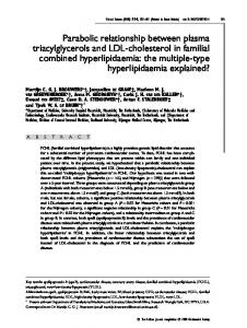
Fig. 1 – Conventional fluorescence microscopy of apoptotic Chang cells submitted to a double staining for actin (phalloidinTRITC) and DNA (Hoechst 33342) labelling. Pa, a’) Same microscopic field of control culture observed in two spectral conditions for actin network (a) and nuclear (a’) examination. Note the well-defined stress fibers (a) of normal cells in comparison to the apoptotic cell (arrowheads) focused to underline the karyorrhexis (a’). b-b’) Same microscopic field, after the culture treatment, showing the actin arrangement as perinuclear ring (arrows) in early apoptotic cells; the arrowheads label the weak actin staining (b) of a karyorrhetic apoptosis (b’). c, d) Presence of actin ring in the membrane blebs (arrows); in the advanced apoptosis (arrowhead) no blebbing is detectable. Original magnification: x 1000.
After VP16 treatment (50 µM/24 h), in some cells the reduction/disorganisation of stress fibers was
258
evident in the whole cell body (Fig. 3 a, b). The simultaneous fluorochromization with Annexin V-
05. Spano
18-02-2002
13:54
Pagina 259
periphery of the cell. Moreover, apoptosis was associated with the cell rounding and subsequent detachment from the growth substrate and to the formation of blebs on the cell surface (Fig. 1 c, d). In general, it was possible to demonstrate a remarkable lower labelling of actin in the advanced stages of apoptosis (karyorrhexis) (Fig. 1 b-b’, c) with persistence in the sites of cell contact (focal adhesion) to the substrate of wellorganised stress fibers (Fig. 2 a). At this stage the perinuclear actin ring and the cell surface blebs were no longer found. In figure 3 different morphological aspects of apoptotic cells are shown. They indicate the relationship between Annexin V binding and actin redistribution during the degenerative phenomenon. In figure 3 c-c’ apoptotic cells that lose their contacts with the growth substrate are represented; a strong positivity for Annexin V is evident especially in some areas of the cell surface. In other cells, possibly representing later stages of apoptosis, the PS exposure was related to a marked storage of actin at the cell periphery (Fig. 3 d-d’; f-f’). In addition, it was shown that the Annexin V binding occurred especially in spotted areas of the plasma membrane that could represent points of possible formation and release of blebs (Fig. 3 e-e’).
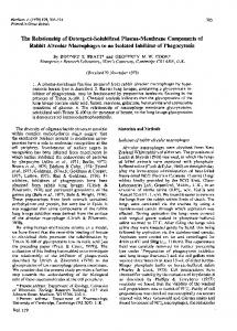
Fig. 2 – Persistence of actin, organised in stress fiber form, in the sites of cell contacts (focal adhesion) to the growth substrate during an advanced stage (karyorrhexis) of a Chang apoptotic cell. The specimen, stained with phalloidinTRITC/Hoechst 33342, after the analysis in fluorescence microscopy of actin (a) was processed for scanning electron microscopy observation (b). The arrowheads point the flat focal adhesions; into the rounded cell body (circle labelling), the nucleus appears in fragmented form (a). Original magnification: x 1000 (a); x 4000 (b).
FITC, localized in defined areas of the cell surface, seems to indicate that these cells represent early stages of apoptosis. During the progression of apoptosis, besides the disappearance of the stress fibers, it was possible to show an accumulation of actin in a ring-like structure surrounding the nucleus, before nuclear fragmentation (Fig. 1 b-b’). This situation was related to an appreciable protein staining, at the
Tridimensional distribution analysis of actin fibers in apoptotic cells The study performed by confocal microscopy was particularly interesting during cell rounding and disappearing of cell-substrate contacts. These changes were found to be associated with apoptosis induced on adherent culture (VP16-treated Chang cells) and were often related to blebbing phenomena of the cell surface (Fig. 4). The analysis of the first optical sections showed the presence of an organization of actin in stress fibers in defined cellular areas, due to the presence of focal adhesions (Fig. 4 a-d). Optical sections referred to higher cell levels displayed a remarkable actin concentration at cell periphery, associated with cell surface areas involved in bleb formation (Fig. 4 e-k). The top levels were characterised by a lower labelling of actin in the cortical area; moreover, this aspect appeared to be related to the release of blebs from the cell surface (Fig. 4 l-o).
259
05. Spano
18-02-2002
260
13:54
Pagina 260
05. Spano
18-02-2002
13:54
Pagina 261
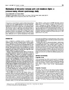
Fig. 4 – Gallery of laser scan images of an apoptotic Chang cell (phalloidin-TRITC staining) with rounding morphology and detaching from the growth substrate. The optical sections (0.4 µ thickness) show, near the substrate (a-c), the presence of actin in form of stress fibers related to the persistence of adhesive contacts (see also figure 2). In the following optical sections the actin filaments are accumulated in the cortical regions of the cell and in surface blebs, in form of dense spherical network. Original magnification: x 630.
Flow cytometry evaluation of actin expression and PS exposure in double-stained HL-60 cells In order to quantify actin content changes and the exposure of phosphatidylserine on the cell surface, we performed multivariate flow cytometric measurements on double stained (phalloidinTRITC/Annexin V-FITC) HL-60 cells after 20 µM VP16 treatment for different times (Fig. 5). As far as Annexin V binding is concerned, this assay revealed that the treatment with 20 µM VP16 for 5h (Fig. 5) induced approximately 80% of cells with PS exposure. The global analysis of the fluorescence histograms showed a progressive reduction of cells positive for such fluorochromization during the progression of treatment (68% after 12 h
treatment, 48% after 24 h treatment) with the apoptogenic drug. This aspect was also evident by the analysis of the cytograms relative to the bivariate fluorescence (Fig. 5), in which a progressive decrease of phalloidin/Annexin V positive elements was present (see Q2 area of figure 5). This was correlated to the increase of cells with weak signals of fluorescence for both labelling, indicative of altered elements, incapable of Annexin V binding and with highly degraded actin (see Q0 area in figure 5 h). As far as actin quantitative aspects are concerned, the analysis of fluorescence histograms (Fig. 5 a, b, c, d) indicated that VP16 treatment elongation induces a progressive increase of cells with weak
Fig. 3 – Conventional fluorescence microscopy of apoptotic Chang cells stained for the co-labelling of actin (phalloidin-TRITC), DNA (Hoechst 33342) and PS exposure (Annexin V-FITC). a) Early apoptotic cell showing the reduction of actin stress fibers (red fluorescence) associated with PS exposure (green-yellow fluorescent spots); b, c-c’) further Annexin V binding to an apoptotic cell characterised by a scarce presence of actin filaments; d-d’, f-f’) persistence of Annexin V labelling in presence of actin residues in the cortical area; e-e’) release of membrane blebs positive to Annexin V labelling. Original magnification: x1000.
261
05. Spano
18-02-2002
13:54
Pagina 262
Fig. 5 – Flow cytometric histograms relative to actin fluorochromization (a, b, c, d), Annexin V binding (i, l, m, n) and cytograms of the bivariate analysis of the two fluorescences (e, f, g, h) in HL-60 cells. In comparison to the control cells (a, e, i), after different times of treatment (f, g, h) as a consequence of an intense apoptotic death, a significant increase of green fluorescence intensity (GR-FL), as expression of Annexin V binding, is detectable. The decrease of red fluorescence (RD-FL), as expression of actin labelling, indicates a loss of this cytoskeleton component (Q1, Q0 areas). The low signals for both green and red fluorescence (Q0 area) could represent advanced stages of the apoptotic progression.
262
05. Spano
18-02-2002
13:54
Pagina 263
staining for this protein. This aspect seems to be related to the fluorescence microscopy observations, relative to the disappearance of actin network in the late stages of apoptosis of Chang cells treated with 50 µM VP16 (Fig. 1 b-b’, c; Fig. 2). Flow cytometry evaluation of mitochondrial activity During the apoptotic progression of HL-60 cells, the mitochondrial activity was estimated using the fluorochromatic cationic dye JC-1, as a probe for monitoring by flow cytometry the changes of the inner transmembrane potential (∆Ψm). Actually this fluorescent carbocianine dye emits an orange fluorescence when mitochondria are highly active, but in condition of low mitochondrial function, the fluorescence becomes green and can be related to the chondrioma mass. In the untreated cells, two subpopulations emerged from the cytograms (Fig. 6). The first, with a major size (about 70% of cells), was characterised by high values of ∆Ψm (orange fluorescence); the second one, of a smaller size, was characterised by low values of ∆Ψm (increased green fluorescence and decreased orange fluorescence). After 5 h treatment with VP16, it was possible to show a marked reduction (34% of positive cells) of orange fluorescence signals (Fig. 6 b). This late aspect can be related to the prevalence of cells in the early stages of apoptosis (strongly positive to Annexin V). A further decrease of orange fluorescence signals appeared evident with the exposure time to the drug: 24% and 22% of positive cells, respectively after 12 h and 24 h. In regard to the green fluorescence emission, it was possible to note that the VP16 stimulation at various times causes the appearance of low signal that remains substantially constant during the treatment. This fact seems to indicate that during apoptosis a partial depletion could also occur in the mitochondrial mass. Moreover, the lowest intensities of orange and green fluorescence as a whole may be also attributed to the presence of apoptotic bodies. DISCUSSION In this study, we examined the structural reorganization of the actin cytoskeleton component during apoptosis. We have also considered the loss of
membrane phospholipid asymmetry and a possible relationship between these two phenomena in two human tumor cell lines: in Chang cell adherent culture, mostly from the morphocytochemical point of view, and in the HL-60 cell suspension culture, also with quantitative assays. In Chang cell cultures, the treatment with VP16 induced a remarkable incidence of apoptotic cells after 24 h treatment, resulting in dramatic changes of the microfilament actin component. The morphological analysis showed that when an adherent cell undergoes apoptotic degeneration in vitro, specific sequential changes occur in actin microfilament network. Concomitantly with the actin reorganization at both the perinuclear region and cytoplasm periphery in collapsed form (Kulkarni and McCulloch, 1994), the cell looses its contacts with the neighbouring cells, detaches from the growth substrate, by disassembling the stress fibers and, finally, reorganising its actin residual. The simultaneous staining of PS exposure, performed by using Annexin V-FITC, indicates that this molecular aspect is related to cytoskeletal network modifications: in fact, the stress fiber disorganisation and the actin redistribution at the cell periphery are events that occur concomitantly with the early PS translocation. Moreover, it is possible to hypothesise that these phenomena are not only structurally, but also functionally related, with an involvement of the disconnection between cytoskeleton and plasma membrane, as a possible consequence of actin redistribution as well as changes on the regulatory proteins (e.g. proteolysis of α-actinin and vinculin) of the microfilament system (Brancolini et al., 1997). The accumulation of actin at the cortical region, observed in several cells, can be related to the dynamic phenomena of the plasma membrane (blebbing), which occur in the early stages of apoptosis. Huot et al. (1998) and Bonanno et al. (2000) showed that the inhibition of actin polymerisation results in the blockade of the surface blebbing phenomenon. On the basis of our data, the actin accumulation below the plasma membrane could be then related to changes of plasma membrane asymmetry as well as bleb formation, probably by the interaction with myosin (Mills et al., 1998). Moreover, the contraction of the cortical thin filament (actin/myosin) ring may produce centripetal forces responsible for cytoplasm condensation and round-
263
05. Spano
18-02-2002
13:54
Pagina 264
Fig. 6 - Flow cytometry of mitochondrial changes in JC-1 fluorochromized HL-60 cells (e) treated with VP16 for different times: (f, g h). The orange fluorescence emission (OR-FL) is expression of the mitochondrial activity and the green fluorescence (GR-FL) represents the mitochondrial mass. The shift from high (Q2 area) to low (Q1 area) values of the orange fluorescence emission, during the treatment, indicates a reduction of the mitochondrial membrane potential (DCm), typical of apoptosis progression, with the appearance of well-distinct subpopulations (d, h, n).
264
05. Spano
18-02-2002
13:54
Pagina 265
ing up of the apoptotic cell. The successive event could be the release of blebs composed by cell membrane areas in which the outer PS appears particularly accumulated. This mechanism could be responsible for the loss of this cell surface signal involved in the identification of early apoptotic cell by the scavenger system (Savill, 1997). The apoptotic phenomenon progression over the early stages causes, beyond the progressive loss of stress fibers, an accumulation of the protein at the perinuclear region. When the nuclear degeneration does not coincide with the karyorrhexis, the actin is organised in a ring-like structure surrounding the nucleus. This fact suggests that the accumulation of the actin at the nuclear periphery could be an important co-factor for the chromatin hypercondensation, in association with other mechanisms (e.g. osmotic piknosis). The modifications at nuclear level involve other cell components as well as lamins, one of the better-characterised targets for caspases. They constitute the main structural components of the lamina underlying the nuclear envelope. Apoptosis induced by different stimulation results in the proteolytic cleavage of lamins to facilitate the nuclear collapse and cleavage that characterises different models of apoptosis (Rao et al., 1996). From this point of view, the morphological and biochemical changes occurring at nuclear, cytoplasmic and plasma membrane level are considered by some Authors as events connected in a single program, in which the caspase activation and the proteolysis of different cell substrates represent a final common way, leading to cell death, even though, by means of mitochondrial inhibitors, it has been possible to demonstrate the existence of a discoupling between plasma membrane modifications and other apoptotic changes (Zhuang et al., 1998). Nevertheless, the complex reorganization of the cell structure that occurs during apoptosis seems to be related to a specific cleavage of different regulators of the microfilament system (Brancolini et al., 1997). The cell fragmentation requires a cytoskeletal rearrangement, the fusion between opposite cytoplasmic portions of the plasma membrane, a massive redistribution of the plasma membrane phospholipids and finally a localised collapse of the membrane asymmetry, which occurs during PS exposure. As far as our flow cytometric data are concerned, referring to the correlation between quantitative
expression of actin and plasma membrane asymmetry changes in HL-60 cells, the analysis of both fluorescence histograms and cytograms allows us to obtain information that partially confirm the morphological analysis of the microfilaments and PS exposure performed on the Chang cells. PS exposure is a transient event with an expression peak during the early stages of apoptosis, later the cells tend to lose this signal. Apoptosis progression is also related to a progressive decrease of actin expression: this could be the consequence of both a lower synthesis of this protein and an increase of proteolytic activity. These two concurrent apoptotic events appeared especially evident in the flow measurements relative to 24 h VP16 treatment, in which an accumulation of cells negative for Annexin V-FITC and phalloidin-TRITC could represent late stages of apoptotic degeneration. In the same experimental model, the functionality of chondrioma has been evaluated cytofluorometrically by means of the supravital fluorochromatic probe JC-1. Recently, a number of evidences showed a role of the mitochondria in transducing pro-apoptotic stimuli; furthermore, these organelles can be considered as important sensors of cellular damage (Susin et. al., 1999; Green and Reed, 1998). Our cytofluorometric data indicate that the treatment conditions that induce the prevalence of early stages of apoptosis (higher percentage of Annexin V positive cells) cause a dramatic reduction of the orange fluorescence frequency values, as expression of mitochondrial transmembrane electric potential (∆Ψm) dissipation. These data seem to support the view of the apoptotic phenomenon dynamics that considers PS exposure and dissipation of ∆Ψm concomitant phenomena. In fact, at the moment, although it is becoming clearer that mitochondria play an important role in programmed cell death, especially on the evidence that the release of pro-apoptotic factors from the mitochondria can initiate the caspase cascade, there is much conflicting data concerning the role of mitochondria upstream of this event (CamilleriBröet et al., 1998). The further analysis of the orange fluorescence histograms shows that the extension of VP16 treatment induces a further decrease of high fluorescence signals, after 12 h as well as after 24 h treat-
265
05. Spano
18-02-2002
13:54
Pagina 266
ment. This trend is also deductible by cytogram analysis, in which it is shown a progressive accumulation of cell subpopulations with weak, or low, orange fluorescence signals, related to the treatment progression. These subpopulations could represent cells in different stages of apoptosis. The analysis of the green fluorescence histograms showed, after 5 h treatment, the appearance of weak fluorescent signals, whose frequency does not increase during the prolonged treatment conditions. This fact would indicate that during the early stages of apoptosis a decrease also in terms of mitochondrial mass could occur. Moreover, the lowest intensities of the orange and green fluorescence as a whole may be also attributed to the presence of apoptotic bodies. Moreover, it must be considered that the low value of green fluorescence can be also attributed to the fluorochrome trend to spread from the cell because of the various membrane permeability alterations. Furthermore, the drastic loss of the orange fluorescence with the apoptosis progression can be partly compensated for a relative increase of the green fluorescence. The complex findings presented in this paper support the conclusions that some changes of cell membrane (PS exposure, surface blebbing), cytoskeleton (actin rearrangement and loss) and chondrioma (mitochondrial transmembrane electric potential dissipation) are common features of apoptosis induced in conditions of suspension or monolayer cell growth. ACKNOWLEDGEMENTS
caspase-dependent proteolytic cleavage of β-catenin. J. Cell Biol. 139, 759-771, 1997. Buja L.M., Eigenbrodt M.L., and Eigenbrodt E.H.: Apoptosis and necrosis. Basic types and mechanisms of cell death. Arch. Pathol. Lab. Med. 117, 1208-1214, 1993. Camilleri-Bröet S., Vanderwerff H., Caldwell E., Hockenbery D.: Distinct alterations of mitochondrial mass and function characterize different models of apoptosis. Exp. Cell Res. 239, 277-292, 1998. Dini L.: Recognizing death: liver phagocytosis of apoptotic cells. Eur. J. Histochem. 44, .......2000 Ellis R.E., Yuan J., and Horvitz H.R.: Mechanisms and functions of cell death. Annu. Rev. Cell Biol. 7, 663-698, 1991. Enari M., Sakahira H., Yokoyama H-, Okawa K., Iwamatsu A. and Nagata S.: A caspase-activated DNase that degrades DNA during apoptosis, and its inhibitor ICAD. Nature 391, 43-50, 1998. Green D.R., and Reed J.C.: Mitochondria and apoptosis. Science 281, 1309-1312, 1998. Guénal I., Risler Y., and Mignotte B.: Down-regulation of actin genes precedes microfilament network disruption and actin cleavage during p53-mediated apoptosis. J. Cell Sci. 110, 489-495, 1997. Huot J., Houle F., Rousseau S., Deschesnes R.G., Shah G.M., and Landry J.: SAPK2/p38-dependent F-actin reorganization regulates early membrane blebbing during stress-induced apoptosis. J. Cell Biol. 143, 1361-1373, 1998. Ijiri K., and Potten C.S.: Further studies on the response of intestinal crypt cells of different hierarchical status to eighteen different cytotoxic agents. Br. J. Cancer 55, 113-123, 1987. Ireland C.M., and Pittman S.M.: Tubulin alterations in taxolinduced apoptosis parallel those observed with other drugs. Biochem. Pharmacol., 49, 1491-1499, 1995. Kerr J.F.R., Wyllie A.H., and Currie A.R.: Apoptosis: A biological phenomenon with wide ranging implications in tissue kinetics. Br. J. Cancer 26, 237-243, 1972
The Authors thank Dr. Elio Negri (Zeiss–Italia, Milan) for technical assistance in the image acquisition by confocal laser scanning microscope.
Kothakota S., Azuma T., Reinhard C., Klippel A., Tang J., Chu K., McGarry T.J., Kirschner M.W., Koths K., Kwiatkowski D.J., and Williams L.T.: Caspase-3-generated fragment of gelsolin: effector of morphological change in apoptosis. Science 278, 294-298, 1997.
REFERENCES
Kulkarni G.V., McCulloch C.A.G.: Serum deprivation induces apoptotic cell death in a subset of Balb/c 3T3 fibroblasts. J. Cell Sci. 107, 1169-1179, 1994.
Alnemri E.S., Livingston D.J., Nicholson D.W., Salvesen G., Thornberry N.A. Wong W.W., and Yuan J.: Human ICE/CED3 protease nomenclature. Cell 87, 171, 1996.
Kumar S. and Lavin M.F.: The ICE family of cysteine proteases as effectors of cell death. Cell, Death Diff. 3, 255-267, 1996.
Bonanno E., Ruzittu M., Carlà E.C., Montinari M.R., Pagliara P. and Dini L.: Cell shape and organelles modifications in apoptotic U937 cells. Eur. J. Histochem. 44, ..... , 2000
Liu X., Zou H., Slaughter C., and Wang X.: DFF, a heterodimeric protein that functions downstream of caspase-3 to trigger DNA fragmentation during apoptosis. Cell 89, 175184, 1997.
Brancolini C., Lazarevic D., Rodriguez J., and Schneider C.: Dismantling cell-cell contacts during apoptosis is couplet to a
Martin S.J., and Green D.R.: Protease activation during apoptosis: death by a thousand cuts? Cell 82, 349-352, 1995.
266
05. Spano
18-02-2002
13:54
Pagina 267
Martin S.J, O’Brien G.A., Nishioka W.K., McGahon A.J., Mahboubi A., Saido T.C., and Green D.R.: Proteolysis of fodrin (non-erythroid spectrin) during apoptosis. J. Biol. Chem. 270, 6425-6428, 1995. Mills J.C., Stone N.L., Erhardt J., Pittman R.N.: Apoptotic membrane blebbing is regulated by myosin light chain phosphorylation. J. Cell Biol. 140, 627-636, 1998. Pittman S.M., Strickland D., and Ireland C.M.: Polymerization of tubulin in apoptotic cells is not cell cycle dependent. Exp. Cell Res. 215, 363-372, 1994. Rao L., Perez D., and White E.: Lamin proteolysis facilitates nuclear events during apoptosis. J. Cell Biol. 135, 1441-1455, 1996. Rudel T., and Bocock G.M.: Membrane and morphological changes in apoptotic cells regulated by caspase-mediated activation of PAK2. Science 276, 1571-1574, 1997. Savill J.: Recognition and phagocytosis of cells undergoing apoptosis. Br. Med. Bull. 53, 491-508, 1997. Scovassi AI, Denegri M, Donzelli M, Rossi L, Bernardi R, Mandarino A, Frouin I, Negri C.: Poly(ADP-ribose) synthesis in cells undergoing apoptosis: an attempt to face death before PARP degradation. Eur J Histochem. 42, 251-258, 1998.
Susin S.A., Lorenzo H.K., Zamzami N., Marco I., Snow S.E., Brothers G.M., Mangion J., Jacotot E., Costantini P., Loeffler M., Larochette N., Goodlett D.R., Aebersold R., Siderovski D.P., Penninger J.M., and Kroemer G.: Molecular characterization of mitochondrial apoptosis-inducing factor. Nature 397, 441-446, 1999. Thompson C.B.: Apoptosis in the pathogenesis and treatment of disease. Science 267, 1456-1462, 1995 Tinnemans M.M.F.J., Lenders M.H.J.H., ten Velde G.P.M., Ramaekers F.C.S., and Schutte B.: Alterations in cytoskeletal and nuclear matrix-associated proteins during apoptosis. Eur. J. Cell. Biol. 68, 35-46, 1995. Wyllie A.H., Kerr J.F.K and Currie A.R.: Cell death: the significance of apoptosis. Int. Rev. Cytol. 68, 251-306, 1980. Wyllie A.H.: The genetic regulation of apoptosis. Curr. Opin. Genet. Dev. 5, 97-104, 1995. Zhuang J., Ren Y., Snowden R.T., Zhu H., Gogvadze V., Savill J.S., and Cohen J.M.: Dissociation of phagocyte recognition of cells undergoing apoptosis from other features of the apoptotic program. J. Biol. Chem. 273, 15628-15632, 1998.
267