Frigeri et al. Nanoscale Research Letters 2011, 6:189 http://www.nanoscalereslett.com/content/6/1/189
NANO EXPRESS
Open Access
Relationship between structural changes, hydrogen content and annealing in stacks of ultrathin Si/Ge amorphous layers Cesare Frigeri1*, Miklós Serényi2, Nguyen Quoc Khánh2, Attila Csik3, Ferenc Riesz2, Zoltán Erdélyi4, Lucia Nasi1, Dezső László Beke4, Hans-Gerd Boyen5
Abstract Hydrogenated multilayers (MLs) of a-Si/a-Ge have been analysed to establish the reasons of H release during annealing that has been seen to bring about structural modifications even up to well-detectable surface degradation. Analyses carried out on single layers of a-Si and a-Ge show that H is released from its bond to the host lattice atom and that it escapes from the layer much more efficiently in a-Ge than in a-Si because of the smaller binding energy of the H-Ge bond and probably of a greater weakness of the Ge lattice. This should support the previous hypothesis that the structural degradation of a-Si/a-Ge MLs primary starts with the formation of H bubbles in the Ge layers. Introduction Hydrogenated a-Si and a-Ge layers are key materials for employment in (nano) structures used, e.g., in the technology of multi-junction solar cells as a-Ge acts as the lowband gap absorber while a-Si acts as the high-band gap one, thus allowing a better exploitation of the solar spectrum and the achievement of higher efficiencies [1]. However, the a-SiGe alloy is now the material of choice as the low-band gap absorber [2-4]. It allows a higher degree of freedom as regards the choice of the band gap, as the latter one can be tailored over some range by changing the Si/Ge ratio [2,4]. The a-SiGe alloy can be realized from a sequence of thin a-Si and a-Ge layers by intermixing them [1,5,6], which is obtained by heat treatments. The latter treatments are often also used for activating dopants. Previous studies have shown that the hydrogen content and annealing conditions can dramatically influence the structural stability of the a-Si/a-Ge multilayers (MLs) produced by sputtering and then annealed to produce intermixing [7-9]. It was reported that surface bumps formed, size and height of which increased with increasing H content and/or annealing temperature and time [7-9] (see Figure 1). Craters also formed subsequent to the explosion * Correspondence:
[email protected] 1 CNR-IMEM Institute, Parco Area delle Scienze 37/A, 43100 Parma, Italy Full list of author information is available at the end of the article
of the bumps. The bumps were ascribed to the formation of bubbles of hydrogen in the MLs [7,8]. The formation of H bubbles was also suggested by Acco et al. [10] in single layers of a-Si. It was hypothesized that H could be first released from the Ge layers because of the lower binding energy of the Ge-H bond with respect to the Si-H one [7,8]. To check this hypothesis, the MLs were additionally investigated by IR absorbance and an analysis of the structural behaviour of single films of a-Si and a-Ge, submitted to the same annealing as for the MLs previously studied, was performed. The results are reported in this article.
Experiment The investigated samples were MLs of alternating layers of a-Si and a-Ge and single layers of a-Si and of a-Ge. The latter ones had a thickness of 40 nm. In the former structure, the 2 × 50 alternating layers were 3 nm thick each. Both types were sputtered from high-purity crystalline silicon and germanium targets in a conventional high-vacuum sputtering apparatus (Leybold Z400) pumped to a base pressure better than 5 × 10⁻⁵ Pa. The target was coupled to a RF generator (13.56 MHz) by a network for impedance matching between the generator and its load. As substrate, polished (100) Si wafers mounted on a water-cooled stage 50 mm away from the target were used. The substrate temperature was ≤60°C.
© 2011 Frigeri et al; licensee Springer. This is an Open Access article distributed under the terms of the Creative Commons Attribution License (http://creativecommons.org/licenses/by/2.0), which permits unrestricted use, distribution, and reproduction in any medium, provided the original work is properly cited.
Frigeri et al. Nanoscale Research Letters 2011, 6:189 http://www.nanoscalereslett.com/content/6/1/189
Page 2 of 6
10 100
6
60
4
40
2
20 0
Bumps size (Pm)
Bumps height (nm)
8 80
0
1
2
3 4 5 H flow rate (ml/min)
6
7
0
Figure 1 Bumps height (dash blue line) and size (solid black line) as a function of the H flow rate in a-Si/a-Ge ML samples annealed at 350°C for 10 h.
It was estimated from measurements of the shift of the emission spectra of InP or GaAs during the deposition of AR (anti reflection) coating for laserdiode, carried out under identical conditions as those used here, by applying the rule 4 nm = 10°C. The temperature increase was always ≤40°C. Sputtering was done with a mixture of high-purity argon and hydrogen gases. No pressure fluctuation was observed. Plasma pressure of 2 Pa and a 1500 V dc wall potential were applied to sputter the targets, yielding a sputtering rate of 6.3 and 13.5 nm/min for a-Si and a-Ge, respectively. Hydrogenation was carried out by letting hydrogen flow into the deposition chamber at flow rates of 0.4, 0.8 and 1.5 ml/min. These values correspond to the measured 0.38, 0.78 and 1.46% partial of total pressure, respectively (all gauge readings were corrected by gas-sensitivity factors). The samples were annealed in high-purity (99.999%) argon at 350 or 400°C for 1, 4 and 10 h. The choice of such temperatures as the optimal ones for the purpose of this experiment was suggested by the findings of previous studies [7-9]. In fact, it was observed that annealing at 450°C causes such a great degradation of the surface that nearly 53% of it was covered by bumps and craters as large as 9 μm, with the craters as deep as the whole ML [7,8]. On the other hand, for annealing temperature of 250°C the formation of the craters rarely occurs only for very high H flow rate (6 ml/min) [7,8], thus making it difficult to evaluate whether the decrease of H content in the samples can be associated with craters for the lower H flow rates considered here. No crater was ever detected for annealing temperatures lower than 250°C. Non-hydrogenated samples were also sputtered to use as reference samples; they were annealed under the same conditions as the hydrogenated ones. The samples were analysed by elastic recoil detection analysis (ERDA), atomic force microscopy (AFM), infrared
(IR) absorption and Makyoh topography (MT). For ERDA, the 1.6 MeV 4He+ beam available at the 5 MeV Van de Graaff accelerator (Research Institute for Nuclear and Particles Physics, Budapest, Hungary) had been applied to measure the hydrogen in the samples. The hydrogen recoiled from the sample by He ions was collected by surface-biased Si detector placed at a detecting angle of 10° with regard to the beam direction, while the sample was tilted to 85° from the normal. Mylar foil with thickness of 6 μm was placed in front of the ERDA detector to stop the forward-scattered He ions. Therefore, the ERDA spectra of the H are almost background-free. Low ion current (ca. 6 nA) has been used to avoid beam heating, i.e. the escape of H from the sample at a high temperature. Evaluation of ERDA spectra was done by the RBX program developed by Kótai [11]. The in-depth spatial resolution of ERDA is approximately 20 nm [12]. Since ERDA is applied here only to the single layers and the Si substrates do not contain H, such error on the depth where the ERDA signal comes from does not impair the results regarding the presence and concentration of H. The region between channels 120 and 100 (see next section) corresponds to a depth of 40 nm from the surface. The relative error on concentration is a few per cent. Therefore, the method is suitable with regard to detecting the tendency of the H change in the samples as presented in the next section. However, the absolute error is worse because of the lack of a calibration sample having a well-known H content. A carbon layer containing H was thus used as a calibration sample. Owing to the small cross section of C for He ion, the error on the absolute H content calculated by this method is about 25%. As stated earlier, it should be noticed that such an error on the absolute concentration does not affect any interpretation of the tendency of the H changes. A VEECO Dimension 3100 in tapping mode was employed for the AFM analysis. IR absorbance gave information on how H bonds to Si and Ge before and after annealing. An Oriel Cornerstone instrument was used. Makyoh topography [13] was employed to measure the mean curvature radius of the films to evaluate their stress status using the Stoney formula [13-15]. A Young’s modulus and Poisson ratio of 130 GPa [16,17] and 0.28 [16,17], respectively, were assumed for the (100) Si wafer.
Results and discussion The calibration of the sputtering apparatus as regards the incorporation of H was done using ERDA by means of the single layers of a-Si and a-Ge. Figure 2a shows the ERDA spectra for the non-annealed a-Si layers hydrogenated at different flow rates. The signal of the recoiled H atoms from the sample surface locates at channel 120. Behind the surface, the distribution of H seems to be reasonably homogeneous in the whole layer. Small peaks at channels 97 and 120 can be
Frigeri et al. Nanoscale Research Letters 2011, 6:189 http://www.nanoscalereslett.com/content/6/1/189
Page 3 of 6
300 4
250 YIELD (counts/ch)
+
1.6 MeV He ERD ANALYSIS, 4=10 o Tilt=85
200
#15 #8 #4
150
o
Q
H
100 50 0
80
a)
100
120
140
H conct. [at %]
CHANNEL NUMBER
20 18 16 14 12 10 8 6 4
a-Si
a-Ge
1
0,5
0
b)
0
0,2
0,4
0,6 0,8 1 H flow rate (ml/min)
1,2
1,4
1,6
Figure 2 Calibration of H incorporation with ERDA. (a) 1.6 MeV 4 He+ ERDA spectra of H in the a-Si single layers hydrogenated at flow rates of 0.4, 0.8 and 1.5 ml/min (#4, #8 and #15, respectively, in the plot). (b) Total H concentration in a-Si (solid black line) and aGe (dash blue line) layers as a function of the H flow rate as determined by ERDA.
associated with the contamination at the surface either of the deposited layer or of the substrate. The tail behind the H peak is due to the multiple scattering, which the RBX code is not yet able to simulate. Similar spectra were obtained for a-Ge. By using the simulation program of ref. [11], the calibration curves of Figure 2b giving the incorporated at.% of H as a function of the H flow rate were obtained. The increase in H concentration in a-Si already tends to slow down significantly between 1 and 1.5 ml/min flow rate (0.78 and 1.46% partial of total pressure), reaching a maximum value of 17 at.%. In a-Ge, the same slowing down trend is observed for the same flow rate values reaching a maximum value of only about 7 at. % (Figure 2b). A typical set of IR absorbance spectra in the stretching mode range of the wave number is shown in Figure 3. The spectra refer to MLs hydrogenated with a H flow rate of 0.8 ml/min: B1 is the spectrum of the as-deposited layer, B2 the spectrum of the ML annealed at 400°C for 1 h and B3 the one of the sample annealed at 400°C
Figure 3 IR absorbance spectra in the stretching mode range of the wave number for a-Si/a-Ge MLs sputtered under H flow rate of 0.8 ml/min. B1 is the spectrum of the as-deposited layer, B2 the spectrum after annealing at 400°C for 1 h and B3 the one after annealing at 400°C for 10 h.
for 10 h. Spectrum B1 shows the peaks at 1880 and at 2010 cm-1, which are the fingerprints of the monohydride bonds of H to Ge and Si, respectively [10,18-20]. The shape of the Si-H peak indicates that the peak of the Si di-hydride bond, Si-H2, at about 2140 cm-1 could also exist hidden in the tail of the Si-H peak at high wave numbers. The shift with respect to the standard value of 2100 cm-1 can be due to the presence of (SiH2)n poly-hydrides [10,18] or to a possible contamination of the hydrides by oxygen [18]. The latter contamination, if any, may come from oxygen residues in the sputtering chamber. The presence of the Ge di-hydride on the high wave number side of the Ge-H peak is not certain. The possible existence of Si-H2 bonds could be suggested by spectrum B2 also showing a peak around 2140 cm-1. Figure 3 shows that, upon annealing, the SiH and Ge-H bonds break with consequent release of H. H has totally been released from Ge already after 1 h annealing, while it still remains somewhat bound to Si as mono- and di-hydride. After 10 h annealing, H is totally released from Si as well. The different release efficiencies of H in a-Si and a-Ge were also studied with ERDA by using the 40-nm-thick single films. The results are summarized in Figure 4 for the case of annealing at 350°C for times of 1 and 4 h.
Frigeri et al. Nanoscale Research Letters 2011, 6:189 http://www.nanoscalereslett.com/content/6/1/189
Page 4 of 6
Si: as-deposited Si: 350 C, 1 hour Si: 350 C, 4 hours
18
H conct. [at%]
15 12 9 6 3
Ge: as-deposited Ge: 350 C, 1 hour Ge: 350 C, 4 hours
0,9 0,6 0,3 0,0 0,0
0,2
0,4
0,6
0,8
1,0
1,2
1,4
1,6
H 2flow rate [ml/minute]
Figure 4 H concentration, as determined with ERDA, as a function of the H flow rate in a-Si and a-Ge single layers before and after annealing at 350°C for 1 and 4 h.
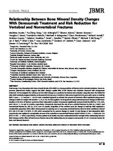
Irrespective of the initial H content (i.e. H flow rate) in the as-deposited films, a decrease in the H concentration upon annealing is observed, which is greater for longer annealing time. However, such a decrease is more effective in the case of the a-Ge film, as can be seen in Figure 5 that compares the decreases in H concentration in the two types of material (sputtered with H flow rate of 0.8 ml/min) as a function of the annealing time at 350°C. In a-Ge, the decrease is 85% with respect to the nonannealed reference sample (from 5.5 to 0.8 at.%) just after 1 h, whereas it is only 35% for a-Si (from 14.7 to 9.6 at.%). By annealing for 4 h only a small further decrease in the H concentration of 3-4% is observed in both Si and Ge. This indicates that the release of H in the a-Ge layer was highly effective, and that its escape from the layer was very fast. Evidence for this is given in Figure 6 which shows the surface morphology of the two types of layer after annealing. For the same annealing time, either 1 or 4 h, the Si layer mainly exhibits surface bumps after 14
H conct. [at %]
12 10
a-Si
8 6 4 2 a-Ge 0
0
1
2 3 Annealing time (h)
4
5
Figure 5 H concentration, extracted from Figure 4, as a function of the annealing time at 350°C in a-Si (solid black line) and a-Ge (dash blue line) single layers hydrogenated at 0.8 ml/min.
annealing, indicating that H is still in the film, though partially gathered in bubbles, whilst the Ge layer exhibits mostly craters, i.e. exploded H bubbles, as deep as the layer, suggesting that nearly all H has escaped in agreement with the ERDA results (Figures 4 and 5). The Si film also contains some broken bumps (one is visible in Figure 6a) which would explain the 35-38% decrease of the H concentration detected by ERDA. The structure degradation that produces craters is caused by two mechanisms in succession. First, the release of H and the formation of the H bubbles. Second, the creation of craters if the initial H content is very high and/or the annealing conditions are very severe [7-9]. As to the release of H, the above mentioned results are evidence that it is more efficient and faster in the a-Ge layers. This is in agreement with the previous literature according to which the binding energy of the Ge-H bond is smaller than that of the SiH bond [4,21-24]. In particular, Tsu et al. [24] found that it is 69 kcal/mole for Ge-H and 76 kal/mole for SiH. The faster release of H in a-Ge would cause a faster increase in the size of the H bubbles to the critical value for their explosion and formation of craters. The results of this study would confirm that the origin of the structural degradation of the MLs of a-Si/a-Ge observed in previous studies (Figure 1 and refs. [7-9]) very likely primarily starts in the Ge layers mostly because of the lower binding energy of H-Ge with respect to H-Si bonds. It should be noticed that crater formation could also be favoured by intrinsic stresses. The sample stress as measured by MT was always compressive, as found by others [25-27], with values of about 1, 0.15 and 0.33 GPa for as-deposited, non-hydrogenated single layers of a-Si, a-Ge and a-Si/a-Ge MLs, respectively. For a-Si, this result is in reasonable agreement with the literature data [27,28]. Not much is known for a-Ge. As expected, the stress for the MLs is in between those of the single layers. Nickel and Jackson [29] have speculated that the strain released as a consequence of the break of the Hhost atom bonds can be re-created by its propagation through the amorphous network to the neighbouring atoms and reconstruction of strained Si-Si bonds. They concluded that the average network strain remains independent of the H concentration and annealing as well [29]. It might thus be assumed that the annealed hydrogenated samples do not change their stress significantly with respect to that measured in the as-deposited ones. Other findings suggest that annealing causes stress relieve in hydrogenated amorphous Si/Ge MLs [30]. Should there be changes in these samples, it is very likely that the intrinsic stress of a-Ge always remains smaller than the one of a-Si upon annealing owing to the great difference between the values of the as-
Frigeri et al. Nanoscale Research Letters 2011, 6:189 http://www.nanoscalereslett.com/content/6/1/189
Page 5 of 6
a)
b)
Figure 6 AFM amplitude image of (a) a-Si and (b) a-Ge single layer after annealing at 350°C for 4 h. H flow rate 1.5 ml/min. In (a) bumps (close bubbles with H still inside) are predominant (only one is broken). In (b) large craters, i.e. exploded bumps with escape of H, are by far predominant. Very tiny bumps are also present.
deposited samples. The contribution of stress should thus play a minor role in differentiating the formation rate of the craters in a-Si and a-Ge. Further investigations are underway to better clarify this point. Abbreviations AFM: atomic force microscopy; ERDA: elastic recoil detection analysis; IR: infrared; MLs: multilayers; MT: Makyoh topography. Acknowledgements This study was supported by the Scientific Cooperation Agreement between MTA (Hungary) and CNR (Italy) under the contract MTA 1102, as well as by OTKA grant Nos. K-67969, CK-80126, K 68534 and TAMOP 4.2.1-08/1- 2008003 project (implemented through the New Hungary Development Plan cofinanced by the European Social Fund, and the European Regional Development Fund). Z. Erdélyi is a grantee of the ‘Bolyai János’ scholarship. Author details 1 CNR-IMEM Institute, Parco Area delle Scienze 37/A, 43100 Parma, Italy 2 Research Institute for Technical Physics and Materials Science, Hungarian Academy of Sciences, P.O. Box 49, H-1525 Budapest, Hungary 3Institute of Nuclear Research of the Hungarian Academy of Sciences, P.O. Box 51, H4001 Debrecen, Hungary 4Department of Solid State Physics, University of Debrecen, P.O. Box 2, H-4010 Debrecen, Hungary 5Institute for Materials Research (IMO), Hasselt University, Diepenbeek, Belgium Authors’ contributions CF coordinated the interpretation of the results and wrote the manuscript, MS grew the samples by sputtering and suggested the experiment, NQK performed the ERDA measurements, ACs carried out the sample heating experiments, FR did the Makyoh topography measurements, ZE participated in the coordination-realisation of the IR measurements, LN made the AFM work, DLB participated in the design of the study, H-GB performed the IR measurements. Competing interests The authors declare that they have no competing interests.
Received: 9 September 2010 Accepted: 1 March 2011 Published: 1 March 2011 References 1. Arrais A, Benzi P, Bettizzo E, Damaria C: Characterization of hydrogenated amorphous germanium compounds obtained by x-ray chemical vapor deposition of germane: Effect of the irradiation dose on optical parameters and structural order. J Appl Phys 2007, 102:104905. 2. Bouizem Y, Belfedal A, Sib JD, Kebab A, Chahed L: Hydrogen-bonding configuration effects on the optoelectronic properties of glow discharge a-Si1-xGex:H with large x. J Phys Condens Matter 2007, 19:356215. 3. Jobson KW, Wells J-PR, Schropp REI, Carder DA, Philips PJ, Dijkhuis JI: Relaxation processes of the Ge-H stretch modes in hydrogenated amorphous germanium. Phys Rev B 2006, 73:155202. 4. Cohen JD: Light-induced defects in hydrogenated amorphous silicon germanium alloys. Sol Energy Mater Sol Cells 2003, 78:399. 5. Sameshima T, Watanabe H, Kanno H, Sadoh T, Miyao M: Pulsed laser crystallization of silicon-germanium films. Thin Solid Films 2005, 487:67. 6. Abo Ghazala MS: Composition and electronic properties of a-SiGe:H alloys produced from ultrathin layers of a-Si:H/a-Ge:H. Physica B 2000, 293:132. 7. Frigeri C, Nasi L, Serényi M, Csik A, Erdélyi Z, Beke DL: AFM and TEM study of hydrogenated sputtered Si/Ge multilayers. Superlatt Microstruct 2009, 45:475. 8. Frigeri AC, Serényi M, Csik A, Erdélyi Z, Beke DL, Nasi L: Structural modifications induced in hydrogenated amorphous Si/Ge multilayers by heat treatments. J Mater Sci Mater Electron 2008, 19:S289. 9. Csik A, Serényi M, Erdélyi Z, Nemcsics A, Cserhati C, Langer GA, Beke DL, Frigeri C, Simon A: Investigation of thermal stability of hydrogenated amorphous Si/Ge multilayers. Vacuum 2010, 84:137. 10. Acco S, Williamson DL, Stolk PA, Saris FW, van den Boogaard MJ, Sinke WC, van der Weg WF, Roorda S, Zalm PC: Hydrogen solubility and network stability in amorphous silicon. Phys Rev B 1996, 53:4415. 11. Kótai E: In Proceedings of the 14th International Conference on the Application of Accelerators in Research and Industry, 1996, Denton, USA. Edited by: Duggan JL, Morgan IL. New York: AIP Press; 1997:631. 12. Szilágyi E, Pászti F, Amsel G: Theoretical approximations for depth resolution calculations in IBA methods. Nucl Instrum Methods B 1995, 100:103.
Frigeri et al. Nanoscale Research Letters 2011, 6:189 http://www.nanoscalereslett.com/content/6/1/189
Page 6 of 6
13. Riesz F: Makyoh topography for the morphological study of compound semiconductor wafers and structures. Mater Sci Eng B 2001, 80:220. 14. Stoney GC: The Tension of Metallic Films Deposited by Electrolysis. Proc R Soc Lond A 1909, 32:172. 15. Nix WD: Mechanical properties of thin films. Metall Trans A 1989, 20:2217. 16. Wertman JJ, Evans RA: Young’s Modulus, Shear Modulus, and Poisson’s Ratio in Silicon and Germanium. J Appl Phys 1965, 36:153. 17. [http://www.ioffe.ru/SVA/NSM/Semicond/Si/mechanic.html]. 18. Daouahi M, Zellama K, Bouchriha H, Elkaïm P: Effect of the hydrogen dilution on the local microstructure in hydrogenated amorphous silicon films deposited by radiofrequency magnetron sputtering. Eur Phys J AP 2000, 10:185. 19. Manfredotti C, Fizzotti F, Pastorino M, Polesello P, Vittone E: Influence of hydrogen-bonding configurations on the physical properties of hydrogenated amorphous silicon. Phys Rev B 1994, 50:18046. 20. Soukup RJ, Ianno NJ, Darveau SA, Exstrom CL: Thin films of a-SiGe:H with device quality properties prepared by a novel hollow cathode deposition technique. Sol Energy Mater Sol Cells 2005, 87:87. 21. Beyer W: Incorporation and thermal stability of hydrogen in amorphous silicon and germanium. J Non-Cryst Solids 1996, 198-200:40. 22. Chou YP, Lee SC: Structural, optical, and electrical properties of hydrogenated amorphous silicon germanium alloys. J Appl Phys 1998, 83:4111. 23. Walther T, Humphreys CJ, Cullis AG, Robbins DJ: A study of interdiffusion and germanium segregation in low-pressure chemical vapour deposition of SiGe/Si quantum wells. Inst Phys Conf Ser 1997, 157:47. 24. Tsu R, Martin D, Gonzales-Hernandez J, Ovshinsky SR: Passivation of dangling bonds in amorphous Si and Ge by gas adsorption. Phys Rev B 1987, 35:2385. 25. Friesen C, Thompson CV: Reversible Stress Relaxation during Precoalescence Interruptions of Volmer-Weber Thin Film Growth. Phys Rev Lett 2002, 89:126103. 26. Chason E, Sheldon BW, Freund LB, Floro JA, Hearne SJ: Origin of Compressive Residual Stress in Polycrystalline Thin Films. Phys Rev Lett 2002, 88:156103. 27. Tzanetakis P: Metastable volume changes of hydrogenated amorphous silicon and silicon-germanium alloys produced by exposure to light. Sol Energy Mater Sol Cells 2003, 78:369. 28. Gotoh T, Nonomura S, Nishio M, Nitta S, Kondo M, Matsuda A: Experimental evidence of photoinduced expansion in hydrogenated amorphous silicon using bending detected optical lever method. Appl Phys Lett 1998, 77:2978. 29. Nickel NH, Jackson WB: Hydrogen-mediated creation and annihilation of strain in amorphous silicon. Phys Rev B 1995, 51:4872. 30. Tripathi S, Brajpuriya R, Sharma A, Shripathi T, Chaudhari SM: Structural characterization of annealed Si/Ge nanostructures using Raman spectroscopy, XRR and AFM. J Phys D Appl Phys 2006, 39:4848. doi:10.1186/1556-276X-6-189 Cite this article as: Frigeri et al.: Relationship between structural changes, hydrogen content and annealing in stacks of ultrathin Si/Ge amorphous layers. Nanoscale Research Letters 2011 6:189.
Submit your manuscript to a journal and benefit from: 7 Convenient online submission 7 Rigorous peer review 7 Immediate publication on acceptance 7 Open access: articles freely available online 7 High visibility within the field 7 Retaining the copyright to your article
Submit your next manuscript at 7 springeropen.com