Arq Neuropsiquiatr 2008;66(4):868-871
RELATIONSHIP BETWEEN THE CORONAL SUTURE AND THE CENTRAL LOBE How important is it and how can we use it in surgical planning? Stênio Abrantes Sarmento1, Danielle Cartaxo Jácome2, Emerson Magno F. de Andrade3, Alessandra V. Albuquerque Melo4, Osleuse Rocha de Oliveira5, Helder Tedeschi6 Abstract – The surgical treatment of the lesions located in the central lobe is a very difficult task for the neurosurgeon. The overall aim of this study is to verify the correlation of the coronal suture and the structures of the central lobe in 32 cadaver hemisphere brains and the importance of this information in surgical planning. The measurement of the nasion to the coronal suture ranged from 11.5 to 13.5 cm. The distance between the coronal suture in the midline to the central, precentral and paracentral sulcus ranged from 5.0 to 6.6, 2.5 to 4.5 and 1.3 to 4.0 cm respectively. Particularly in the normal cortex these measurements can be used to guide the surgical access. However, the identification of the central sulcus is not easy when the anatomical pattern is distorted or displaced by a lesion or edema. In cases such as these the use of other tools becomes crucial for good surgical planning and cortical mapping or awake craniotomy for a safer resection of the lesion as well. Key words: coronal suture, central lobe, central sulcus, motor cortex, preoperative planning. Relação entre a sutura coronária e o lobo central: qual a sua importância e como podemos usá-la no planejamento cirurgico? Resumo – O tratamento cirúrgico de lesões localizadas no lobo central é difícil para o neurocirurgião. O objetivo deste estudo é verificar a relação da sutura coronária com as estruturas do lobo central utilizandose de dissecção realizada em 32 hemisférios cerebrais de 16 cadáveres, assim como, a importância desta informação no planejamento cirúrgico. A medida da distância entre o nasion e a sutura coronária variou entre 11,5 e 13,5 cm. A distância da sutura coronária na linha média para os sulcos central, pré-central e paracentral variou de 5,0 a 6,6 cm, 2,5 a 4,5 cm e 1,3 a 4,0 cm respectivamente. O conhecimento destas medidas pode ser usado no planejamento cirúrgico principalmente num córtex normal. Porém, a identificação do sulco central é difícil quando as estruturas anatômicas estão deslocadas pela lesão ou quando há edema. Nestes casos a utilização de outros meios diagnósticos para o planejamento cirúrgico torna-se necessária, como também a estimulação cortical ou a craniotomia com o paciente acordado pode proporcionar uma ressecção mais segura da lesão. Palavras-chave: sutura coronal, lobo central, sulco central, córtex motor, planejamento pré-operatório.
The central lobe is an eloquent area of the central nervous system (CNS), limited anteriorly by the pre-central sulcus and posteriorly by the post-central sulcus. The central sulcus, which separates the pre from the post-central gyri, is one of the most important anatomical landmarks of the cerebral cortex1-3. The central lobe is a site of a wide variety of lesions and sometime surgeons need to approach it directly, for
example in the resection of gliomas. However, manipulation in an eloquent area increases the risk of pos surgical complications and deficits. A knowledge of the morphological relationship of this lobe and its relationship to the craniometric points and sutures facilitates the approach to the lesion preserving neurovascular structures1-6. Exact and correct localization of the central sulcus becomes crucial.
1
Neurosurgeon, Professor of Neuroanatomy and Neurosurgery, Nova Esperança Medical School (FAMENE), João Pessoa PB, Brazil, Postgraduate, State University of Campinas (UNICAMP), Campinas SP, Brazil; 2Medical Student, FAMENE; 3Neurosurgery Resident, Hospital Beneficência Portuguesa de São Paulo, São Paulo SP, Brazil; 4Radiologist, Ecoclínica, João Pessoa PB, Brazil; 5Anesthesiologist, Hospital Memorial São Francisco, João Pessoa PB, Brazil; 6 Neurosurgeon, Associate Professor, UNICAMP. Received 18 April 2008, received in final form 18 August 2008. Accepted 10 September 2008. Dr. Stênio A. Sarmento – Rua Major Ciraulo 433 / 2501 - 58038-290 João Pessoa PB - Brasil. E-mail:
[email protected] 868
Coronal suture and the central lobe Sarmento et al.
Arq Neuropsiquiatr 2008;66(4)
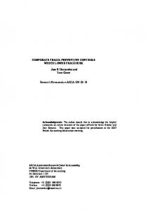
Fig 1. (A) A superior view of the cranium after scalp removal; continuous line – measurement of the distance between the nasion and the coronal suture in the midline and the mark of the bilateral craniotomy to expose the central lobe. (B) After bilateral removal of the piamater and vessels for a better visualization of the sulcus the measurement procedures were undertaken. The line 1 corresponds to the distance between the coronal suture to the beginning of the central sulcus on the midline (6.0 cm in this case) and the line 2 corresponds to the same distance, but just paramedian on the high convexity (5.2 cm in this case).
Table 1. Coronal suture and sulcal points related measurements. Average
Standard deviation
R
L
Total
R
L
Total
Right × left (p value)
CoSut – CS distance
5.85
5.97
5.91
0.51
0.31
0.37
1.11
CoSut – preCS distance
3.67
3.66
3.67
0.47
0.53
0.41
0.09
CoSut – posCS distance
2.60
2.91
2.76
0.68
0.71
0.62
1.90
CoSut – CS inf ext distance
2.90
3.04
2.97
0.70
0.68
0.62
0.91
ParacLob length
3.76
3.77
3.76
0.44
0.42
0.36
0.05
R, right; L, left; CoSut, coronal suture; CS, central sulcus; preCS, precentral sulcus; posCS, poscentral sulcus; CS inf ext, central sulcus inferior extremity; ParacLob, paracentral lobe. Measurements are in centimeters.
Table 2. Frequencies of coronal suture - nasion distance related measurements. Distance
Frequencies
Percentage (%)
11.5
1
15
12
13
75
12.5
1
5
13.5
1
5
Total
16
100
Average=12.02; standard deviation=0.41; measurements are in centimeters.
The objective of this anatomical study is to verify the correlation between the coronal suture and the structures of the central lobe and discuss how to use this information to plan the craniotomy and surgical access preserving the structures as well. The pattern of the course of the sulcus and veins were not an aim of this study because these structures have been well studied in the literature.
METHOD A total of 16 adult cadaver brains were studied by a craniotomy, totaling 32 hemispheres. The study was performed in the anatomy laboratory of the Faculdade de Medicina Nova Esperança (FAMENE). The scalp was removed by a biauricular incision to expose the external cranial surface. Both coronal sutures were identified (Fig 1A). A bilateral craniotomy was performed to expose the entire central lobe leaving the coronal suture as a landmark. The posterior extension of the craniotomy was about 10 cm behind the coronal suture. The opening of the dura was performed including part of the superior sagittal sinus, followed by removal of the piamater and veins (Fig 1B). In addition to measurement between the nasion to the coronal suture, the following measurements were taken in all of the hemispheres: the distance between the coronal suture and the central sulcus in the midline; the distance between the coronal suture and pre-central sulcus in the midline; the distance between the coronal suture and paracen869
Coronal suture and the central lobe Sarmento et al.
Arq Neuropsiquiatr 2008;66(4)
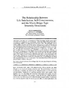
Fig 2. An ilustrative case: 42 year old, male, presented a motor deficit on the left side (grade 4 on the upper and lower limbs). (A) Pre-operative sagittal MRI, (B) axial and (C) coronal showing a subcortical anaplastic astrocytoma in the right motor area with perilesional edema causing a distortion of the sulcus. (D) Tractography shows a close relationship between the tumor (T) and the pyramidal tract (some fibers were interrupted by the tumor). In cases such this, the risk of damage in the motor cortex or descendent tract is high. The knowledge of the relationship between the coronal suture and structures of the central lobe helps the neurosurgeon to estimate the site of the incision and craniotomy based on the MRI data. But, for a safer resection of the tumor preserving the normal structures we decided to operate on the patient by an awake craniotomy. (E) Surgical cavity post resection.
tral sulcus; the length of the paracentral gyrus and the distance between the coronal suture in the pterion to the central sulcus. Our data were submitted to statistical analysis using t Student test; p-values