American Journal of Botany 92(11): 1836–1852. 2005.
REPRODUCTIVE DEVELOPMENTAL COMPLEXITY IN THE AFRICAN OIL PALM (ELAEIS GUINEENSIS, ARECACEAE)1 HE´LE`NE ADAM,2 STEFAN JOUANNIC,2 JACQUES ESCOUTE,3 YVES DUVAL,2 JEAN-LUC VERDEIL,3,4 AND JAMES W. TREGEAR2,4 IRD/CIRAD Palm Biology Laboratory, UMR 1098, Centre IRD Montpellier, BP 64501, 911, Avenue Agropolis, 34394 Montpellier, France; and 3CIRAD-AMIS, UMR 1098, Avenue Agropolis, 34398 Montpellier Cedex 5, France
2
Species of the palm family (Arecaceae) are remarkably diverse in their inflorescence and floral morphologies, which make them a particularly interesting group for studies of reproductive development and its evolution. Using light and scanning electron microscopy, we describe inflorescence and flower development in the African oil palm Elaeis guineensis from the initiation of the inflorescence meristem to flower maturity. In mature palms, the inflorescence develops over 2–3 years and is characterized by individual stages within which differentiation may be either relatively slow, as in the case of early inflorescence meristem development, or rapid, as in the case of flower organogenesis. The female inflorescence bears floral triads composed of single pistillate flowers flanked by two abortive staminate flowers, whereas the male inflorescence contains single functional staminate flowers. This suggests a possible evolutionary movement from an ancestral hermaphrodite inflorescence form containing fully functional floral triads to the situation of temporal dioecy observed at present. Wild type flowers are compared to those bearing an epigenetic homeotic abnormality, known as mantled, involving an alteration of the identity of the organs in the fertile and sterile androecium. Key words:
development; floral triad; flower; inflorescence; mantled; oil palm.
Despite practical constraints associated with their often large size, the palm family (Arecaceae, sole member of the order Arecales) is a group of plants of considerable interest in developmental biology studies. A key feature of palms, which lends itself to developmental analysis, is the existence usually of a single vegetative shoot apical meristem, which initiates the entire aboveground structure of the plant. Palms also have a number of interesting features in their reproductive development, such as variable modes of sex determination and single- or mixed-sex flower clusters, which may define specific clades. Indeed, studies of inflorescence and floral ontogenesis are key elements to understanding evolutionary relationships both within the palm family and with respect to other monocot groups, as well as in angiosperms as a whole. The Arecaceae were recently restructured into five subfamilies (Dransfield et al., 2005) as opposed to the six subfamily structure of the previous classification (Uhl and Dransfield, 1987). The new nomenclature is adopted here. Two main and contrasting types of flowering behavior within a single shoot may be seen in palm species (Tomlinson, 1990). Hapaxanthy, in general, refers to a single shoot in which there is an abrupt transition to the flowering state, with flowering branches expanding in larger numbers after vegetative extension has ceased. The flowering phase is thus short. Hapaxanthic shoots occur in about 5% of palms. Pleonanthy refers to a single shoot in which flowering branches (inflorescences) appear in the axils of vegetative leaves and continue to be produced as the palm continues its vegetative extension. Such shoots are termed pleonanthic. The flowering phase is extended and indeterminate. Pleonanthic shoots occur in about 95% of all palm species. Manuscript received 9 December 2004; revision accepted 21 July 2005. The authors gratefully thank Jean-Christophe Pintaud, Francis Halle´, and three anonymous referees for helpful suggestions in the writing and improvement of this paper, FELDA Agricultural Services (Malaysia) and ASD (Costa Rica) for the generous supply of plant material, and Axel Labeyrie for photography and logistical help. 4 Authors for correspondence (e-mail:
[email protected];
[email protected]) 1
A wide range of floral ontogenetic studies has been reported for palms; notable examples include Ptychosperma species (Uhl, 1976); several genera of the tribe Phytelepheae (Palandra, Phytelephas, Ammandra, and Aphandra; Uhl and Moore, 1977; Uhl and Dransfield, 1984; Barfod and Uhl, 2001); the chamaedoreoid species Hyophorbe indica (Uhl and Moore, 1978); a number of polyandrous genera (Uhl and Moore, 1980); the calamoid genus Eugeissona (Uhl and Dransfield, 1984); two members of the tribe Cocoseae (Beccariophoenix madagascariensis and Polyandrococos pectinatus; Uhl, 1988); the species Geonoma interrupta in the tribe Geonomateae (Stauffer et al., 2002); and the genus Dypsis of the tribe Areceae (Rudall et al., 2003). The flowering apparatus of palm species is a useful indicator of phylogenetic relationships and therefore evolutionary events. Within the family as a whole, a number of general trends in the evolution of floral characters have been noted, including a progression from bisexual to unisexual flowers and from monoecy to dioecy (Moore and Uhl, 1982). We describe here a detailed study of reproductive development in the African oil palm (Elaeis guineensis Jacq.), a pleonanthic species belonging to the subfamily Arecoideae, in the tribe Cocoseae, and the subtribe Elaeidinae. The Arecoideae form the largest subfamily of the Arecaceae and have been classified into 112 genera (Dransfield et al., 2005). Oil palm is the second most important world vegetable oil crop plant after soya and is one of the most important perennial crops in the tropics. Despite its economic importance, only partial studies of oil palm inflorescence and flower development have been reported (Beinaert, 1935; Van Heel et al., 1987). Oil palm is perennial and relatively long-lived, sometimes attaining more than 100 years in age. The oil palm is a single-stemmed palm, which bears, like the majority of palm species, a single vegetative shoot apical meristem maintained throughout the lifetime of the plant and localized at the center of the leaf crown. Under favorable climatic conditions, this meristem is continuously active, producing a new leaf primordium approximately every 2 weeks in mature palms; at the
1836
November 2005]
ADAM
ET AL.—REPRODUCTIVE DEVELOPMENT IN
ELAEIS GUINEENSIS
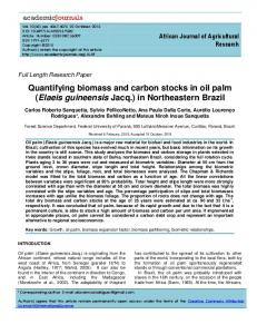
Fig. 1. Features of reproductive development in oil palm. (A) Oil palm inflorescence structure and axis organization. The inflorescence unit axis (axis 1) is in the axil of a leaf. Abbreviations: F, flower; fb, flower bract; L, leaf; p, prophyll; pb, peduncular bract; rab, rachilla bract. (B) Chronology of the principal phases of inflorescence and flower development in oil palm. Note that this is a generalized scheme applying to inflorescences of both sexes. (C) Diagram showing the structure of the floral triad of the female inflorescence (after Beirnaert, 1935; Van Heel et al., 1987).
juvenile stage, the plastochron may be as low as 9 days (Corley and Gray, 1976). The leaf takes 2–3 years to develop from initiation to the time when leaflets unfold in the center of the palm crown. Inflorescences are formed throughout the year in an acropetal sequence in the axils of their subtending leaves. The immature inflorescence is protected within the crown by its subtending leaf. Moreover, as in many palms of the Cocoseae, the expanding inflorescence axes are themselves protected by a prophyll and a peduncular bract, which become thick and lignified. Elaeis guineensis is monoecious, a character that predominates in the Arecaceae (Dransfield and Uhl, 1998). Oil palm produces separate male and female inflorescences on the same palm in an alternating cycle of variable duration depending on genetic factors, age, and particularly environmental conditions, with the production of male inflorescences generally favored by water stress (Corley, 1976a). Occasionally mixed sex inflorescences are produced at the transition between the male and female cycles (Biradar, 1978). Mature inflorescences are visible typically 32–36 months after seed germination (Corley, 1976b). As for all monocotyledoneous species, oil palm has trimerous flowers. In the Arecaceae, the perianth is usually clearly distinguished into sepals and petals (Dransfield and Uhl, 1998). In the case of oil palm however, the sepals and petals have a similar petalloid appearance and are often referred to as tepals. The reproductive organs of staminate flowers are composed of six stamens (two whorls of three) with connate filaments surrounding a pistillode, whereas female flowers have rudimentary stamens (staminodes) and a gynoecium of three carpels. The aim of the studies described here was to provide a complete description at the histological and cellular levels of all stages of inflorescence development from the initiation of the primordium from the central axis through to the completion
Fig. 1.
Continued.
1837
1838
AMERICAN JOURNAL
Fig. 1.
of flower development. The study was extended to include the floral variant known as mantled, commonly observed in palms produced by in vitro micropropagation and characterized by homeotic transformation of the fertile or sterile androecium (Corley et al., 1986). To our knowledge, this is the first indepth study of this type reported for palms. Mantled palms resemble B class mutants of model monocot and dicot species (Coe¨n and Meyerowitz, 1991; Zahn et al., 2005) with which comparisons can be made to obtain a fuller understanding of floral homeotic functions in relation to phylogeny. MATERIALS AND METHODS Plant material—Inflorescence material for the characterization of stages of reproductive development was collected in Coto, Costa Rica at the plantation of the ASD Company. All palms were 9 years old and of the tenera variety, i.e., obtained from a cross between pisifera and dura palms (Hartley, 1988). For studies of the mantled floral phenotype, normal and variant flowers were collected in Tun Razak, Malaysia at the plantation of the FELDA Company. The source palms for the mantled and normal pistillate flowers were of identical genotype and age. Palms of similar genetic origin and age were also used as the source of staminate flowers displaying either a mantled or a normal phenotype. The numbering attributed to inflorescences corresponds to that of their axillary leaves (Fig. 1A, B). The youngest expanding leaf was numbered as leaf 0. The leaf produced immediately before leaf 0 is thus leaf F11 and the leaf produced subsequently is leaf F21 (this leaf is not yet expanded). In this study, the inflorescences collected individually were located between leaf 227 (the earliest stage at which inflorescence isolation was possible) and leaf 118 (flower maturity; Fig. 1B). Earlier stages were observed without prior isolation along with the subtending partly formed leaves. Oil palms were felled in order to dissect the least accessible primordia near the shoot apical meristem. Inflorescences 227 to 26 were directly fixed. For inflorescences 25 to 14, the prophyll and peduncular bract were removed and the entire inflorescence fixed (as described in next section). For inflorescences 15 to 116, rachillae were dissected from the rachis and fixed. For inflorescences 116 to 118, floral triads and staminate and pistillate flowers of normal and mantled phenotypes were dissected individually from the center of the rachilla and fixed. We used the nomenclature of Tomlinson (1990) to describe the morphological features of the oil palm material. Fixation of samples—After dissection, sampled material was fixed for 1 h under vacuum and thereafter for 8 h at 48C in fixation buffer (4% paraformaldehyde, 0.1 M phosphate buffer, pH 7). Samples were then dehydrated
OF
BOTANY
[Vol. 92
Continued.
through a graded ethanol series (30, 70, and 100%, v/v) for 2 h and stored at 48C. Scanning electron microscopy—For scanning electron microscopy (SEM), material was passed through three changes of absolute ethanol, critical point dried, mounted on stubs, and coated with platinum in a Baltec (Manchester, New Hampshire, USA) SCD 50 sputter coater. Samples were visualized with a JEOL (Tokyo, Japan) 6300F scanning electron microscope at 20 kV. Histological studies and transmission light microscopy—For histological analysis, samples were transferred to 100% butanol for 2 days and embedded in Technovit resin (LKB Pharmacia, Uppsala, Sweden) using a previously described protocol (Buffard-Morel et al., 1992). Blocks were sectioned at 4– 5 mm thickness using a HISTORANGE microtome (LKB Broma, Uppsala, Sweden). Slides were double stained with periodic acid–Schiff’s reagent (PAS) stain (Sigma-Aldrich, Lyon, France) to detect carbohydrate compounds (Buffard-Morel et al., 1992) and naphthol blue-black to detect proteins (Fisher, 1968).
RESULTS Development and structure of the inflorescences—The inflorescence is initiated in the axil of each leaf of the oil palm from an early stage shortly after germination and continues throughout the lifetime of the plant. The development of the inflorescence takes over 2 years; the organ is completely enclosed at the base of the subtending leaf for most of this time. The reproductive development of oil palm is represented schematically in Fig. 1, which details the structural organization of the inflorescence (Fig. 1A), the key stages of inflorescence and flower development (Fig. 1B), and the structure of mature flowers (Fig. 1C). The inflorescence is a compound rachis composed of 100–300 rachillae in the case of the male inflorescence or approximately 150 rachillae in the case of the female inflorescence (Jacquemard, 1995). In each case, the rachis is borne by a peduncle with a typical length at maturity of either 20–30 cm for the female inflorescence or around 40 cm for the male inflorescence (Beirnaert, 1935). Rachillae (axis 2 in Fig. 1A) are arranged spirally around a central rachis (axis 1) emanating from the stem (axis 0). A prophyll, along with the peduncular bract, tightly encloses the inflorescence until approximately 6 weeks before flower maturity. The prophyll attains a final length of approximately 45 cm, whereas
November 2005]
ADAM
ET AL.—REPRODUCTIVE DEVELOPMENT IN
the peduncular bract, with which it forms an essentially continuous layer of protection, is typically 2–4 cm shorter. Both layers become fibrous as their development progresses and finally undergo necrosis before being ruptured by the elongating inflorescence. Male and female inflorescences differ in their rachilla morphology (Figs. 2, 4), notably with respect to the numbers of flowers that they bear. The female inflorescence reaches a length of 35 cm or more when fully developed (Fig. 2), and each of its rachillae bears 5–30 floral triads (Fig. 3). The triads are arranged spirally around the rachilla axis, and each is subtended by a spiny floral triad bract. The functional pistillate flower develops within a floral triad between two nonfunctional staminate flowers (Beirnaert, 1935). The male inflorescence reaches a length similar to that of the female inflorescence at maturity and is composed of rachillae, each bearing 400–1500 staminate flowers (Figs. 4 and 5). The staminate flowers are subtended by floral bracts, which are smaller than those of the female inflorescence. The earliest stage at which the inflorescence bud is visible by light microscopy is in the axil of the fourth-youngest leaf close to the vegetative shoot apical meristem. At this stage, the inflorescence consists of a group of a few cells localized in the axil of the partly formed leaf (Fig. 6). These cells display a low cytoplasm to nucleus ratio. The inflorescence bud is visible to the naked eye from the stage corresponding to the 12th-youngest leaf onwards. Figure 7 shows an inflorescence meristem at a very young stage (leaf 227), corresponding to the initiation of the rachis (Fig. 1A; axis 1). This meristem, also named the rachis meristem, is composed of a central dome, which is flanked by the primordium of the future prophyll (or outer spathe). Within the inflorescence dome, composed of isodiametric meristematic cells, a distinct layer of cells on the surface (L1; Vaughan, 1955) will give rise to the epidermal cells (Fig. 8). About 10 cell layers under the L1 layer, cells with a large nucleus and small cytoplasm are apparent. Few divisions are visible in this zone, suggesting that they might perform a ‘‘stem cell’’ function. A third zone of cells is visible under the two previously mentioned layers; in this case, the cells are vacuolated and may later contribute to the pith of the inflorescence. At this stage, the center of the inflorescence (rachis) is not vascularized. Periclinal and anticlinal divisions contribute respectively to the growth in depth and width of the dome. Figure 9 shows the inflorescence meristem at the 226 leaf stage, when the prophyll is more elongated along the meristem. Moreover, many more cells under the L1 layer are actively dividing at this point. At the 224 leaf stage, the prophyll entirely envelops the inflorescence meristem, next to which the peduncular bract (or inner spathe) primordium is visible around the dome (Fig. 10). The prophyll and peduncular bract elongate until they entirely envelop the inflorescence meristem. During its subsequent development, the meristem grows in depth along with the rachis, the number of vacuolate cells in the center of rachis increasing. In this phase of development, the inflorescence meristem (Fig. 1A; axis 1) displays only one functional axis, which gives rise to the prophyll and peduncular bract. The activity of the rachis meristem is maintained and allows the production of rachilla bracts, which subtend an axillary rachilla. At the 220 leaf stage, the rachis meristem (axis 1) is completely enclosed by the prophyll and peduncular bract (Fig. 11). At the flanks of the rachis meristem, anticlinal divisions are visible under the L1 cells. These
ELAEIS GUINEENSIS
1839
divisions will give rise to the rachilla bracts (Fig. 12). The basal and apical bracts are the first to develop with a slight difference in their rates of development (Fig. 13). The inflorescence meristem maintains the same form and cellular organization as in the previous stage; however, more vascular cells are observed in the center of the rachis and starch may be seen in these cells. During the development and growth of the rachis, vascularization and starch accumulation become progressively more visible. The rachilla bracts of the inflorescence develop in a parastichial and acropetal manner along the rachis axis (Figs. 14–16), the tissues at the center of lateral bracts gradually developing a vascularized structure (Fig. 14). At this stage, no differences are visible between male and female inflorescences (leaf stage 26). Rachilla initiation and development—The lateral rachis meristem (rachilla meristem) is initiated when ;10–15 rachilla bracts have been formed (Fig. 17; 26 leaf stage). Rachilla meristems (Fig. 1A; axis 2) are initiated at the bases of lateral bracts by periclinal divisions in 2 or 3 cell layers underlying the L1 layer (Fig. 17). At the initiation stage of the rachillae, the cells that give rise to these structures possess a large nucleus, as illustrated by their intense and homogeneous naphthol blue-black coloration. This staining, which is specific to proteins, marks the presence of histones and suggests that the chromatin of these cells might be condensed (Fig. 17). When the rachilla primordium is formed (Fig. 18), the cells of the primordium have a higher nucleus to cytoplasm ratio. The nuclei are larger, with a blue, less-homogeneous coloration, suggesting a decondensation of the chromatin in the nucleus. The cells underlying the L1 cells thus seem to be active, but do not divide, during the initiation stage, despite their apparent potential to do so (Fig. 17). The growth of the rachilla axis is achieved by anticlinal divisions in cells of the L2 layer (Fig. 19; Leaf stage 22). Rachillae develop in a basipetal manner (Fig. 20). Each rachilla will produce a series of floral bracts (in the male inflorescence), or floral triad bracts (in the female inflorescence), each containing a floral meristem in its axil. The rachilla meristem (axis 2) has the same basic organization as the rachis meristem (axis 1), namely a well-organized L1 cell layer, overlying cell layers with a high division potential. The elongation of the rachilla is achieved by the activity of an adjoining region of vacuolating cells. The rachilla meristem has a conical shape, the meristematic zone of the rachilla being more substantial in size than that of the rachis observed previously, suggesting that the rachilla is at this point undergoing an active growth phase (Fig. 21). The periphery of the rachilla displays some periclinal divisions in cells underlying the L1 layer. These divisions will later give rise to floral bracts or floral triad bracts. For both sexes, bracts are more advanced in their development at the abaxial side of the inflorescence axis, and the development is parastichial and acropetal (Figs. 21, 22). During the development of the rachilla, tissues in the center undergo vascularization (Fig. 22; leaf stage 14). At this point, the sex of the inflorescence can be identified, the future male rachilla displaying a larger number of bracts than the future female rachilla (data not shown). Development and structure of the floral triad—On the female inflorescence, the rachillae bear triads of flowers, each consisting of a functional pistillate flower flanked by two nonfunctional staminate flowers. The latter normally undergo ab-
1840
AMERICAN JOURNAL
OF
BOTANY
[Vol. 92
Figs. 2–5. Macroscopic view of mature female and male inflorescences of oil palm. 2. Female inflorescence at anthesis after opening of the peduncular bract and prophyll. 3. Detail of female rachillae carrying pistillate flowers at maturity with exposed stigmas. 4. Male inflorescence at anthesis. 5. Detail of male rachilla and flowers at anthesis. Abbreviations: f, flower; p, prophyll; ra, rachilla; St, stigma.
November 2005]
ADAM
ET AL.—REPRODUCTIVE DEVELOPMENT IN
ELAEIS GUINEENSIS
1841
Figs. 6–10. Longitudinal sections (LS) of early inflorescence development in oil palm visualized by LM. 6. Inflorescence primordium at the fourth leaf after the shoot apical meristem. 7. Inflorescence meristem at 227 leaf stage. 8. Detail of inflorescence meristem at 227 leaf stage. 9. Inflorescence meristem at 226 leaf stage. 10. Early initiation of peduncular bract of inflorescence meristem at 224 leaf stage. Abbreviations: pbp, peduncular bract primordium; Im, Inflorescence meristem; Ip, Inflorescence primordium; L, leaf; L1, overlying cell Layer; p, prophyll; pp, prophyll primordium; PV, provascular strands. Bar 5 0.1 mm.
1842
AMERICAN JOURNAL
OF
BOTANY
[Vol. 92
Figs. 11–18. Light and SEM micrographs of rachilla development up to and including leaf stage 24. 11–16. Rachilla bract development. 17–18. Rachilla initiation and development. 11. Longitudinal section (LS) of developing inflorescence at 220 leaf stage. 12. Detail of rachilla bract initial. 13. Upper view of inflorescence meristem. Development of anterior and posterior rachilla bracts. 14. Longitudinal section of developing inflorescence at 210 leaf stage. 15. Side view of developing inflorescence at leaf stage 210. 16. LS of developing rachilla at leaf stage 210. 17–18. LS of inflorescence to reveal successive phases of rachilla development. 17. Initiation of rachilla primordium at 26 leaf stage. Arrow points to cells at the origin of rachilla. 18. Rachilla primordium at leaf stage 24.
November 2005]
ADAM
ET AL.—REPRODUCTIVE DEVELOPMENT IN
ELAEIS GUINEENSIS
1843
Figs. 19–22. Rachilla and floral bract development at 19. leaf stages 22, 20. 0, 21. 12 and 22. 14 respectively. Abbreviations: ant rab, anterior rachilla bract; Im, Inflorescence meristem; Fb, floral bracts; fpb, peduncular bract; p, prophyll; post rab, posterior rachilla bract; r, rachis; rab, rachilla bract; rabi, rachilla bract initium; rabp, rachilla bract primordium; Ram, rachilla meristem; Vb, vascular bundles. Bar 5 0.1 mm, except for Fig 15, bar 5 1 mm.
scission before complete maturity is reached (discussed later). The floral triad is the most common flowering unit in palms (Moore and Uhl, 1982) and is designated as a cincinnus (Uhl, 1976). In the case of oil palm, the developmental sequence observed illustrates that the floral triad is a sympodial flower cluster, i.e., involving a branching system where the main axis is determinate and side branches are produced successively via axillary meristems. At leaf stage 16, a new meristem arises in the axil of the floral triad bract initially formed by the rachilla meristem (Fig. 1A, axis 2; Fig. 23). This new meristem forms an apex which, after producing bracteole 1, develops into staminate flower 1 (Fig. 24). Triad development continues in the axil of bracteole 1, where a new apex emerges and gives rise to bracteole 2 before developing into staminate flower 2 (Fig. 25). Note that the section in Fig. 25 does not include bracteole 2. A group of meristematic cells is present between asf1 and asf2. These cells will subsequently give rise to bracteole 3 and the pistillate flower in the same manner as observed for the staminate flowers (Fig. 26). During their development, the peduncles of the
staminate flowers elongate, until they are above the pistillate flower (Fig. 26). Floral meristems have a similar form to that of the rachis and rachilla meristems, consisting of a L1 layer on the surface, overlying 5 or 6 layers of active cells (Fig. 24). The two staminate flowers are asynchronous in their development (Figs. 25, 26). Structure of normal flowers—The trimerous flowers of oil palm contain a perianth consisting of three petals surrounded by three sepals. Since these two whorls are similar in appearance, they are frequently referred to as tepals (Hartley, 1988). In staminate flowers, the perianth surrounds an androecium, usually consisting of two whorls of three stamens, themselves enclosing a rudimentary gynoecium. In the case of pistillate flowers, the perianth surrounds a sterile androecium of usually six rudimentary stamens (staminodes) enclosing a syncarpous gynoecium composed of three carpels. Development and structure of normal flowers on the female inflorescence—At leaf stage 110, organ development
1844
AMERICAN JOURNAL
OF
BOTANY
[Vol. 92
Figs. 23–31. Light and SEM micrographs of floral triad development. 23. Longitudinal section (LS) showing the initiation of the triad meristem (i.e., the apex that will produce the first staminate flower) in the axil of floral bracts. 24. Transverse section of first staminate flower primordium surrounded by bracteole 1. 25. LS of staminate flowers 1 and 2 showing sepal development. 26. LS of floral triad of flowers at leaf stage 110. 27. LS of accompanying staminate flower and initiation of stamens. 28. LS of pistillate flower development of sepals at leaf stage 112. 29. LS showing development of staminate flower 1. 30. LS of triad of flowers at leaf stage 115.
November 2005]
ADAM
ET AL.—REPRODUCTIVE DEVELOPMENT IN
begins in the accompanying staminate flowers with the initiation and development of sepals (Fig. 25). The elongation of these organs is achieved by anticlinal divisions at their base. The cells localized at the distal end of sepals are already vacuolate at this stage (Figs. 26, 27). After the initiation and development of the three petals, the floral meristem rapidly initiates stamen primordia through periclinal cell divisions in the L2 cells at the flanks of the floral meristem (Fig. 27). While the organs of the staminate flowers are initiated, the pistillate flower meristem does not develop any organs. However, the cells of the pistillate flower meristem have a high nucleus to cytoplasm ratio, suggesting that they remain active (Fig. 26). The development of the floral triad accelerates after leaf stage 110. At leaf stage 112, the sepals of pistillate flowers are elongating by anticlinal division of the L2 cells. Floral organs undergo elongation through cell divisions at their bases (Fig. 28). At the same stage, the third whorl of the staminate flowers shows the first signs of differentiation of the future stamens (Fig. 29). The development of stamens is achieved by the division of active cells in their upper regions, which will give rise to anthers. The accompanying staminate flowers are larger than the pistillate flower at this point; however, during the next phase of development, the pistillate flower will increase in size, whereas the accompanying staminate flower will stay the same (Figs. 34, 35). At leaf stage 115 (Figs. 30, 31), the floral triads are composed of accompanying staminate flowers bearing six initiated stamens entirely enveloped by the perianth. The pistillate flower at this stage is composed of only the perianth. At leaf stage 116, the reproductive organs of pistillate flowers are developing, the androecium and gynoecium forming concomitantly (Fig. 32). Organs are initiated by periclinal divisions in L2 layer cells (not shown). Three free carpels are formed in the innermost whorl. In contrast to the perianth, the development of floral organs in the inner whorls is achieved by cell divisions in their distal regions (not shown). The development of accompanying staminate flowers continues with the differentiation of the filament and anther regions at leaf stage 116 (Fig. 33). The cells of the filaments are seen to be vacuolate, unlike those at the distal end of anthers, which have a high nucleus to cytoplasm ratio. The meristematic zone of the staminate flower is completely exhausted (Fig. 33); the cells are vacuolate and will not give rise to a gynoecium. By leaf stage 117, the anthers of the accompanying staminate flowers are completely developed and vacuolar polyphenol accumulation is seen within the cells of the petals. The anther consists of a pollen sac surrounded by the tapetum (not shown). The pollen sac contains a mass of dividing cells, the microsporocytes. However, the anther will not give rise to pollen because the microsporocytes will degenerate in the pollen sac. Subsequently, the stalk of the staminate flowers will abscise, and they will shed before anthesis. During the following stages (Fig. 36), the reproductive organs of pistillate flowers develop. Organs in the sterile androecium, known as staminodes, are arrested in their development and do not develop into stamens. The carpels formed in the innermost whorl are joined at their bases. At the base of the carpels, a group of meristematic cells (Fig. 36) will give rise to the ovary. Subsequently, the stigma of the carpel grows and the region in the center of the stigma becomes vascularized. Some polyphenols accumulate at the distal end in the center and in the lateral faces. The ovary is localized at the base of the carpel. As previously indicated by Uhl and Drans-
ELAEIS GUINEENSIS
1845
field (1987), the ovule appears to be orthotropous. Although the ovules of many palm species contain two integuments, in our studies we observed only one. Nevertheless, because none of our sections cut perfectly through the micropyle, the existence of two integuments cannot be excluded. Within the oil palm ovule, a nucellus surrounds a megaspore mother cell (Fig. 37). Just before anthesis, the staminodes accumulate polyphenols at their distal end, and the stigmatic region becomes lignified. A canal is formed in the carpels between their distal end and base by modification of the middle lamella of the epidermal cells (Fig. 36). This canal may represent the future path taken by the pollen tube prior to fertilization as observed in Geonoma interrupta (Stauffer et al., 2002). At maturity (leaf stage 118), the prophyll and peduncular bract protecting the inflorescence rupture. The length of the mature female inflorescence is about 40 cm by this time. The perianth and stigmas of the mature pistillate flower are essentially white, becoming progressively pink or violet after pollination. Subsequent to fertilization, fruit development and ripening take place over a period of approximately 6 months. Development and structure of normal flowers on the male inflorescence—The floral meristem of functional staminate flowers is visible in the axil of the floral triad bracts at leaf stages 13 or 14 (Fig. 38). As for the pistillate flower, the meristem is elliptical and all cells display a high nucleus to cytoplasm ratio. Whorl initiation appears to follow the same developmental pattern as that in the accompanying staminate flowers. The first sepal is initiated at the base of the floral meristem by periclinal divisions (Fig. 39) and is localized on the external side of the rachilla. The elongation of the sepals is achieved by anticlinal divisions at the base of these organs. Later the petals are initiated as observed for the sepals. Perianth organs completely envelop the meristematic zone (Fig. 40). During the development of the staminate flowers, the rachilla undergoes elongation. At about the leaf stage 115/116, stamens are initiated on the flanks of the floral meristem by periclinal cell divisions in the L2 cell layers (Fig. 40). Stamen development and elongation is relatively rapid compared with the development of the perianth organs. The functional staminate flower of oil palm carries six stamens, each consisting of a bilobed anther supported by a filament. Each stamen is composed of four pollen sacs grouped in pairs on each side of the filament (Fig. 41). Anthers are formed from three concentric layers: an epiderm, a transitory middle layer, and an inner zone representing the future microspore mother cells (Fig. 42). Polyphenols accumulate in cells of the conducting tissues of the stamens (Figs. 42, 43). Figure 43 shows the formation of microspores, arranged in tetrads, after the second meiotic division at the 118 leaf stage. Once stamen development is complete, the floral meristem no longer contains dividing cells, and the pistillodes remain rudimentary. The male inflorescence matures in same way as the female inflorescence. The mature sepals of the functional staminate flower are scariose and yellow or brown, whereas the petals are colorless. Staminate flowers, like their pistillate counterparts, produce an anise-like odor at maturity, probably through the action of papillae (Beirnaert, 1935; Genty et al., 1986). No septal nectaries have been observed in flowers of either sex. Development and structure of mantled flowers—Homeotic transformation of oil palm flowers has been observed in sev-
1846
AMERICAN JOURNAL
OF
BOTANY
[Vol. 92
Figs. 31–37. 31. LS of pistillate flower at leaf stage 115. 32. LS showing development of the pistillate flower at leaf stage 116. 33. LS showing developing stamens in staminate flower at leaf stage 115. 34. Side view of floral triad at leaf stage 112. 35. Side view of floral triad at leaf stage 117. 36. LS of pistillate flower at leaf stage 117. 37. LS of ovary of pistillate flower at 118 leaf stage. Abbreviations: a, anther; asf, accompanying staminate flower; B, bracteoles; B1, B2, B3, bracteole 1, 2 and 3; c, carpel; Fb, floral bract, ff, female flower; Fm, floral meristem; m, megaspore mother cell; mi, micropyle; n, nucellus; ov, ovary; ovp, ovary primordium; pd, peduncle; pe, petal; ps, pollen sac; Ra, rachilla; rg, rudimentary gynoecium; s, sepal; sc, stylar canal; sta, staminodia; stp, stamen primordium; tg, integument; Bar 5 0.1 mm for LM, 5 1 mm for SEM.
November 2005]
ADAM
ET AL.—REPRODUCTIVE DEVELOPMENT IN
ELAEIS GUINEENSIS
1847
Figs. 38–43. Staminate flower development. 38. Longitudinal section (LS) of staminate flower meristem enclosed by floral bracts. 39. LS showing elongation of first sepals. 40. LS showing initiation of stamens. 41. Transverse section (TS) of elongating stamen. 42. TS showing detail of pollen sac. 43. TS of microspore after meiosis. Abbreviations: ep, epiderm; Fb, floral bract; fm, floral meristem; ml, mechanical layer; msp, microspore; pe, petal; ps, pollen sac; s, sepal; st, stamens; stp, stamen primordium; te, tetrad; vt, vascular tissue. Bar 5 0.1 mm.
1848
AMERICAN JOURNAL
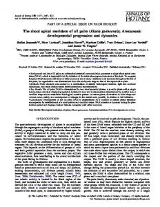
eral cases, notably in the case of the somaclonal variant mantled induced during micropropagation in vitro (Corley et al., 1986), but also in seed-derived palms (Hartley, 1988). In mantled palms, the staminodes of the pistillate flowers and the stamens of staminate flowers develop as carpel-like structures (Figs. 44–47). The organ of the innermost whorl does not undergo a homeotic transformation; thus the carpel is conserved in the pistillate flower, and the gynoecium remains vestigial in staminate flowers (Fig. 47). In the case of the mantled pistillate flower, floral triad initiation is similar to that in normal flowers, and the perianth organs are not visibly altered (Fig. 48). As in normal palms, the reproductive organs of pistillate flowers are initiated at about leaf stage 115. No difference is visible at this stage, organs in the third whorl being initiated in the same way as for normal pistillate flowers (Fig. 49). However, at the next stage observed, organs resembling carpels (Fig. 50) are developing in the mantled pistillate flowers in place of staminodes. Ovules do not develop in these structures, which are thus sterile. In the case of the mantled staminate flower, the floral meristem develops in a similar way to the normal flower, as does the perianth (Fig. 51). At this stage, it is not possible to distinguish the identity of the organ in the androecium (Fig. 52). During their subsequent elongation, the organs of the androecium develop the central vascularization characteristic of carpels (Fig. 53), as opposed to that of the peripheral vascularization of the stamens. As for the mantled pistillate flower, ovules do not develop in the carpelloid structures of mantled staminate flowers. In some cases, not all stamens are observed to be transformed into carpels. Interestingly, the homeotic transformation of stamens to carpel-like structures may be observed not only in staminate flowers of the male inflorescence, but also in the accompanying staminate flowers of floral triads on the female inflorescence (data not shown). DISCUSSION Studies of the reproductive development of palm species are often hampered by the morphological characteristics that typify the family, notably the large size of the adult plant and the deep embedding of the inflorescence in the palm crown during its development. Consequently, relatively few developmental studies have covered the entire sequence of events during inflorescence and flower organogenesis. We report here the first comprehensive analysis at the cytological level of inflorescence and flower development in the oil palm. Using scanning electron micrographs and sectioned material, we have identified a series of morphological landmarks that define each stage of reproductive development. Inflorescence development—A summary of the time course of reproductive development in oil palm, as revealed in this study, is shown in Fig. 1B. Inflorescence initiation was observed in the axils of leaves only a few plastochrons after their separation from the shoot apical meristem. A small group of cells is visible along the base of the leaf at this stage. It is not known if these cells will give rise directly to the rachis or if they are the precursor cells of the inflorescence meristem. It will be interesting to study the development of these cells more closely in order to identify individual cell lineages. This may be possible with the aid of in situ hybridization in order to detect mRNA transcripts of genes which regulate the shoot
OF
BOTANY
[Vol. 92
apical meristem. Our results show that the longest phase in oil palm reproductive development is that involving the development of the inflorescence meristem (Fig. 1B). For about 20 months, the inflorescence meristem expands and gives rise to rachilla bracts, then the rachilla meristem (Fig. 1B). The subsequent development of staminate and pistillate flowers takes place more rapidly during a further 10 months. Our results show that oil palm reproductive development consists of several growth phases, which are characterized by slow and fast growth rates. The two distinct types of growth rates are associated with two different cell types. The first is characterized by a small nucleus and a low nucleus to cytoplasm ratio. These cells are prevalent in the slow growth phases, when they allow the conservation of the meristematic potential of the cells. The second type of cell is characterized by a large nucleus and a higher nucleus to cytoplasm ratio, suggesting a decondensation of the chromatin. These cells are associated with the reactivation of meristematic potential and thus the development of the inflorescence and floral organs. A key feature highlighted in this study is the degree of protection afforded to the inflorescence, which is initially protected within the crown, especially by its subtending leaf. The expanding inflorescence axes are themselves protected by the prophyll and a peduncular bract, which enlarge to become massive and lignified. Direct protection of the reproductive organs is provided by the imbricated bracteoles, sepals, and petals. The ovules are additionally protected by the ovary walls, which are lignified and contain some polyphenol and idioblasts of calcium oxalate crystals in the apical parts (Uhl and Moore, 1973). The extreme protection afforded to the reproductive apparatus is likely to constitute an adaptation to the long period of development of the inflorescence. In more derived Arecoideae such as Geonoma interrupta, the perianth and the staminodial tube are considered to constitute the most important structural protective devices for the gynoecium (Stauffer et al., 2002). Even though palm inflorescences are often elaborate and massive structures, relatively few terms are needed to describe them, in that they are largely made up of repeating units, each with the same construction, and with gradual but progressive changes along any given axis (Tomlinson and Moore, 1968; Moore and Uhl, 1982). Palm inflorescences are panicles with up to six orders of branching, each branch subtended by a bract. As with many other palm species, the oil palm inflorescence has a reduction in the number of its branch orders compared with the basic inflorescence unit described by Tomlinson (1990), reflecting morphological changes during the evolutionary diversification within Arecaceae. The oil palm inflorescence may be considered to have a fundamental two-order structure on the basis of its two distinct types of meristem. The first order of complexity is governed by the rachis meristem, which produces the prophyll and peduncular bract and afterwards the rachilla bracts. The structure and form of the rachis meristem is always the same, rachilla bracts being continuously produced. However, it is interesting to note that during the development of this unit, the functioning of the rachis meristem changes in terms of the organ phyllotaxies that it produces (opposite in the case of the prophyll and peduncular bract to spiral in the case of the rachillae). The second order of complexity is governed by the rachilla meristem formed in the axil of the rachilla bract. The rachilla meristem has the same form and structure as the rachis meristem and has the same form in both male and female inflorescences. However,
November 2005]
ADAM
ET AL.—REPRODUCTIVE DEVELOPMENT IN
ELAEIS GUINEENSIS
1849
Figs. 44–53. Micrographs showing whorl structure of developing and mature oil palm flowers (light micrographs unless otherwise stated). 44. Side view of normal pistillate flower revealed by SEM. 45–53. Mantled flowers. 45. Side view of mantled pistillate flower revealed by SEM. 46. Upper view of mantled staminate flower revealed by SEM. 47. Longitudinal section (LS) of staminate mantled flowers with stamen and pseudocarpels. The fourth whorl is not developed. 48. LS of pistillate mantled flower with sepals developed. 49. LS of pistillate mantled flower at initiation of inner whorls. No difference with the normal pistillate flower is visible. 50. Transverse section showing supernumerary carpels of mantled pistillate flower. 51. TS of staminate mantled flower with perianth organs developed. 52. LS of staminate mantled flower at stage of initiation of reproductive organs. 53. Transverse view of staminate mantled flower with pseudocarpels developed. Abbreviations: c, carpel; fm, floral meristem; pc, pseudocarpel; pe, petal; rg, rudimentary gynoecium; s, sepals; sc, surpernumerary carpel; st, stamen; stap, staminodia primordia; stp, stamen primordium. Bar 5 0.1 mm for LM, 5 1 mm for SEM.
1850
AMERICAN JOURNAL
the divergent modes of functioning of the male and female rachilla meristems is evident in the differences in the number and form of rachilla bracts and floral meristems they produce. Sexual specialization—Female inflorescences of oil palm display floral triads with a central pistillate and two lateral staminate flowers. The triad characterizes the subfamily Arecoideae in which it represents the ultimate unit of the rachilla (Uhl and Dransfield, 1987). Floral triads have been reported within 138 of the 212 palm genera listed by Moore (1973). In oil palm the floral triads are borne in groups consisting of lateral staminate flowers and a central pistillate flower with three associated bracteoles. Elaeis guineensis displays, like species of the genus Ptychosperma (Uhl, 1976), the initiation of the two staminate flowers first, followed by the pistillate flower. It has been suggested that the functional staminate flowers of the male oil palm inflorescence are equivalent to the first monoaxial part of the female inflorescence triad (Van Heel et al., 1987). Our observations support the conclusion that the triad is a sympodial flower cluster originating from a single meristem. It is interesting to note that the accompanying staminate flower is borne upon a pedicel, whereas the functional staminate flower is sessile. The floral triad is considered to represent the ancestral configuration of the Cocoseae (Tomlinson, 1990). Within this tribe, a number of different modes of inflorescence organization have been characterized. Of particular interest is Beccariophoenix madagascariensis, which belongs to a genus presumed to constitute the least specialized group within the tribe (Uhl, 1988). Inflorescences are of mixed sex, with triads of flowers nearly throughout except for a few pairs of staminate flowers at the distal end. A second and more specialized member of the Cocoseae tribe characterized by Uhl (1988) was Polyandrococos pectinatus. The latter species has a more marked spatial separation of staminate flowers (at the distal end of the inflorescence) and floral triads (at the basal end). Polyandrococos pectinatus is protandrous, thus enabling allogamy. A similar situation is seen in coconut (Cocos nucifera; Tomlinson, 1990), in which staminate and pistillate flower function are separated both spatially and temporally within a mixed sex inflorescence. Oil palm might be considered to have evolved further than the aforementioned Cocoseae species in that its functional dioecy is achieved by the separate production of single sex inflorescences. Nevertheless, it is interesting to note the presence of the nonfunctional accompanying staminate flowers on the female inflorescence of E. guineensis. One possible explanation of their presence is that they represent an intermediate stage in an evolutionary progression involving the loss of the ancestral floral triads to give fully unisexual inflorescences. This evolutionary tendency appears to occur in several palm groups (Moore and Uhl, 1982). Ontogenesis and structure of male and female reproductive organs—In common with about 30% of all angiosperms, oil palm produces unisexual (single sex) flowers rather than hermaphrodite ones (Richards, 1986). The ontogenesis of unisexual flowers may be classified into two general patterns corresponding to different plant species. In the first group, both stamens and gynoecial primordia are initiated and then arrested at different stages of development. This mechanism is observed in the Leguminosae (Tucker, 1992), in Asparagus officinalis (Bracale et al., 1991), Silene sp. (Ye et al., 1991), Zea mays and Tripsacum sp. (De Long et al., 1993; Dellaporta and
OF
BOTANY
[Vol. 92
Calderon-Urrea, 1993; Li et al., 1997). In staminate flowers of A. officinalis, which is dioecious, ovule development in the sterile gynoecium resembles that of the fertile gynoecium of pistillate flowers until degeneration starts in the cells of the nucellus and integuments. Anther development is initially the same in pistillate as in staminate flowers, but the tapetum degenerates precociously followed by the abortion of the microspore mother cells (Lazarte and Palser, 1979). In the second group of species that produce unisexual flowers, one or other of the sex organs is never initiated. For example, in staminate flowers of Cannabis sativa (Mohan Ram and Nath, 1964) and in both staminate and pistillate flowers of the dioecious species Mercurialis annua (Durand and Durand, 1991), Spinacia oleracea (Sherry et al., 1993), Rumex acetosella (Ainsworth et al., 1995), and Philondendron acutatum (Boubes and Barabe´, 1996), either the gynoecium or the androecium is initiated but not both. Amongst those species that produce unisexual flowers, E. guineensis falls into the first group mentioned; nevertheless, the primordia of reproductive organs lose their meristematic potential very early just after their initiation. As with date palm (De Mason et al., 1982), the early development of staminate and pistillate flowers of oil palm is identical up to and including the stage of staminode/stamen initiation, divergence being observable for the first time when carpel initiation occurs. The mantled homeotic variant—Our results provide the first clear demonstration at the cytological level that the mantled phenotype involves a homeotic transformation of the floral organs of the sterile or fertile gynoecium. Homeosis may be defined as the complete or partial replacement of one part of an organism with another (Lehmann and Sattler, 1992). The variant phenotype mantled is regularly observed amongst clonal palms (Corley et al., 1986). In mantled oil palms, staminodes and stamens of pistillate and functional staminate flowers develop respectively as pseudocarpels. In severe cases, the flowers are sterile, although lesser-affected female flowers may be fertilized to give mantled fruits. Although some observations of the mantled phenotype have been reported (Corley et al., 1986), no studies at the histological and cellular level necessary to obtain an in-depth understanding of this homeotic variant have been described until now. Phenotypes resembling that of mantled palms have been described in model plants such as Arabidopsis thaliana or Antirrhinum majus. They are most frequently due to a disruption of the activity of B class genes of the ABC flower development model (Coen and Meyerowitz, 1991; Zahn et al., 2005) whereby petals are transformed into sepals and stamens are replaced by carpels. Given the similar appearance of sepals and petals in oil palm, it is difficult to determine whether or not the identity of whorl 2 is affected by the mantled abnormality. Nevertheless, it is interesting to note that the expression of a key homeotic gene distinguishes whorls 1 and 2 in Asparagus officinalis (Park et al., 2003), a species which displays, like oil palm, morphologically similar sepals and petals. Thus, although the mantled abnormality does not involve any major changes to whorl 2 at the morphological level, there may perhaps be molecular alterations. Ovules are not found in the developed pseudocarpels. This suggests that the homeotic C and D functions found to specify carpel and ovule identity, respectively, in Petunia hybrida (Angenent and Colombo, 1996) might also be independent in oil palm. Some other naturally occurring floral abnormalities of a similar nature, named diwakkawakka (Hartley,
November 2005]
ADAM
ET AL.—REPRODUCTIVE DEVELOPMENT IN
1988) and coronado (unpublished data in our laboratory) have been observed. In diwakkawakka palms, staminate flowers are normal while pistillate flowers have the same phenotype as those of mantled palms. This character appears to be inherited monofactorially and to be dominant. In coronado flowers, both pistillate and functional staminate flowers are affected in the same way as for the mantled abnormality. The origin of the coronado phenotype, which has been observed in both E. guineensis and E. oleifera, is unknown. It will clearly be of interest to investigate the molecular causes of these three distinct abnormalities. Studies of palm inflorescence ontogenesis of the type described here are aimed at improving our understanding of the function and evolution of the flowering apparatus within and beyond the Arecaceae. This approach should help to provide a meaningful classification of the reproductive development of palm species as we incorporate our knowledge about the growth, development, and functioning of inflorescences and flower structures. These reference observations will be useful in studies of the molecular determination of normal and variant modes of flower development in oil palm. They also provide a useful starting point for studies on the functioning of the vegetative shoot apical meristem and the mechanisms by which it gives rise to a regular succession of inflorescence meristems throughout the life of the plant. LITERATURE CITED AINSWORTH, C., S. CROSSLEY, V. BUCHANAN-WOLLASTON, M. THANGAVELU, AND J. PARKER. 1995. Male and female flower of the dioecious plant sorrel show different patters of MADS box gene expression. Plant Cell 7: 1583–1598. ANGENENT, G. C., AND L. COLOMBO. 1996. Molecular control of ovule development. Trends in Plant Science 1: 228–232. BARFOD, A. S., AND N. W. UHL. 2001. Floral development in Aphandra (Arecaceae). American Journal of Botany 88: 185–195. BEIRNAERT, A. 1935. Introduction a` la biologie florale du palmier a` huile (Elaeis guineensis Jacquin). Publications de l’Institut National pour l’Etude Agronomique du Congo Belge (I.N.E.A.C.), Brussels, Belgium. BIRADAR, N. V. 1978. An unusual inflorescence in Elaeis guineensis. Principes 22: 115. BOUBES, C., AND D. BARABE´. 1996. De´veloppement de l’inflorescence et des fleurs du Philodendron acutatum Schoot (Araceae). Canadian Journal of Botany 74: 909–918. BRACALE, M., E. CAPORALI, M. G. GALLI, C. LONGO, G. MARZIANILONGO, G. ROSSI, A. SPADA, C. SOAVE, A. FALAVIGNA, F. F. RAFFALDI, E. MAESTRI, F. M. RESTIVO, AND F. TASSI. 1991. Sex determination and differentiation in Asparagus officinalis L. Plant Science 80: 67–77. BUFFARD-MOREL, J., J. L. VERDEIL, AND C. PANNETIER. 1992. Embryogene`se du cocotier (Cocos nucifera L.) a` partir de tissus foliaires: e´tudes histologiques. Canadian Journal of Botany 70: 735–741. COEN, E. S., AND E. M. MEYEROWITZ. 1991. The war of the whorls: genetic interactions controlling flower development. Nature 353: 31–37. CORLEY, R. H. V. 1976a. Germination and seedling growth. In R. H. V. Corley, J. J. Hardon, and B. J. Wood [eds.], Developments in crop science, vol. 1, Oil palm research, 23–36. Elsevier, Amsterdam, Netherlands. CORLEY, R. H. V. 1976b. Inflorescence abortion and sex differentiation. In R. H. V. Corley, J. J. Hardon, and B. J. Wood [eds.], Developments in crop science, vol. 1, Oil palm research, 37–54. Elsevier, Amsterdam, Netherlands. CORLEY, R. H. V., AND B. S. GRAY. 1976. Growth and morphology. In R. H. V. Corley, J. J. Hardon, and B. J. Wood [eds.], Developments in crop, vol. 1, Oil palm research, 7–21. Elsevier, Amsterdam, Netherlands. CORLEY, R. H. V., C. H. LEE, I. H. LAW, AND C. Y. WONG. 1986. Abnormal flower development in oil palm clones. Planter (Kuala Lumpur) 62: 233– 240. DELLAPORTA, S. L., AND A. CALDERON-URREA. 1993. Sex determination in flowering plants. Plant Cell 5: 1241–1251.
ELAEIS GUINEENSIS
1851
DE LONG, A., A. CALDERON-URREA, AND S. L. DELLAPORTA. 1993. Sex determination gene TASSELSEED2 of maize encodes a short chain alcohol dehydrogenase required for stage specific floral organ abortion. Cell 74: 757–768. DE MASON, D. A., K. W. STOLTE, AND B. TISSERAT. 1982. Floral development in Phoenix dactylifera. Canadian Journal of Botany 60: 1439– 1446. DRANSFIELD, J., AND N. W. UHL. 1998. Palmae. In K. Kubitzki [ed.], Families and genera of vascular plants, flowering plants: monocotyledons, vol. 4, 306–389. Springer-Verlag, Berlin, Germany. DRANSFIELD, J., N. W. UHL, C. B. ASMUSSEN, W. J. BAKER, M. M. HARLEY, AND C. E. LEWIS. In press. An outline of a new phylogenetic classification of the palm family, Arecaceae. Kew Bulletin. TSO Publications, Norwich, UK. DURAND, B., AND R. DURAND. 1991. Sex determination and reproductive organ differentiation in Mercurialis. Plant Science 80: 49–65. FISHER, D. B. 1968. Protein staining of ribboned epon sections for light microscopy. Histochemie 16: 92–96. GENTY, P., A. GARZON, F. LUCCHINI, AND G. DELVARE. 1986. Polinizacio´n entomofila de la palma africana en America tropical. Ole´agineux 41: 99– 112. HARTLEY, C. W. S. 1988. The oil palm, 3rd ed. Longman Agriculture Series. Longman, London, UK. JACQUEMARD , J. C. 1995. Le palmier a` huile. Series le technicien d’agriculture tropicale. Editions Maisonneuve & Larose, Paris, France. LAZARTE, J. E., AND B. F. PALSER. 1979. Morphology, vascular anatomy and embryology of pistillate and staminate flowers of Asparagus officinalis. American Journal of Botany 66: 753–764. LEHMANN, N., AND R. SATTLER. 1992. Irregular floral development in Calla palustris and the concept of homeosis. American Journal of Botany 79: 1145–1157. LI, D., A. BLAKEY, C. DEWALD, AND S. L. DELLAPORTA. 1997. Evidence for a common sex determination mechanism for pistil abortion in maize and its wild relative Tripsacum. Proceedings of the National Academy of Sciences, USA 94: 4217–4222. MOHAN RAM, H., AND R. NATH. 1964. The morphology and embryology of Cannabis sativa Linn. Phytomorphology 14: 414–429. MOORE, H. E. JR. 1973. The major groups of palms and their distribution. Gentes Herbarum 11: 27–141. MOORE, H. E. JR., AND N. W. UHL. 1982. Major trends of evolution in palms. Botanical Reviews 48: 1–69. PARK, J. H., Y. ISHIKAWA, R. YOSHIDA, A. KANNO, AND T. KAMEYA. 2003. Expression of AODEF, a B-functional MADS-box gene, in stamens and inner tepals of the dioecious species Asparagus officinalis L. Plant Molecular Biology 51: 867–875. RICHARDS, A. J. 1986. Plant breeding systems. Georges Allen & Unwin, London, UK. RUDALL, P. J., K. ABRANSON, J. DRANSFIELD, AND W. BAKER. 2003. Floral anatomy in Dypsis (Arecaceae-Areceae): a case of complex synorganization and stamen reduction. Botanical Journal of the Linnean Society 143: 115–133. SHERRY, R. A., K. J. ECKARD, AND E. M. LORD. 1993. Flower development in dioecious Spinacia oleracea (Chenopodiaceae). American Journal of Botany 80: 283–291. STAUFFER, F. W., R. RUTISHAUSER, AND P. K. ENDRESS. 2002. Morphology and development of the female flowers in Geonoma interrupta (Arecaceae). American Journal of Botany 89: 220–229. TOMLINSON, P. B. 1990. The structural biology of palms. Oxford Science Publications, Oxford, UK. TOMLINSON, P. B., AND H. E. MOORE JR. 1968. Inflorescence in Nannorrhops ritchiana (Palmae). Journal of the Arnold Arboretum 49: 16–34. TUCKER, S. C. 1992. The development basis of sexual expression in Ceratonia siliqua (Leguminosae: Caesalpiniodeae: Cassieae). American Journal of Botany 79: 318–327. UHL, N. W. 1976. Developmental studies in Ptychosperma (Palmae). I. The inflorescence and flower cluster. II. The staminate and pistillate flowers. American Journal of Botany 63: 82–109. UHL, N. W. 1988. Floral organogenesis in palms. In P. Leins, S. C. Tucker, and P. K. Endress [eds.], Aspects of floral development, 25–44. J. Cramer, Berlin, Germany. UHL, N. W., AND H. E. MOORE JR. 1973. The protection of pollen and ovules in palms. Principes 17: 111–149.
1852
AMERICAN JOURNAL
UHL, N. W., AND H. E. MOORE, JR. 1977. Centrifugal stamen initiation in phytoelephantoid palms. American Journal of Botany 64: 1152–1161. UHL, N. W., AND H. E. MOORE JR. 1978. The structure of the acervulus, the flower cluster of chamaedoreoid palms. American Journal of Botany 65: 197–204. UHL, N. W., AND H. E. MOORE JR. 1980. Androecial development in six polyandrous genera representing five major groups of palms. Annals of Botany 45: 57–75. UHL, N. W., AND J. DRANSFIELD. 1984. Development of the inflorescence, androecium and gynoecium with reference to palms. In R. A. White and W. C. Dickinson [eds.], Contemporary problems in plant anatomy, 397– 449. Academic Press, New York, New York, USA. UHL, N. W., AND J. DRANSFIELD. 1987. Genera palmarum: a classification of palms based on the work of H. E. Moore Jr., L. H. Bailey Hortorium, and the International Palm Society. Allen Press, Lawrence, Kansas, USA.
OF
BOTANY
[Vol. 92
VAUGHAN, J. G. 1955. The morphology and growth of the vegetative and reproductive apices of Arabidopsis thaliana (L.) Heynh., Capsella bursapastoris (L.) Medics and Anagallis arvensis L. Journal of the Linnaean Society of London (Botany) 55: 279–301. VAN HEEL, W. A., C. J. BREURE, AND T. MENENDEZ. 1987. The early development of inflorescences and flowers of oil palm (Elaeis guineensis Jacq.) seen through the scanning electron microscope. Blumea 32: 67– 78. YE, D., M. OLIVEIRA, Y. WU, P. INSTALLE, S. HINNISDAELS, A. T. TRUONG, S. BROWN, A. MOURAS, AND I. NEGRUTIU. 1991. Sex determination in the dioecious Melandrium: the X/Y chromosome system allows complementary cloning strategy. Science 80: 93–106. ZAHN, L. M., J. LEEBENS-MACK, C. W. DE PAMPHILIS, H. MA, AND G. THEISSEN. 2005. To B or not to B a flower: the role of DEFICIENS and GLOBOSA orthologs in the evolution of the angiosperms. Journal of Heredity 96: 225–240.