DOI:http://dx.doi.org/10.7314/APJCP.2013.14.8.4679 ERCC1 Expression Does Not Predict Response in NSCLC Treated with Platinum Based Chemotherapy
RESEARCH ARTICLE ERCC1 Expression Does Not Predict Survival and Treatment Response in Advanced Stage Non-Small Cell Lung Cancer Cases Treated with Platinum Based Chemotherapy Ozer Ozdemir1, Pelin Ozdemir1, Ali Veral2, Hatice Uluer3, Mustafa Hikmet Ozhan1 Abstract Background: ERCC1 is considered as a promising molecular marker that may predict platinum based chemotherapy response in non small cell lung cancer patients. We therefore investigated whether its expression is indeed associated with clinical outcomes in advanced stage NSCLC patients. Materials and Methods: Pretreatment tumor biopsy samples of 83 stage 3B and 4 non-small cell lung cancer patients treated with platinum based chemotherapy were retrospectively analyzed for immunohistochemical ERCC1 expression. None of the patients received curative surgery or radiotherapy. Results: By calculating H- scores regarding the extent and intensity of immunohistochemical staining of tumor biopsy samples, ERCC1 expression was found to be positive in 50 patients (60.2%). ERCC1 positive and negative groups had no statistically significant differences regarding treatment response, progression free survival and overall survival (respectively p=0.161; p=0.412; p=0.823). Conclusions: In our study we found no association between ERCC1 expression and survival or treatment response. The study has some limitations, such as small sample size and retrospective analysis method. There is need of more knowledge for use of ERCC1 guided chemotherapy regimens in advanced stage NSCLC. Keywords: Non-small cell lung cancer - lung cancer chemotherapy - ERCC1 - molecular markers Asian Pac J Cancer Prev, 14 (8), 4679-4683
Introduction Lung cancer became the leading form of cancer in the second half of twentieth century as a result of increased rate of smoking. Lung cancer is responsible for 12.4% of cancer cases and 17.6% of cancer deaths worldwide (Parkin et al., 2005). Non small cell lung cancer (NSCLC) constitutes approximately 80-85% of lung cancer cases. Overall five year survival rate of non small cell lung cancer was found as 8% in 1960’s, had a slight increase to 15% in 1990’s reaching a plateau (Bepler et al., 2002). Median overall survival according to clinical stage is approximately 59 months for stage 1A and decreases to 4 months for those that have stage 4 disease (Groome et al., 2007). Curative surgery is suitable for only 20-30% of cases (van Zandwijk et al., 2001). Recurrences especially metastatic recurrences, reflecting the systemic nature of the disease, are an important problem despite curative surgery in selected patients. This situation raises the importance of systemic chemotherapy even if acceptable curative surgery is performed. Beside the use of the chemotherapy in adjuvant and neoadjuvant setting in early stages, it also remains as a main treatment modality in metastatic disease. However, the response rate to the systemic chemotherapy
is not similar for all patients. In fact, only minority of patients experience complete response to chemotherapy. Response rates to first line chemotherapy and second line single agent chemotherapy in non small cell lung cancer are about 17-37% and 10%, respectively (Simon et al., 2007). Thus, it’s important to predict which patient would have more benefit from systemic therapy to avoid unnecessary toxicities. Likewise, selection of the most suitable chemotherapy regimen for the individual patient is essential. The usage of the molecular markers as a guide to define more efficient chemotherapy regimen is one of the most challenging research fields. More than 100 candidate genes and gene families for prediction of chemotherapy response in lung cancer patients were detected in in-vitro studies (Sekine et al., 2006). One of these markers is excision repair cross complementing group 1 (ERCC1) protein which is a key enzyme in nucleotide excision repair pathway that functions in removal DNA helix distorting lesions including big adducts induced by platinum based cytotoxic chemotherapy (Ludovic et al., 2006). Low ERCC1 is found to be related with more aggressive but more chemosensitive disease especially in adjuvant chemotherapy trials where target lesions are absent (Breen et al., 2008).
Department of Chest Diseases, 2Department of Pathology, 3Department of Biostatistics, Ege University Faculty of Medicine, Izmir, Turkey *For correspondence:
[email protected] 1
Asian Pacific Journal of Cancer Prevention, Vol 14, 2013
4679
Ozer Ozdemir et al
In this study we aimed to investigate the role of ERCC1 expression on tumor cells, in predicting the outcome to platinum based chemotherapy used in advanced stage nonsmall cell lung cancer patients. Primary outcome measure was overall survival and secondary outcome measures were progression free survival and treatment response.
Materials and Methods The study was conducted in Department of Chest Diseases of Ege University Faculty of Medicine Hospital, Bornova, İzmir. The patients with advanced stage non small cell lung cancer diagnosed with tissue biopsies in between 2004 and 2009 were retrospectively analyzed. Patients treated with platinum based chemotherapy combinations were included into the study. The criteria for exclusion were the use of curative local treatment either surgery or radiotherapy for primary tumors and inadequate tissue sample for immunohistochemical analysis. 83 cases of stage 3B and stage 4 non small cell lung cancer were included in study. All patients had transoral or transnasal fiberoptic bronchoscopic examination (with Olympus CLE-10) after local anesthesia (Lidocaine HCl 2%, maximum 10 ml). Pretreatment bronchoscopic biopsy materials were used for immunohistochemical analyses. Seventy-seven patients had diagnosis with bronchoscopic biopsies. Six patients had their diagnosis via pleural biopsy (1 patient), lymph node excision (1 axillary, 1 mediastinal with anterior mediastinotomy), metastasectomy (1 patient) or thoracotomic biopsy (2 patients). Immunohistochemical analysis Sections with a thickness of five microns were retrieved from the paraffin blocks of tumor biopsy samples and mounted on electrostatically charged slides (X-traTM, Surgipath Medical Industries, Richmond, Illinois, USA). Slides were dried in 60°C for at least two hours. Immunohistochemical staining procedure including deparaffination and extraction of antigen process was done on a fully automated immunohistochemical staining device (Ventana BenchMark XT, Ventana Medical Systems, Tucson, AZ). Biotin free, HRP multimer based antibody detection kit including hydrogen peroxide substrate and 3-3’-diaminobenzidine tetrahydrochloride (DAB) chromogen (ultraView™ Universal DAB Detection Kit, Catalog number 760-500, Ventana Medical Systems, Tucson, AZ) was used for the staining procedure. Only primary antibody Anti-ERCC1 (clone SPM243, Spring Bioscience, 1:50 dilution, catalog number: E5674) was dropped manually and incubated at 37°C for 32 minutes. Dehydration of the sections with opposite staining completed with hematoxylin and blue colored solution, clearing with xylene and closure of lamella was done manually and process was terminated. Tonsillar epithelial cells were used for positive control. The sections prepared for positive control without antibody drop was accepted as negative control. ERCC1 immunoreactivity was assessed with the proportion of stained cells and intensity of staining. The extent of staining was visually graded on a scale of 0-3,
4680
Asian Pacific Journal of Cancer Prevention, Vol 14, 2013
0=none, 1=1-9%, 2=10-49%, 3=≥50%. The intensity of staining was also graded 0-3, 0=no staining, 1=weak staining, 2=moderate staining, 3=strong staining. A semiquantitative H score was obtained by multiplying of grades of intensity and extent of staining. Median value of all H-scores was used as a cut off value for dividing the expression of ERCC1 into high and low. Evaluation All of the patients were evaluated with thorax computed tomography (CT), bone scintigraphy and abdominal ultrasonography (US) and also with positron emission tomography (PET) and cranium magnetic resonance imaging (MRI) as needed. Staging of tumors was done according to the 6th TNM staging system (Greene et al., 2002). Treatment response evaluation was done after 2, 4 and 6th cycle of chemotherapy on the initiative of following physician with thorax CT or PET. As needed additional imaging procedures like bone scintigraphy, cranium MRI and abdominal US was obtained for response evaluation. After the completion of treatment, patients were evaluated with chest radiography and thorax CT every 3 months for 1 year and every 6 months thereafter. Statistical analyses The comparison of clinical and pathological characteristics was done with Fisher exact test and X2 test. Overall survival and progression free survival rates was calculated by using Kaplan-Meier method. The duration from the beginning day of chemotherapy to death was used to define overall survival. Similarly, duration from the beginning day of chemotherapy to date of death or detection of progression was used to define progression free survival. For the analyses of the patients survived, the last day of the follow up period was used. The differences between the survival curves were tested by using the logrank test.
Results The characteristics of patients are seen in Table 1. The total number of patients included in the study was 83 and 70 of them (84.3%) were male. The average age of patients were found as 57.9 (±9.1) years. All patients included in the study had diagnosis of clinical stage 3B and 4 non-small cell lung carcinoma. Lungs were the most common site of metastasis (n=35, 42.1%). Seven patients were free of metastasis at the time of diagnosis, 5 of them had mediastinal and vascular invasion, 2 of them had malignant pleural effusion and 1 had contralateral mediastinal lymph node involvement. At least one comorbidity was present in 37 (45%) patients. Mostly seen comorbidities were hypertension (n=13, 16%), chronic obstructive pulmonary disease (n=11, 13%) and type II diabetes mellitus (n=11, 13%). Two patients had additional malignancy history, one patient was operated due to larynx carcinoma 11 years before diagnosis of lung carcinoma, and one other had an operation due to endometrial carcinoma 18 years ago. Both additional malignancies were accepted as cured without any sign of recurrence.
DOI:http://dx.doi.org/10.7314/APJCP.2013.14.8.4679 ERCC1 Expression Does Not Predict Response in NSCLC Treated with Platinum Based Chemotherapy
Table 1. Characteristics of Patients Characteristics
N (%)
Age (years) 57.93 (medium) (range 32-79) Sex (male/female) 70 (84.3)/13 (15.6) Smoking history Active smokers 42 (50.6) Ex-smokers 27 (32.5) Never smokers 13 (15.6) Histology NSCLC 25 (30.1) Adenocarcinoma 32 (38.6) Squamous cell carcinoma 26 (31.3) Performance score (ECOG) 0 13 (15.6) 1 57 (68.7) 2 12 (14.4) 3 1 (1.2) Clinical stage (according to the 6th TNM) IIIB 7 (8.4) IV 76 (91.6) Metastases Brain 13 (15.6) Lung 35 (42.1) Bone 22 (26.5) Adrenal 15 (18.1) Liver 7 (8.4) Other 6 (7.2) Pattern of recurrence Local recurrence 61 (73.5) Metastatic recurrence 23 (27.7) Treatment response Partial response 43 (51.8) Stable disease 11 (13.3) Progressive disease 23 (27.7)
Table 2. Preferred Chemotherapy Regimens Chemotherapy regimen
N
%
Cisplatin/Carboplatin+Gemcitabine Cisplatin/Carboplatin+Etoposide Cisplatin/Carboplatin+Vinorelbine Cisplatin/Carboplatin+Docetaxel/Paclitaxel Cisplatin+Pemetrexed Mitomycin+Ifosfamide+Cisplatin
30 36.1 18 21.7 15 18.1 16 19.3 3 3.6 1 1.2
Table 3. Patient Characteristics According to the ERCC1 Status Characteristics Age (medium) Male Smoking Co-morbidity Weight loss ECOG 0-1 Partial response Adenocarcinoma Squamous cell carcinoma NSCLC A)
ERCC1 (+) ERCC1 (-) (n=50) (n=33) 58.72 43 (86%) 43 (87.8%) 25 (50%) 11 (23.9%) 43 (86%) 29 (63%) 16 (32%) 14 (28%) 20 (40%)
56.73 27 (81.8%) 26 (78.7%) 12 (36.4%) 12 (38.7%) 27 (81.8%) 14 (40.3%) 16 (48.5%) 11 (33.3%) 6 (18.2%)
p value 0.382 0.759 0.304 0.263 0.207 0.657 0.161 0.1
B)
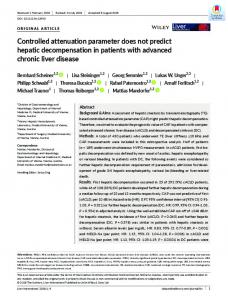
Figure 1. Progression Free Survival and Overall Survival Curves of Patients According to the ERCC1 Status are Seen on (A) and (B) All patients were treated with platinum based chemotherapy. None of the patients were treated with surgery or curative radiotherapy. Sixty one patients (73.5%)
had 4 or more cycles of chemotherapy. Dose reduction was applied in 13 patients (15.6%) because of infectious complications and toxic effects of chemotherapy. The most common choice of first line chemotherapeutic regimen was cisplatin/carboplatin and gemcitabine combination (n=30, 36.1%). Preferred chemotherapy regimens are seen in Table 2. In 46 patients (55%) second line chemotherapy was used after recurrence, mostly preferred regimen was single agent docetaxel (n=27, 32.5%). In 51 patients (61.4%) palliative radiotherapy was applied. Most common indication of palliative radiotherapy was cranial metastasis (n=24, 27%) and bone metastasis in 16 (19.3%) of patients. And another 19.3% (n=16) had palliative radiotherapy to thorax because of central airway obstruction. Treatment responses were evaluated after 2, 4 or 6th cycle of chemotherapy according to the opinion of physician in charge, mainly by thorax computed tomography and in minority of patients by positron emission tomography. In 36 patients (43%) response evaluation was done after 2 cycles of chemotherapy, whereas in 41% of patients it was done after four cycles of chemotherapy. None of the patients had complete response and 43 patients (51.8%) partial response was seen. Stable disease was found in 13.3% of the patients and in 27.7% of the patients (n=23) progressive disease was detected. The recurrence was a metastatic disease in 27.7% of patients. The ERCC1 expression was found positive in 50 patients (60.2%). ERCC1 positive and negative patients had no statistically significant difference according to the age range, sex, smoking status, comorbidities, weight loss, ECOG performance score, histopathological diagnosis, cancer stage and chemotherapy dosage. Clinical characteristics of patients according to the ERCC1 status are summarized in Table 3. There was no statistically significant difference between both groups of patients according to treatment response. Both groups were similar in partial response, stable disease and progressive disease. Overall mean time to progression was 5.79 months. At the time of the analysis of study, only 5 patients were alive. Average overall survival time was 13,12 months. Overall survival and progression free survival curves of patients are seen. As a primary outcome of study, overall survival of patients according to the ERCC1 status are compared by Kaplan Meier method and no statistically significant difference was detected (p=0.823). Also progression free survival of both groups did not differ significantly (p=0.412) (Figure 1). Other factors were evaluated for their effects on prognosis of patients. Patients with weight loss and ECOG performance score 2 and 3 patients had significantly worse prognosis (respectively p=0.006 and p=0.032).
Discussion In this study, we evaluated the role of ERCC1 expression of tumor cells on progression free survival and overall survival in advanced stage non-small cell lung cancer patients treated with platinum based chemotherapy combinations. No significant difference was detected
Asian Pacific Journal of Cancer Prevention, Vol 14, 2013
4681
Ozer Ozdemir et al
regarding two endpoints as well as treatment response. ERCC1 is one of the most promising molecular markers for individualized chemotherapy in lung cancer patients. Largest study population was in a cohort of adjuvant chemotherapy trial in patients with stage 1A3B non small cell lung cancer patients (Olaussen et al., 2006). High expression of ERCC1 was related with higher survival rates in patients treated with curative surgery alone, whereas adjuvant chemotherapy, over observation, improved survival in ERCC1 negative tumors. In advanced stage non-small cell lung cancer patients treated with platinum based palliative chemotherapy, studies also suggest that low ERCC1 correlates with better survival, although robust clinical evidence does not exist (Lord et al., 2002; Ceppi et al., 2006; Lee et al., 2009). Two recent metaanalyses support the role of ERCC1 as a guide to individualized chemotherapy. Roth et al, analyzed 8 studies in their metaanalysis and both overall survival and treatment response were significantly better in patients with low ERCC1 tumors (HR=2.04; 95% CI: 1.48, 2.80 for overall survival, and RR=0.80; 95% CI: 0.66, 0.98 for treatment response) (Roth et al., 2011). Chen et al. (2010) investigated 11 studies in their metaanalysis, and also found significantly different treatment response and overall survival, favoring ERCC1 negative tumors (Chen et al., 2010). Majority of the studies in these metaanalyses were from Asia, and in Caucasian subgroup of patients the difference was lower. In contrast to these findings, in a recent study Gao et al. (2011) randomized 190 patients of advanced nonsmall cell lung cancer into either individualized therapy group or standard therapy group at a ratio of 2:1, according to their ERCC1 status (Gao et al., 2011). Although median survival time was better in the individualized study group, there was not any significant difference of overall survival, treatment response or time to progression according to ERCC1 status. Also, Bepler et al evaluated individualized chemotherapy in 275 advanced stage lung cancer patients using ERCC1 and RRM1 in their prospective study (Bepler et al., 2013). They found no significant difference concerning overall and progression free survival between groups, only the patients with low levels of both proteins showed better progression free survival, compared with the control group. So far, the studies examining the predictive and prognostic role of ERCC1 expression in advanced stage non-small cell lung cancer patients have conflicting results. Our study showed no association between ERCC1 expression and survival or treatment response. This may have several reasons. Firstly, ERCC1 may not be the only marker that is thought to be related with survival. Many other markers like RRM1, BRCA1, thioredoxin, p53 and β-tubulin are being investigated in studies in decision of individualized chemotherapy and chemosensitivity (Bepler et al., 2008). Another point is that nucleotide excision repair pathways where ERCC1 has an essential role, may not be the only mechanism of platinum resistance (Martin et al., 2008). Other mechanisms like mismatch repair defects may be responsible, either through inherited defects or epigenetic silencing of related genes (Fink et al., 1997).
4682
Asian Pacific Journal of Cancer Prevention, Vol 14, 2013
Also other mechanisms of platinum resistance are BRCA1 and BRCA2 tumor suppressor genes, ATP-depended pump systems and detoxification by glutathione/glutathione acetyl transferases (Rosell et al., 2003). Also, in advanced stage patients, primary tumoral lesions and metastases may have different genetic profiles. In our study, tumoral biopsies were taken from primary lesions except three patients. In NSCLC patients it was shown that primary tumor and metastatic sites may have different epithelial growth factor receptor (EGFR) mutations (Italiano et al., 2006). Gomez-Roca et al. (2009) investigated the differential expression of EGFR, ERCC1, Ki67 and vascular endothelial growth factor receptor (VEGF) in 49 NSCLC patients (Gomez-Roca et al., 2009). Sixty-one percent of patients had metachronous metastases and one third of patients were treated with adjuvant chemotherapy before occurrence of metastases. There were 41% discordance between primary and metastatic sites regarding ERCC1 status towards a higher expression in metastases, especially in brain and adrenals. Metastases are important determinants of mortality in stage 4 NSCLC patients and data derived from primary sites regarding tumor biology may lead to misclassification of patients. These differences between primary and metastatic sites suggest two probabilities; the differential expression of biomarkers in tumor itself and biological change in the progress of metastatic disease. Diagnosis of patients is mostly obtained by examination of the small tissue samples derived from bronchoscopic procedures. In the study of Taillade et al, preoperative tissue biopsies and surgical samples had a discordance rate of 9% regarding ERCC1 positivity and this ratio was higher for expression of EGFR and pAkt (Taillade et al., 2007). So, it should be reminded that tumors may be heterogeneous in structure and small biopsy samples may be misleading. Total excision of tumors in adjuvant chemotherapy studies provides more accurate measure of ERCC1 expression. Whereas in metastatic disease, these findings raise the question about the requirement of metastatic tissue sampling in molecular analysis directed individualized therapy and the inadequacy of biopsy procedures. Another point, beside intratumoral and metastatic heterogeneity, is the ethnical differences in expression of ERCC1 expression. To our knowledge, there was not any study published from Turkey regarding this topic. As seen in metaanalyses of the role of ERCC1 in advanced stage lung cancer, most of the knowledge comes from Asian population. Another important challenge of this area of research is the methodology used to detect immunohistochemical staining and determination of the cut off value of ERCC1 positivity. In our study we used the same visual grading scale of intensity and extend of staining used by Lee et al. (2009) and cut off value was derived from calculated H-scores (Lee et al., 2009). This method makes it harder to compare results of different studies, as a consistent evaluation method does not exist. ERCC1 expression level was first measured by mRNA analyses using reference genes like β-actin and 18SrRNA. But these analyses are relatively complicated and not
None
Remission
Persistence or recurrence
Newly diagnosed with treatment
Newly diagnosed without treatment
DOI:http://dx.doi.org/10.7314/APJCP.2013.14.8.4679 ERCC1 Expression Does Not Predict Response in NSCLC Treated with Platinum Based Chemotherapy Cancer Res, 57, 1841-5. feasible in every center. Immunohistochemical analyses Friboulet L, Olaussen KA, Pignon JP, et al (2013). ERCC1 are more suitable for large population studies and more isoform expression and DNA repair in non-small-cell lung easily applied. It was also the preferred way of analysis cancer. N Engl J Med, 368, 1101-10. in our study. But, is that true, that immunohistochemical Gao Z, Han B, Shen J, et al (2011). ERCC1 protein as a guide analysis of protein expression really reflects the function for individualized chemotherapy of late stage advanced non of nucleotide excision repair pathway and correlates with small cell lung cancer. Exp Ther Med, 2, 811-15. mRNA analyses? Zheng et al. (2007) investigated the Gomez-Roca C, Raynaud CM, Penault-Llorca F, et al (2009). prognostic role of ERCC1 and RRM1 in 187 NSCLC Differential expression of biomarkers in primary non-small patients (Zheng et al., 2007). They measured ERCC1 and cell lung cancer and metastatic sites. J Thorac Oncol, 4, 1212-20. RRM1 protein levels by automated quantitative analysis Greene FL, Page DL, Fleming ID, et al (2002). AJCC Cancer 100.0 100.0 and also by mRNA analysis in 44 patients. RRM1 levels Staging Manual. 6th ed. Springer: New York. correlated well between both techniques (RHO=0.41; 6.3 10.1 20.3 Groome PA, Bolejack V, Crowley JJ, et al (2007). The IASLC p=0.004) but this was not true for ERCC1 expression Lung Cancer Staging Project: validation of the proposals (RHO=0.1; p>0.30). Beside that in their recently published 25.0and consequent75.0 30.0 75.0 for revision of the T, N, and M descriptors papers Friboulet et al. (2013) observed that none of stage groupings in the forthcoming (7th) edition of the TNM the antibodies used for immunohistochemical analysis classification of46.8 malignant tumours. J Thorac Oncol, 2, 56.3 of ERCC1 could distinguish functional isoforms, and 694-705. 54.2 concluded that currently available methodology is of50.0Italiano A, Burel-Vandenbos F, Otto J (2006).31.3 Comparison of the50.0 30.0 epidermal growth factor receptor gene and protein in primary limited value for therapeutic guidance. nonsmall-cell lung cancer and metastatic sites: implications Our study has several limitations. It is a single-center retrospective study, with a limited number of cases. This25.0 for treatment with EGFR inhibitors. Ann Oncol, 17, 981-5.25.0 Lee HW, Choi YW, Han JH, et al (2009). Expression of excision decreases the power of study. All patients were treated 38.0 repair31.3 cross-complementation group 1 protein 31.3 predicts poor 30.0 with platinum based regimens, but these regimens were 23.7 cell lung cancer patients outcome in advanced non-small various. As mentioned before, analyses depend on the treated with platinum based doublet chemotherapy. Lung 0 small bronchoscopic biopsy samples and may not reflect 0 Cancer, 65, 377-82. the whole characteristics of tumor and metastases. Lord RV, Brabender J, Gandara D, et al (2002). Low ERCC1 In conclusion, the need for more multi-centered expression correlates with prolonged survival after cisplatin plus gemcitabine chemotherapy in non-small cell lung prospective studies investigating molecular analysis of cancer. Clin Cancer Res, 8, 2286-91. the tumor tissue to guide treatment options, especially in Ludovic C, Gillet J, Scharer OD (2006). Molecular mechanisms advanced stage lung cancer patients is still present. The of mammalian global genome nucleotide excision repair. intratumoral and metastatic heterogeneity should be taken Chem Rev, 106, 253-76. into concern for the evaluation of treatment response and Martin LP, Hamilton TC, Schilder RJ (2008). Platinum survival in metastatic disease, differently from adjuvant resistance: The role of DNA repair pathways. Clin Cancer treatment approaches. Res, 14, 1291-5. Olaussen KA, Dunant A, Fouret P, et al (2006). DNA repair by ERCC1 in non-small cell lung cancer and cisplatin-based References adjuvant chemotherapy. N Engl J Med, 355, 983-91. Bepler G, Gautam A, McIntyre LM, et al (2002). Prognostic Parkin DM, Bray F, Ferlay J, Pisani P (2005). Global cancer significance of molecular genetic aberrations on chromosome statistics, 2002. CA Cancer J Clin, 55, 74-108. segment 11p15.5 in non-small-cell lung cancer. J Clin Oncol, Rosell R, Taron M, Barnadas A, et al (2003). Nucleotide excision 20, 1353-60. repair pathways involved in cisplatin resistance in non-smallBepler G, Begum M, Simon GR (2008). Molecular analysiscell lung cancer. Cancer Control, 10, 297-305. based treatment strategies for non-small cell lung cancer. Roth JA, Carlson JJ (2011). The prognostic role of ERCC1 in Cancer Control, 15, 130-9. advanced non-small cell lung cancer: a systematic review Bepler G, Williams C, Schell MJ, et al (2013). Randomized and meta-analysis. Clin Lung Cancer, 12, 393-401. international phase III trial of ERCC1 and RRM1 expressionSekine I, Minna JD, Nishio K, Tamura T, Saijo N (2006). A based chemotherapy versus gemcitabine/carboplatin in literature review of molecular markers predictive of clinical advanced non-small-cell lung cancer. J Clin Oncol, 31, response to cytotoxic chemotherapy in patients with lung 2404-12. cancer. J Thorac Oncol, 1, 31-7. Breen D, Barlési F (2008). The place of excision repair cross Simon GR, Ismail-Khan R, Bepler G (2007). Nuclear excision complementation 1 (ERCC1) in surgically treated non-small repair-based personalized therapy for non-small cell lung cell lung cancer. Eur J Cardiothorac Surg, 33, 805-11. cancer: From hypothesis to reality. Int J Biochem & Cell Ceppi P, Volante M, Novello S, et al (2006). ERCC1 and RRM1 Biology, 39, 1318-28. gene expressions but not EGFR are predictive of shorter Taillade L, Penault-Llorca F, Boulet T, et al (2007). survival in advanced non-small-cell lung cancer treated with Immunohistochemichal expression of biomarkers: a cisplatin and gemcitabine. Ann Oncol, 17, 1818-25. comparative study between diagnostic bronchial biopsies Chen S, Zhang J, Wang R, Luo X, Chen H (2010). The platinum and surgical specimens of non-small-cell lung cancer. Ann based treatments for advanced nonsmall cell lung cancer, is Oncol, 18, 1043-50. low/negative ERCC1 expression beter than high/positive van Zandwijk N (2001). Neoadjuvant strategies for non-small ERCC1 expression? A metaanalysis. Lung Cancer, 70, cell lung cancer. Lung Cancer, 34, 145-50. 63-70. Zheng Z, Chen T, Li X, et al (2007). DNA synthesis and repair Fink D, Zheng H, Nebel S (1997). In vitro and in vivo resistance genes RRM1 and ERCC1 in lung cancer. N Engl J Med, to cisplatin in cells that have lost DNA mismatch repair. 356, 800-8. Asian Pacific Journal of Cancer Prevention, Vol 14, 2013
4683
6. 1
56 5
3 31