Journal of Immunological Methods 391 (2013) 146–153
Contents lists available at SciVerse ScienceDirect
Journal of Immunological Methods journal homepage: www.elsevier.com/locate/jim
Research paper
Usage of standardized antigen-presenting cells improves ELISpot performance for complex protein antigens Marcelo A. Navarrete a, b,⁎, Cristina Bertinetti-Lapatki a, Ines Michelfelder a, Hendrik Veelken b a b
Department of Hematology/Oncology, University Medical Center Freiburg, 79106 Freiburg, Germany Department of Hematology, Leiden University Medical Center, 2300 RC Leiden, The Netherlands
a r t i c l e
i n f o
Article history: Received 17 October 2012 Received in revised form 5 February 2013 Accepted 7 March 2013 Available online 16 March 2013 Keywords: ELISpot Dendritic cell Protein Antigen
a b s t r a c t The enzyme-linked immunospot (ELISpot) assay is a widely used method for immune monitoring in cancer immunotherapy trials. In the ELISpot assay, peripheral blood mononuclear cells (PBMC) are stimulated with specific antigens, and cytokines of interest produced by individual cells are detected. In the standard procedure, T cells rely for antigen presentation on other cells like the monocyte/macrophage population present among the PBMC. Whereas oligopeptides can be added directly to the ELISpot assay without the necessity of a preincubation step, protein antigens must be internalized and processed by antigen-presenting cells to accomplish efficient presentation via HLA class I or II. We have studied the impact of sources for different antigen-presenting cell (i.e. PBMC-resident monocytes and monocyte-derived dendritic cells maturated with Poly I:C and PGE-2 based cocktails) on ELISpot assay performance and defined an optimized dendritic cell-based ELISpot protocol. This protocol is suitable for monitoring immune responses directed to protein antigens with higher sensitivity than the standard procedure. © 2013 Elsevier B.V. All rights reserved.
1. Introduction The enzyme-linked immunospot (ELISpot) assay allows the detection of individual cells secreting a cytokine of interest, such as interferon gamma (IFN-γ) (Czerkinsky et al., 1988). This method has shown to be reliable and reproducible for monitoring immune responses in cancer vaccine trials. However, accurate measurement of immune responses can be adversely affected by the assay conditions.
Abbreviations: ELISpot, enzyme-linked immunospot assay; DC, dendritic cells; APC, antigen-presenting cells; PBMC, peripheral blood mononuclear cells ⁎ Corresponding author at: Department of Hematology, C2-R, Leiden University Medical Center, P.O. Box 9600, 2300 RC Leiden, The Netherlands. Tel.: +31 71 5262267; fax: +31 71 5266755. E-mail address:
[email protected] (M.A. Navarrete). 0022-1759/$ – see front matter © 2013 Elsevier B.V. All rights reserved. http://dx.doi.org/10.1016/j.jim.2013.03.004
As a result, recommendations for the standardization of the assay have been developed by joint efforts of the European Cancer Immunotherapy (CIMT) and the U.S. Cancer Vaccine (CVC) consortia (Britten et al., 2008; Janetzki et al., 2008; Singh et al., 2011). Our laboratory has actively participated in the harmonization process proposed by the CIMT monitoring panel. In the referred ELISpot assay, peripheral blood mononuclear cells (PBMC) are stimulated with specific antigens and the cytokines produced by individual cells are trapped on the filters of microtiter wells that were pre-coated with a specific anti-cytokine antibody. However, under these assay conditions, T cells present among the PBMC rely on other cells like the monocyte/macrophage population for antigen presentation. Whereas HLA-binding oligopeptides can be added directly to the ELISPOT assay without the necessity of a pre-incubation step, complex protein antigens must be internalized and processed by professional antigen-presenting cells (APC) for
M.A. Navarrete et al. / Journal of Immunological Methods 391 (2013) 146–153
147
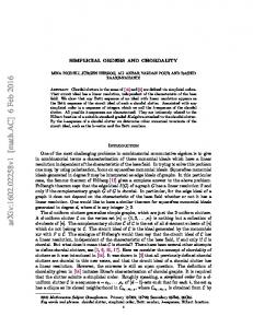
A
Filled shapes: Antigen Stimulation
White shapes: No Antigen Stimulation
B
C
Fig. 1. APC source and ELISpot performance. A) IFN-γ spots in 3 proposed ELISpot protocols. Direct ELISpot included no external APC source. In PGE-2 DC-ELISpot and αtype1 DC-ELISpot, autologous dendritic cells maturated with PGE-2 and TNFα or with a Poly I:C-based cytokine cocktail respectively were introduced as APC source. White symbols represent the background spots in each replicate, black filled symbols are spots upon antigen stimulation. In the left graph analysis of tetanus toxoid reactivity in 5 donors (donors #02, #03, #04, #05 and #06). Right graph shows 4 donors with negative, low, intermediate and high reactivity to CMV-pp65 protein (donors #12, #13, #15, #16). B) Comparison of tetanus toxoid induced IFN-γ spots/background ratio in 5 donors with similar precursor frequencies (donors #02, #03, #04, #05 and #06) at the left and the absolute number of background spots at the right. In the direct ELISpot protocol the background is represented by PBMC stimulated without a specific antigen. For DC-ELISpot protocol is represented by PMBC stimulated with unpulsed dendritic cells. C) Receiving operating characteristics of the proposed ELISpot protocols calculated on antigen-induced spots and background spots.
efficient presentation via HLA class I or II (Scheibenbogen et al., 1997). Therefore, the proportion of APC, predominantly represented by monocytes within the PBMC compartment, can influence T-cell frequencies detected by this assay when
proteins represent the target antigens of interest (Keilholz et al., 2002). Compared with monocytes and macrophages, dendritic cells (DC) are more proficient in antigen-presenting function.
148
M.A. Navarrete et al. / Journal of Immunological Methods 391 (2013) 146–153
Further, DC could improve the sensitivity of detection and the quantitative analyses of antigen-specific T cells of ELISpot (Nehete et al., 2003). Monocytes can be differentiated into mature DC under serum-free conditions by stimulation with appropriate cytokines. However, depending on the selected combination of cytokines, the DC acquire distinctive functional characteristics (Moller et al., 2008). Therefore, we tested the influence of Poly I:C and PGE-2 serum-free DC maturation cocktails on the performance of an ELISpot assay based on autologous dendritic cells to present protein antigens.
2. Materials and methods 2.1. Blood donors Heparinized blood samples were taken from healthy donors and from lymphoma patients enrolled in the Freiburg University idiotype vaccination program (Bertinetti et al., 2006b; Navarrete et al., 2011). Healthy donors were selected for prior immunization against tetanus toxoid (TT) within one to five years (n = 7). For cytomegalovirus reactivity HLAA*02.01 healthy donors were selected on basis of the frequency of pp65 (NLVPMVATV) tetramer positive T-cell and CMV serological status (n = 4). Idiotype vaccination patients received a vaccination schedule as previously reported (n = 20) (Navarrete et al., 2011). PBMC were isolated by density gradient centrifugation using Ficoll-Hypaque 1.077 (Pharmacia, Freiburg, Germany) and cryopreserved in liquid nitrogen until use. The protocol was approved by the Institutional Review Board of Freiburg University, and all probands gave informed consent.
2.2. Generation of antigen-presenting monocyte-derived dendritic cells PBMC were let to adhere to culture flasks for 2 h at 37 °C in RPMI supplemented with 0.5% human serum albumin (HSA). Adherent cells were cultured in serum-free medium (CellGro DC; CellGenix, Germany) with 100 ng/ml GM-CSF and 50 ng/ml interleukin-4 (GellGenix, Germany) for 5 days. At day 4 protein antigen was added in the following concentrations: 40 μg/ml tetanus toxoid (Sigma-Aldrich, Germany), 40 μg/ml recombinant Fab protein (Bertinetti et al., 2006a) or 10 μg/ml pp65-recombinant protein (Miltenyi Biotech, Germany). Final DC maturation was induced with 20 ng/ml tumor necrosis factor-α (TNF-α) and 1 μg/ml prostaglandin E2 for 48 h (“PGE-2 DC”) or with a cocktail including 50 ng/ml TNFα, 20 μg/ml polyI:C, 25 ng/ml IL 1β, 3000 U/ml IFN-α, and 1000 U/ml IFN-γ (“α-type 1DC”) (Mailliard et al., 2004).
2.3. Interferon-γ ELISpot assay Thawed PBMC (4 × 10 5) were stimulated in triplicates with 1 × 10 4 dendritic cells (DC) loaded with TT, recombinant Fab or medium alone in 96-well multiScreen plates (Millipore). Phorbol myristate acetate (PMA; Sigma-Aldrich) stimulation was used as viability control. IFN-γ-secreting cells were detected with streptavidine–alkaline phosphatase and substrate solution (BioRad, USA). Spots were counted semi-automatically with an enzyme-linked immunospot (ELISpot) reader (AID, Strassberg, Germany). The background was defined as the number of spots resulting in the stimulation of PBMC with DC maturated in the absence of a given antigen. A MART-1 HLA-A2 restricted peptide (AAGI GILTV) was introduced as an irrelevant control for HLA-A2 negative samples. The optimal DC/PBMC ratio was established in preliminary experiments. Ratios between 1:20 and 1:40 DC:PBMC showed the best performance. A 1:40 ratio was used in subsequent experiments. Acceptance criteria for analysis were defined as 200 to 400 spots per well in the positive controls and a intra-replicate variation [variance of the triplicate / (median of the triplicate + 1)] lower than 10 (Moodie et al., 2010). Intra and inter-individual variability was measured with the variance mean ratio (VMR) dispersion index. Unless otherwise specified, results were confirmed in two independent experiments. 2.4. Cytokine ELISA Secreted interleukins culture supernatants of DC were measured after maturation. IL-10 and IL-12p70 were quantified using commercially available ELISA kits (OptEIA kits, BD, USA). 2.5. Flow cytometry staining For immunophenotyping, 104 DC were stained with monoclonal antibodies (eBiosciences, USA) for 20 min at 4 °C in the dark. The samples were acquired in a Dako Cyan flow cytometer and .fcs files were exported and analyzed by Flow Jo software. 2.6. Generation of antigen-specific T-cell line PBMC from a TT vaccinated donor were stimulated with autologous DC pulsed with 40 μg/ml of TT. PBMC were cocultured with TT pulsed DC under serum free conditions in X-vivo 15 medium. After 5 days of culture, the medium was supplemented with 60 IU/ml IL2. After 28 days of culture and weekly re-stimulation with TT-pulsed DC and IL-2, the proliferating T-cells were examined for antigen specificity
Fig. 2. Sensitivity and background induction in DC-ELISpot.A) For a donor with weak TT reactivity (donor #08), an anti-tetanus toxoid cell line was established. TT-reactive cells were serially diluted into the original PBMC and tested in direct and PGE-2 DC-ELISpot protocols. The background was defined as the number of spots obtained with fresh PBMC from which the TT-reactive line was established. The grid lines represent the detection limit of the 2 ELISpot protocols.B) Detection of low frequency TT reactivity with PGE-2 DC-ELISpot (donor #09): TT reactivity was tested by direct ELISpot protocol and PGE-2 DC-ELISpot ex-vivo and after 1 week in vitro stimulation with tetanus toxoid loaded DC. MART-1 HLA-2 restricted peptide was used as specificity control.C) Intra-replicate and intra-individual variability of background induction in DC-ELISpot. The upper graph represents the mean and range of spots induced by PGE-2 maturated DC in absence of specific antigen. Each section delimited by a dashed line depicts 3–5 samples taken at different time points from 1 individual. The lower graph indicates the intra-individual dispersion (VMR: variance mean ratio) for each case.
M.A. Navarrete et al. / Journal of Immunological Methods 391 (2013) 146–153
A
1st read-out above detection limit No Ag TT D ir e c t E L IS p o t
DCE L IS p o t
Detection Limits: - DC ELISpot - Direct ELISpot
B
X
X X X
C
X X
X X
X
X X
X
No Antigen Tetanus Toxoid MART-1
149
}
150
M.A. Navarrete et al. / Journal of Immunological Methods 391 (2013) 146–153
with IFN-γ-ELISpot assay, and for proliferative capacity with CFSE proliferation assay as previously described (Angulo and Fulcher, 1998).
induced IFN-γ spots/background ratio compared with the Direct ELISpot. 3.2. Detection limit of proposed ELISpot protocols
2.7. Statistical analysis Appropriate statistical tests were performed using GraphPad Prism 5 (GraphPad Software Inc., San Diego, CA). Differences with a p value b 0.05 were considered statistically significant. Limit of detection of the assays was calculated based on the signal-to-noise ratio method described in the guideline on the validation of analytical procedures reported by the International Conference on Harmonization of Technical Requirements for Registration of Pharmaceuticals for Human Use (ICH) (available at http://www.ich.org). 3. Results 3.1. Impact of antigen-presenting cell source on ELISpot assay performance In order to evaluate the impact of the APC source on ELISpot assay performance for detecting responses to protein antigens, first the response to tetanus toxoid (TT) was assessed in five TT-vaccinated donors with different ELISpot protocols. The standard ELISpot procedure involved direct stimulation of 4 × 10 5 PBMC with TT protein for 24 h. For DC-ELISpot additional sample wells were set up simultaneously with co-cultures consisting of protein pulsed DC and lymphocytes. Two different maturation cocktail were used for DC generation: a PGE-2 based (PGE-2 DC-ELISpot) and a poly I:C based cocktail (αtype1 DC-ELISpot). The background was defined as the number of spots resulting in the stimulation of PBMC with DC maturated in the absence of a given antigen. The number of spots upon stimulation increased when including DC for antigen presentation. This increase is accompanied by a higher background; the median of spots observed without addition of antigen was 8.8 spots/4 × 10 5 PBMC for the direct ELISpot, 18 spots/4 × 105 PBMC for PGE-2 DC-ELISpot, and 98 spots/4 × 105 PBMC for αtype1 DCELISpot (Fig. 1A, B). However, despite this background increase, the DC-ELISpot protocols showed a higher antigeninduced IFN-γ spots/background ratio (Fig. 1B). Receivingoperator curves (ROC) of antigen-induced spots and background spots demonstrate the highest performance of PGE-2 DC-ELISpot with an AUC of 0.98 (Fig. 1C). Secondly, PBMC from HLA-A*0201 donors were screened for the presence of pp65-(NLVPMVATV) specific T-cells by tetramer staining and CMV serology. Four samples with negative, low, intermediate and high precursor frequency (Fig. 1A) were subsequently selected for testing using the direct and PGE-2 DC-ELISpot protocols using recombinant pp65 protein as antigen. As observed for TT protein, PGE-2 DC-ELISpot upon pp65 stimulation shows a higher antigen-
The higher background in proposed DC-ELISpot protocols adversely influences the limit of detection of the assay, which increases from 18–27 spots/4 × 10 5 PBMC in direct ELISpot to 36–54 spots/4 × 10 5 PBMC in PGE-2 DC-ELISpot. However, more efficient stimulatory properties of PGE-2 DC may also result in more reliable activation of antigen-specific T cells, thereby potentially increasing the sensitivity of the assay. To address this question, a T-cell line with specificity for tetanus toxoid was established, and TT-reactive cells were serially diluted into autologous non pre-stimulated PBMC and tested in standard and PGE-2 DC-ELISpot protocols. The background was set at the base-line TT response, i.e. the number of spots obtained with fresh PBMC from which the TT-reactive line was established. The detection limit was recalculated according to this background. For the standard ELISpot protocol the minimal frequency of antigen specific T cells for detecting a response was 5 reactive cells in 4 × 10 5 PBMC vs. 15 reactive cells in 4 × 10 5 PBMC in PGE-2 DC-ELISpot (Fig. 2A). To evaluate whether a previous in vitro stimulation can further enhance the detection of low-frequency antigenspecific cells, PBMC were pre-stimulated in vitro during 1 week with antigen-pulsed DC. The low frequency of TT reactive cells using the direct ELISpot protocol can be detected only after 1 week of pre-stimulation in vitro pre-stimulation, whereas the PGE-2 DC-ELISpot allowed ex vivo enumeration of reactive cells (Fig. 2B). 3.3. Background induction in DC-ELISpot Further experiments were conducted searching for factors that could explain the higher number of spots and background in αtype1 DC-ELISpot. The DC showed no significant differences in the expression of DC membrane markers HLA-DR, CD83 and CD86. Both types of DC showed similar antigen uptake capacity as assessed by immunofluorescence microscopy using FITC-dextran and FITC labeled tetanus toxoid (data not shown). However, culture supernatants of α-type 1 DC showed higher levels of IL-12p70 and IL-10 as assessed by ELISA (p b 0.001 and p = 0.02, respectively). IL-12 can be responsible for inducing IFN-γ release. However, the background stimulation appears to be strictly dependent on cell contact, since the physical separation of DC and PBMC with transwell membranes during ELISpot assay completely abrogated induction of spots (data not shown). Independent DC preparations from individual donors may differ in the background spot induction (Fig. 1). In order to estimate the intra and inter individual variability (i.e. DC preparations from samples taken a different time points and preparation from different individuals respectively) we analyzed the background induction of 80 DC preparations
Fig. 3. PGE-2 DC-ELISpot for monitoring immune responses.Immune monitoring of idiotype-reactive cells in an idiotype vaccination setting in 3 lymphoma patient samples (#103, #115 and #117). Samples were taken before and at several time points after vaccination. PBMC were tested for vaccine-induced IFN-γ releasing cells with PGE-2 DC-ELISpot and direct ELISpot. PGE-2 DC-ELISpot shows increased sensitivity in detecting low frequency antigen specific IFN-γ releasing cells. Arrow heads represents vaccination time points. Bellow each graph an exemplary filter plate well image from different time points is depicted.
M.A. Navarrete et al. / Journal of Immunological Methods 391 (2013) 146–153
151
152
M.A. Navarrete et al. / Journal of Immunological Methods 391 (2013) 146–153
obtained form 20 individuals from our anti-lymphoma idiotype vaccination clinical trial (Navarrete et al., 2011). From each individual DC where generated from blood samples taken at 3–5 different time points, DC where maturated with PGE-2 and TNFα. The PBMC stimulation with unloaded DC showed a median of 14 spot/4 × 10 5 PBMC (range: 0–167). The mean intra-individual dispersion index (VMR) was 9.3 with an inter-individual VMR of 34.5 (Fig. 2C). 3.4. PGE-2 DC-ELISpot for monitoring immune responses The PGE-2 DC-ELISpot protocol was selected as the best candidate for monitoring immune responses in our antilymphoma idiotype vaccination clinical trial and was compared with the standard direct ELISpot protocol in 3 vaccinated patients. Fig. 3 shows examples of immune monitoring of T-cell responses in an idiotype vaccination setting, PGE-2 DC-ELISpot showed increased sensitivity in detecting low frequency antigen specific IFN-γ releasing cells than direct ELISpot. 4. Discussion In order to accurately measure T cell frequencies by ELISpot assay with proteins as antigen format we have adapted the standard ELISpot protocol for optimal antigen processing and presentation of exogenously loaded protein antigens. It has been previously described that a pre-incubation in a tube prior to plating the cells in the ELISpot assay allows protein antigen presentation by monocytes (Schmittel et al., 2001). However, fluctuations in the monocytes percentage in peripheral blood may impact on the estimated T-cell frequency in direct ELISpot protocol (Quiding et al., 1993; Schmittel et al., 2001). To overcome this issue, we optimized the ELISpot protocol to allow the detection of low frequency T cells taking advantage of the immunostimulatory function of DC. The use of DC not only avoids fluctuations in the number of APC but also increases the assay performance. Nevertheless, we observed that the maturation cocktail used for DC generation impacts on the ELISpot results. The use of DC maturated with a prostaglandin-E2 (PGE-2) based cocktail compared with direct stimulation or the use of α-type 1 polarized DC enabled more accurate enumeration of cellular immune response. This difference is mainly explained by a cell contact-dependent background induction. It has been recently shown that the presence of PGE-2 in maturation cocktails inhibit DC ability to attract naive T cells (Muthuswamy et al., 2010). These data may suggest that PGE-2 DC may favor the stimulation of antigen experienced T-cells, explaining a lower background and providing a desirable feature when monitoring immune responses upon vaccination. The immunological analysis in cancer vaccination trials requires the detection of low-frequency reactive T-cells. Traditionally, prior in vitro stimulation steps are included to increase the sensitivity of the assay (Czerkinsky et al., 1988; Scheibenbogen et al., 1997; Moodie et al., 2010). This approach has the same effect in our optimized protocol. However, an in vitro pre-stimulation may selectively expand selected antigen specificities, thereby possibly leading to overestimation of the true T-cell frequency. Since the proposed PGE-2 DC-ELISpot
protocol decreases the minimal frequency of antigen-specific T cells required for detecting a response approximately 3 times under ex vivo assay conditions, it may be a more accurate assay to assess the true frequency of low-frequency antigenspecific T cells compared with standard direct ELISpot. The use of standardized DC may increase the time and work load required for the assay compared with the standard ELISpot protocol in approximately 6 days. However, when the detection of low precursor frequencies is required, the higher sensitivity of the proposed DC-ELISpot protocol allows the ex-vivo detection whereas the standard ELISpot protocol would require pre-stimulation in vitro. Therefore under this circumstances the time and work load required in both protocols would be equivalent. Alternatively to PGE-2 DC as APC source the use of activated monocytes (Singh et al., 2012) or dendritic cell generated with shorter protocols (Ramadan, 2011) may further accelerate the procedure reducing costs and the required amount of patient material. Finally, the PGE-2 DC-ELISpot as developed in this paper was applied for monitoring of immune responses in a phase II idiotype vaccination clinical trial. In this clinical setting, high-performance assays are critically required due to a profound lymphopenia of untreated lymphoma patients (Christopoulos et al., 2008, 2011) and low immunogenicity of tumor-derived immunoglobulin as tumor antigen. Indeed, PGE-2 DC-ELISpot proved superior to the conventional ELISpot protocol, thereby facilitating statistical correlations between vaccination-induced cellular immune responses and objective tumor regressions as well as progression free survival (Navarrete et al., 2011). Together, these results recommend the PGE-2 DC-ELISpot protocol as a sensitive and specific test for monitoring immune responses to therapeutic vaccines based on protein antigens. The protocol provides a basis for international harmonization and standardization of immune monitoring for protein vaccines. Acknowledgments We thank Heide Dierbach for her expert technical assistance. References Angulo, R., Fulcher, D.A., 1998. Measurement of Candida-specific blastogenesis: comparison of carboxyfluorescein succinimidyl ester labelling of T cells, thymidine incorporation, and CD69 expression. Cytometry 34, 143–151. Bertinetti, C., Simon, F., Zirlik, K., Heining-Mikesch, K., Pfeifer, D., Osterroth, F., Rosenthal, F.M., Veelken, H., 2006a. Cloning of idiotype immunoglobulin genes in B cell lymphomas by anchored PCR and production of individual recombinant idiotype vaccines in Escherichia coli. Eur. J. Haematol. 77, 395–402. Bertinetti, C., Zirlik, K., Heining-Mikesch, K., Ihorst, G., Dierbach, H., Waller, C.F., Veelken, H., 2006b. Phase I trial of a novel intradermal idiotype vaccine in patients with advanced B-cell lymphoma: specific immune responses despite profound immunosuppression. Cancer Res. 66, 4496–4502. Britten, C.M., Gouttefangeas, C., Welters, M.J., Pawelec, G., Koch, S., Ottensmeier, C., Mander, A., Walter, S., Paschen, A., Muller-Berghaus, J., Haas, I., Mackensen, A., Kollgaard, T., thor Straten, P., Schmitt, M., Giannopoulos, K., Maier, R., Veelken, H., Bertinetti, C., Konur, A., Huber, C., Stevanovic, S., Wolfel, T., van der Burg, S.H., 2008. The CIMT-monitoring panel: a two-step approach to harmonize the enumeration of antigenspecific CD8+ T lymphocytes by structural and functional assays. Cancer Immunol. Immunother. 57, 289–302.
M.A. Navarrete et al. / Journal of Immunological Methods 391 (2013) 146–153 Christopoulos, P., Follo, M., Fisch, P., Veelken, H., 2008. The peripheral helper T-cell repertoire in untreated indolent B-cell lymphomas: evidence for antigen-driven lymphomagenesis. Leukemia 22, 1952–1954. Christopoulos, P., Pfeifer, D., Bartholome, K., Follo, M., Timmer, J., Fisch, P., Veelken, H., 2011. Definition and characterization of the systemic T-cell dysregulation in untreated indolent B-cell lymphoma and very early CLL. Blood 117, 3836–3846. Czerkinsky, C., Andersson, G., Ekre, H.P., Nilsson, L.A., Klareskog, L., Ouchterlony, O., 1988. Reverse ELISPOT assay for clonal analysis of cytokine production. I. Enumeration of gamma-interferon-secreting cells. J. Immunol. Methods 110, 29–36. Janetzki, S., Panageas, K.S., Ben-Porat, L., Boyer, J., Britten, C.M., Clay, T.M., Kalos, M., Maecker, H.T., Romero, P., Yuan, J., Kast, W.M., Hoos, A., 2008. Results and harmonization guidelines from two large-scale international Elispot proficiency panels conducted by the Cancer Vaccine Consortium (CVC/SVI). Cancer Immunol. Immunother. 57, 303–315. Keilholz, U., Weber, J., Finke, J.H., Gabrilovich, D.I., Kast, W.M., Disis, M.L., Kirkwood, J.M., Scheibenbogen, C., Schlom, J., Maino, V.C., Lyerly, H.K., Lee, P.P., Storkus, W., Marincola, F., Worobec, A., Atkins, M.B., 97–138, 2002. Immunologic monitoring of cancer vaccine therapy: results of a workshop sponsored by the Society for Biological Therapy. J. Immunother. 25, 97–138 (Hagerstown, Md.). Mailliard, R.B., Wankowicz-Kalinska, A., Cai, Q., Wesa, A., Hilkens, C.M., Kapsenberg, M.L., Kirkwood, J.M., Storkus, W.J., Kalinski, P., 2004. Alphatype-1 polarized dendritic cells: a novel immunization tool with optimized CTL-inducing activity. Cancer Res. 64, 5934–5937. Moller, I., Michel, K., Frech, N., Burger, M., Pfeifer, D., Frommolt, P., Veelken, H., Thomas-Kaskel, A.K., 2008. Dendritic cell maturation with poly(I:C)based versus PGE2-based cytokine combinations results in differential functional characteristics relevant to clinical application. J. Immunother. 31, 506–519 (Hagerstown, Md.). Moodie, Z., Price, L., Gouttefangeas, C., Mander, A., Janetzki, S., Lower, M., Welters, M.J., Ottensmeier, C., van der Burg, S.H., Britten, C.M., 2010. Response definition criteria for ELISPOT assays revisited. Cancer Immunol. Immunother. 59, 1489–1501. Muthuswamy, R., Mueller-Berghaus, J., Haberkorn, U., Reinhart, T.A., Schadendorf, D., Kalinski, P., 2010. PGE(2) transiently enhances DC
153
expression of CCR7 but inhibits the ability of DCs to produce CCL19 and attract naive T cells. Blood 116, 1454–1459. Navarrete, M.A., Heining-Mikesch, K., Schuler, F., Bertinetti-Lapatki, C., Ihorst, G., Keppler-Hafkemeyer, A., Dolken, G., Veelken, H., 2011. Upfront immunization with autologous recombinant idiotype Fab fragment without prior cytoreduction in indolent B-cell lymphoma. Blood 117, 1483–1491. Nehete, P.N., Gambhira, R., Nehete, B.P., Sastry, K.J., 2003. Dendritic cells enhance detection of antigen-specific cellular immune responses by lymphocytes from rhesus macaques immunized with an HIV envelope peptide cocktail vaccine. J. Med. Primatol. 32, 67–73. Quiding, M., Granstrom, G., Nordstrom, I., Ferrua, B., Holmgren, J., Czerkinsky, C., 1993. High frequency of spontaneous interferongamma-producing cells in human tonsils: role of local accessory cells and soluble factors. Clin. Exp. Immunol. 91, 157–163. Ramadan, G., 2011. Generation of functional monocyte-derived fast dendritic cells suitable for clinical application in the absence of interleukin-6. Cytotechnology 63, 513–521. Scheibenbogen, C., Lee, K.H., Mayer, S., Stevanovic, S., Moebius, U., Herr, W., Rammensee, H.G., Keilholz, U., 1997. A sensitive ELISPOT assay for detection of CD8+ T lymphocytes specific for HLA class I-binding peptide epitopes derived from influenza proteins in the blood of healthy donors and melanoma patients. Clin. Cancer Res. 3, 221–226. Schmittel, A., Keilholz, U., Bauer, S., Kuhne, U., Stevanovic, S., Thiel, E., Scheibenbogen, C., 2001. Application of the IFN-gamma ELISPOT assay to quantify T cell responses against proteins. J. Immunol. Methods 247, 17–24. Singh, S.K., Laske, K., Gouttefangeas, C., Britten, C.M., Welters, M.J., 2011. CIMT 2010: report on the eighth annual meeting of the association for cancer immunotherapy, May 26–28, 2010, Mainz, Germany. Cancer Immunol. Immunother. 60, 443–450. Singh, S.K., Meyering, M., Ramwadhdoebe, T.H., Stynenbosch, L.F., Redeker, A., Kuppen, P.J., Melief, C.J., Welters, M.J., van der Burg, S.H., 2012. The simultaneous ex vivo detection of low-frequency antigen-specific CD4+ and CD8+ T-cell responses using overlapping peptide pools. Cancer Immunol. Immunother. 61, 1953–1963.