The Journal of Immunology
Mechanical Interactions between Dendritic Cells and T Cells Correlate with T Cell Responsiveness Tong Seng Lim,* Alessandra Mortellaro,* Chwee Teck Lim,†,‡,x Gu¨nter J. Ha¨mmerling,{ and Paola Ricciardi-Castagnoli* Ag recognition is achieved through the communication across intercellular contacts between T cells and APCs such as dendritic cells (DC). Despite remarkable progress in delineating detailed molecular components at the intercellular contacts, little is known about the functional roles of physical cross-junctional adhesion between T and DC in shaping T cell responses. In addition, the mechanisms underlying sensitivity and specificity of Ag discrimination by T cells at intercellular contacts remain to be elucidated. In this study, we use single-cell force spectroscopy to probe the mechanical interactions between DC and T cells in response to stimulation with a panel of altered peptide ligands. The results show that intercellular interactions of DC–T cell conjugates exhibited different ranges of interaction forces in peptide-dependent manners that match the ability of the peptides to activate T cells. Elevated calcium mobilization and IL-2 secretion by T cells were only promoted in response to antigenic peptides that induce strong interaction forces, suggesting that mechanically stable DC–T cell contacts are crucial for driving T cell activation. Strong interactions were not solely dependent on cell-surface molecules such as TCRs and the adhesion molecule LFA-1, but were also controlled by cytoskeletal dynamics and the integrity of membrane lipid rafts. These data provide novel mechanical insights into the effect of Ag affinity on intercellular contacts that align with T cell responsiveness. The Journal of Immunology, 2011, 187: 258–265.
A
ctivation of T cells by cognate Ag is the central event in initiating adaptive immune responses. The specificity of T cell activation is achieved by tightly regulated TCR recognition of antigenic peptides in complex with MHC (pMHC) glycoproteins presented by APC such as dendritic cells (DC). Upon recognition of cognate Ag, TCR and its ligand pMHC, together with other coreceptors and costimulatory molecules, are spatially translocated into the two-dimensional contact zone between T cells and APC, resulting in the formation of an immune synapse (IS) (1– 5). For induction of efficient T cell activation, several requirements of the binding kinetics for TCR–pMHC interactions must be satisfied. It is generally believed that the potency of pMHC depends on the strength of its binding to TCR in an IS and that sustained TCR triggering is the primary determinant for T cell activation. In parallel with the efforts to develop model systems for measuring TCR–pMHC interactions, several studies have examined the im*Singapore Immunology Network, Agency for Science, Technology and Research, 138648 Singapore; †Mechanobiology Institute, National University of Singapore, 117411 Singapore; ‡Division of Bioengineering, National University of Singapore, 117576 Singapore; xDepartment of Mechanical Engineering, National University of Singapore, 117576 Singapore; and {Division of Molecular Immunology, German Cancer Research Center, D-69120 Heidelberg, Germany Received for publication January 26, 2011. Accepted for publication May 2, 2011. This work was supported by the Biomedical Research Council from the Agency for Science, Technology and Research. Address correspondence and reprint requests to Dr. Tong Seng Lim and Dr. Paola Ricciardi-Castagnoli, Singapore Immunology Network, Agency for Science, Technology and Research, 8A Biomedical Grove, IMMUNOS Building #4, Biopolis, Singapore 138648. E-mail addresses:
[email protected] and
[email protected] The online version of this article contains supplemental material. Abbreviations used in this article: AFM, atomic force microscopy; APL, altered peptide ligand; Colc, colchicine; CytoD, cytochalasin D; DC, dendritic cell; IS, immune synapse; LatA, latrunculin A; MbCD, methyl-b-cyclodextrin; pMHC, peptide in complex with MHC; SCFS, single-cell force spectroscopy; Taxol, paclitaxel. Copyright Ó 2011 by The American Association of Immunologists, Inc. 0022-1767/11/$16.00 www.jimmunol.org/cgi/doi/10.4049/jimmunol.1100267
munological and biochemical consequences of signaling T cells using different synthetic variant peptides (6–8). The difficulty in interpreting any of these measurements from engineered soluble pMHC complexes or TCRs is that under physiological conditions, T cell activation is initiated at intercellular contact areas where molecular freedom is normally restrained by two-dimensional membrane surfaces and where other components such as coreceptors and lipid rafts are involved. Without the specific features of the synaptic environment at intercellular contacts, the physiological binding kinetics of TCR–pMHC interactions cannot be determined directly from studies using techniques such as surface plasmon resonance (9–11). Recently, two-dimensional kinetics of TCR–pMHC interactions were characterized using single-molecule microscopy and fluorescence resonance energy transfer (12). In another study, micropipette and biomembrane force probes were used to probe two-dimensional kinetics of TCR–pMHC interactions between two cells (13). Collectively, these elegant approaches showed that the two-dimensional binding affinities of TCR–pMHC interactions exhibit far broader dynamic ranges than measurements performed under three-dimensional conditions. It was shown previously that immune synapse formation determines the development of strong physical interaction forces between T cells and Ag-presenting B cells explaining the high stability of T cell–B cell conjugates (14). However, a role of physical interaction forces between T cells and DC for induction of T cell responses has not yet been addressed. In this study, by taking advantage of the OT-I mouse system expressing a transgenic TCR with specificity of an OVA-derived peptide (15, 16), we used single-cell force spectroscopy (SCFS) (14, 17, 18) to probe mechanical interactions between DC and OT-I T cells in response to a panel of altered peptide ligands (APLs) with wellcharacterized binding affinities (13). The advantage of SCFS is that the interaction forces at the DC–T cell contact area are probed in their physiological environment. The results showed that the interaction force of DC–T cell contacts increased significantly in
The Journal of Immunology
259
the presence of antigenic peptides. Moreover, disruption of cytoskeleton and depletion of membrane cholesterol reduced the interaction forces, showing that they are very important in stabilizing DC–T cell interactions. Notably DC–T cell interactions were found to display different strength for different APLs that correspond to their ability to activate T cells. Indeed, efficient calcium mobilization and IL-2 secretion are only promoted in T cells in response to APLs that induce high interaction forces. These data indicate that a mechanically stable environment within the DC–T cell contact area is required for efficient T cell activation and Ag discrimination by T cells.
Materials and Methods Mice OT-I.Rag12/2 mice (16, 19) were provided by Taconic from the National Institute of Allergy and Infectious Diseases Exchange Program (# 004175; Bethesda, MD) and maintained at the specific pathogen-free animal facility of the Biological Resource Centre of Biopolis in Singapore. All animal work was performed according to the Institutional Animal Care and Use Committee protocols approved by the Biological Resource Centre.
Cells and peptides The splenic D1 DC line was cultured as described previously (20). Splenic CD8+ T cells were purified from OT-I mice by negative selection using the CD8a+ T cell isolation kit II (Miltenyi Biotec) according to the manufacturer’s protocol. Freshly isolated OT-I T cells were maintained at 37˚C in endotoxin-free IMDM (Euroclone) containing 10% FBS (100 IU/ml penicillin, 100 mg/ml streptomycin, 2 mM glutamine, and 50 mM 2-ME; all from Life Technologies) before use in atomic force microscopy (AFM) experiments. Peptides OVA (SIINFEKL), A2 (SAINFEKL), E1 (EIINFEKL), R4 (SIIRFEKL), and Y5 (SIINYEKL) were purchased from Anaspec. The irrelevant OVA-II peptide (ISQAVHAAHAEINEAGR) specific for MHC class II (I-Ab) was used as a control.
Flow cytometry DC or freshly isolated OT-I T cells were stained with Abs against cellsurface molecules and measured by BD FACSCalibur Cytometer (BD Biosciences). Abs were: allophycocyanin hamster anti-mouse CD3ε (1452C11), FITC rat anti-mouse CD8a (53-6.7), PE mouse (BALBC/c) antimouse H-2Kb (AF6-88.5), PE hamster anti-mouse CD11c (HL3), FITC rat anti-mouse CD11a (M17/4), allophycocyanin rat anti-mouse CD43 (S7), APC hamster anti-mouse CD54 (3E2), allophycocyanin rat anti-mouse TCR Va2 (B20.1), and FITC mouse (SWR) anti-mouse Vb5 (MR9-4) (all from BD Pharmingen); and anti-mouse OVA–H-2Kb (25-DC.16; eBioscience). Isotype controls were: allophycocyanin hamster IgG1k (A19-3), FITC rat IgG2ak (R35-95), PE mouse IgG2ak (G155-178), PE hamster IgG1, l1 (G235-2356), allophycocyanin rat IgG2ak (R35-95), FITC hamster IgG1k (A19-3), allophycocyanin rat IgG2a, l (B39-4), and FITC mouse IgG1, k (MOPC-31C) (all from BD Pharmingen).
Functionalization of AFM cantilevers Functionalization of AFM cantilever was performed as described previously (14). Briefly, soft, tipless silicon nitride tips (NP-O10; Veeco, Santa Barbara, CA) with a nominal spring constant of 0.06 N/m were coated with biotinylated BSA (0.5 mg/ml in 100 mM NaHCO3; Sigma-Aldrich) overnight at 37˚C. After washing three times with PBS, the tips were incubated in 0.5 mg/ml streptavidin (Sigma-Aldrich) for 1 h. Cantilevers were washed again and incubated with 0.5 mg/ml biotinylated anti-CD43 Ab (BD Pharmingen) for 1 h. Prior to each experiment, the spring constant of the cantilever was verified using the built-in thermal tune module of the AFM.
SCFS The basic principles of SCFS using AFM have been described elsewhere (14, 17, 18). AFM measurements were performed with a MultiMode Picoforce AFM (Veeco) coupled to a microscopy using a fluid cell in cellculture medium at 37˚C on thermally heated plate. One day before the experiment, DC were seeded onto round coverslips and incubated overnight to allow firm adhesion. DC were pulsed with or without peptides 4 h before the AFM experiment. Peptide-pulsed DC were maintained in the continuous presence of the same concentration of peptides in cell-culture medium without washing. Freshly isolated T cells were attached to the anti-CD43 functionalized cantilever. Force-distance curves were obtained
FIGURE 1. Expression of cell-surface molecules on OT-I T cells. Top panel, Splenic CD8+ T cells isolated from OT-I mice were stained with Abs against CD3, CD8, CD11a (LFA-1), CD43, CD54 (ICAM-1), TCR Va5, and Vb2. Data are representative of three independent experiments for three mice. Bottom panel, Unpulsed (black) or 4 h 10 ng/ml OVApulsed (red) D1 cells were stained with Abs against H-2Kb (MHC class I), OVA-bound H-2Kb (Ova/H-2Kb), CD11c, CD11a (LFA-1), CD43, CD54 (ICAM-1), TCR Va2, and Vb5. Filled histograms, isotype controls; unfilled histograms, staining with Ab. Data are representative of three independent experiments.
by positioning the cantilever with the attached OT-I T cells onto the adherent DC on the coverslip and applying contact force of 1 nN with predefined contact duration. The retraction speed was set to 1 mm/s for all measurements in all conditions for comparison purposes. For long contact durations (1, 2, and 3 min), a new T cell was attached to the cantilever for each AFM measurement. At least 10 pairs of D1/T interactions were probed for each experimental point. All data were analyzed with MATLAB (The MathWorks, Natick, MA) to quantify the interaction force of individual DC–T cell conjugates (Fig. 3). For blocking experiments, OT-I T cells were incubated with Abs against LFA-1 (10 mg/ml) or TCR (10 mg/ ml) for 30 min at 37˚C prior to the AFM experiment. AFM measurements were performed in the continuous presence of the same concentration of the blocking Abs in cell-culture medium.
ELISA cytokine assay DC were prepulsed for 4 h with different concentration of the corresponding peptides and then cocultured with OT-I T cells in 96-well plates (5 3 104 DC and 5 3 104 T cells/well) without peptide washing. After 24 h, IL-2 and IFN-g secretion was measured by ELISA.
Pharmacological treatments Freshly isolated OT-I T cells were incubated with 1 mM cytochalasin D (CytoD), latrunculin A (LatA), paclitaxel (Taxol), colchicines (Colc), or 5 mM methyl-b-cyclodextrin (MbCD) (all from Sigma-Aldrich) in cellculture medium at 37˚C for 1 h (CytoD, Taxol, Colc, MbCD) or 30 min (LatA) before they are attached to AFM cantilever. AFM measurements
Table I.
Altered peptide ligands used in SCFS experiments
Peptide
OVA A2 Y5 E1 R4
Peptidea
T Cell Activation
Presumed Contactsb
AcKa (3 1024 mm4)c
SIINFEKL SAINFEKL SIINYEKL EIINFEKL SIIRFEKL
Ag Agonist Agonist Antagonist Antagonist
H-2Kb H-2Kb H-2Kb TCR
11 7.4 NA 0.054 0.017
The table lists all of the altered peptide ligands that are used to probe mechanical interactions between DC and T cell in SCFS experiments. a The bold letter is the single amino acid substitution for the corresponding altered peptide ligand. b The presumed contact site of the substituted residues in wild-type antigenic peptide (OVA) are determined from the crystal structure H-2Kb/OVA complex (43). c Two-dimensional effective affinity (defined as AcKa; Ac is the contact area, Ka is binding affinity) of pMHC–TCR interaction was measured using micropipette and biomembrane force probe (from Table 1 in Ref. 13). H-2Kb, MHC class I; NA, Y5 peptide was not used to characterize 2D kinetics of pMHC–TCR interaction before.
260
DC–T CELL INTERACTIONS CORRELATE WITH T CELL RESPONSIVENESS
FIGURE 2. Cytokine secretion by altered peptides ligands. A and B, Secretion of IL-2 (left panel) and IFN-g (right panel) after 1 d coculturing of CD8+ OT-I T cells with D1 prepulsed with indicated altered peptide ligands (OVA, A2, E1, R4, Y5, and OVA-II) at different concentrations (1 pg/ml–1 mg/ml). C and D, IL-2 (left panel) and IFN-g (right panel) secretion induced by coculturing T cells and D1 prepulsed with 10 ng/ml OVA peptide. Bars indicate mean 6 SEM. OT-I T cells were isolated from six mice in three independent experiments. *p , 0.001, unpaired t test.
were performed in the continuous presence of the same concentration of the pharmacological agent in the medium.
Calcium mobilization Freshly isolated OT-I T cells were incubated with 1 mM fluo-4-AM and 10 mM Fura Red-AM (Invitrogen) at 37˚C for 1 h in cell-culture medium. After washing twice with medium, the T cells were allowed to bind to live D1 cells
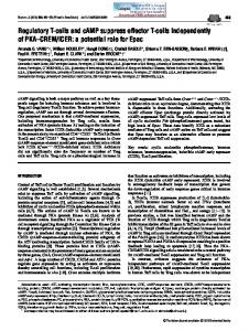
FIGURE 3. Typical force-distance curve of interaction between T cell and DC. A, Schematic illustration of AFM experiments. A T cell-mounted AFM cantilever was placed above a DC that is firmly attached to a glass coverslip. The T cell is then brought into contact with the DC to form DC–T cell conjugate. Interaction forces of DC–T cell conjugate are measured by the deflection of the cantilever after predefined contact time. B, Corresponding force-distance curve of DC–T cell interactions. Steps or jumps usually correlate to breakages of single molecules or whole molecular clusters at intercellular contact. F represents maximal interaction force (double arrow).
grown on glass-bottom dishes (MatTek). Calcium responses in individual T cells were measured using an inverted microscope (Olympus IX81; Olympus) with a 603 objective. The T cells were illuminated at 488 nm, and the fluorescent emission of fluo-4-AM and Fura Red-AM was captured every 5 s by time-lapse confocal imaging (Olympus FV1000; Olympus) and analyzed with Imaris software (Bitplane). Integrated fluo-4/Fura Red ratio was calculated from fluorescent images as a measurement for intracellular calcium concentration.
The Journal of Immunology
261
FIGURE 4. Interaction force of DC–T cell conjugates in the presence of altered peptide ligands. A, Interaction forces of DC–T cell conjugates in the absence (No Ova) or presence of 10 ng/ml OVA peptide (+ Ova) for the indicated contact time duration. The irrelevant OVA peptide (+ Ova-II) was used as a control. B, Interaction force between T cell and DC pulsed with indicated peptides (10 ng/ml) for contact time of 0 (∼1–3 s) and 3 min. Bars indicate mean 6 SEM (n . 10 pairs of cells) for each condition. For each condition, OT-I T cells were isolated from more than three independent experiments. *p , 0.001, unpaired t test.
Results Expression of cell-surface molecules on T cells and DC For the current study, we used homogeneous T cells from the wellstudied OT-I transgenic mouse model (15). FACS analysis showed that the purity of the isolated OT-I T cells is .98% (CD3ε+, CD8a+) and with .99% cells expressing a TCR (Va2; Vb5) specific for the model Ag OVA peptide OVA257–264 (SIINFEKL) presented by MHC class I H-2Kb (Fig. 1, top panel). As a source of homogenous DC, we used the well-characterized D1 cells derived from immature splenic DC (20), which expresses both CD11c and H-2Kb (purity .98%) (Fig. 1, bottom panel). The OVA peptide could be loaded successfully onto the DC, as determined by cytofluorometry using an Ab recognizing OVA-bound H-2Kb (Fig. 1, H-2Kb/OVA). Expression of other surface markers such as ICAM-1, LFA-1, CD3, and CD43 are shown in Fig. 1. The
FIGURE 5. Effects of Ab blocking and drug treatment on interaction forces of DC–T cell conjugates. A, Interaction forces of DC–T cell conjugates in the presence of blocking Abs against LFA-1 and TCR. B, Interaction forces in the presence of reagents that induce cytoskeleton disruption and cholesterol depletion, respectively. All force measurements were conducted with contact time of 3 min between DC and T cells in the presence of OVA peptide. Bars indicate mean 6 SEM (n . 10 pairs of cells) for each condition. For each condition, OT-I T cells were isolated from more than three independent experiments. *p , 0.001, **p , 0.01, unpaired t test.
homogenicity of both OT-I T cells and DC was an important feature that allowed us to investigate their interactions at the single-cell level. Efficient T cell activation is only promoted by Ags with high affinity TCR–pMHC interactions (13, 21–25) were probed with several APLs (A2, E1, R4, and Y5), which are known to possess different affinities for interaction with TCR and MHC (Table I) as well as different potential for T cell stimulation. To evaluate how T cell activation was influenced by TCR–pMHC interaction of different affinities, cytokine secretion was measured by stimulating T cells with DC prepulsed with various APLs at different concentrations (1 pg/ml–1 mg/ml). As shown in Fig. 2, only TCR–pMHC interactions mediated by peptides with high affinities (OVA and A2) were able
262
DC–T CELL INTERACTIONS CORRELATE WITH T CELL RESPONSIVENESS
to promote efficient IL-2 and IFN-g secretion. The Y5 APL, which is known to bind less strongly to H-2Kb MHC class I molecules than the parental OVA peptide (SIINFEKL) (26), was found to induce IL-2 and IFN-g secretion as well, albeit at a lower level. In contrast, DCs pulsed with E1 or R4 APLs with low affinities (AcKa , 0.1 mm4, Table I) failed to stimulate IL-2 and IFN-g secretion, similar to the control OVA-II peptide, which is specific for I-Ab MHC class II molecules (Fig. 2, upper panel). In addition to IL-2 and IFN-g production, we characterized surface expression of CD69 on T cells after coculture with DC for 4 h. In the absence of peptide, 12.5% of T cells showed intermediate level of CD69 expression (CD69+). In the presence of antagonist peptides (E1 and R4), a low fraction of T cells was CD69+ (15.4 and 32.2% for E1 and R4, respectively). In contrast, most T cells (.99%) show high surface expression of CD69 (CD69++) in the presence of agonist peptide (OVA, A2, and Y5) (Supplemental Fig. 3).
To see how increased concentration of peptide would affect DC–T cell interactions, we measured the binding strength of DC–T cell interactions by raising the concentration of peptide by 100-fold to 1 mg/ml. The results are shown in Supplemental Fig. 1. The average adhesion strength of DC–T cell interaction was not much affected by increasing the peptide concentrations from 10 ng/ml to 1 mg/ml (Supplemental Fig. 1). Thus, an increase in concentration of altered peptides did not significantly increase the binding forces, suggesting that a concentration of 10 ng/ml is already optimal.
Mechanical interactions between DC and T cells SCFS (14, 17, 18) was used to investigate the interaction force between DC and T cell using AFM. For this, a single T cell was attached to the AFM cantilever via anti-CD43 Abs (14) (see Materials and Methods for details). To allow DC–T cell conjugate formation, the T cell-mounted cantilever was positioned above a DC and then brought into contact with the DC at a force of 1 nN for establishment of adhesion with a predefined contact duration before subsequent separation (Fig. 3A). Physical interactions between DC and T cells were observed as a negative deflection in the retraction curve (Fig. 3B, red curve). Ruptures of molecular bonds were identified as jumps or force peaks in the retraction curve. For quantification of the mechanical interactions between DC and T cell, the maximal interaction force (14, 17, 27) was determined for individual DC–T cell conjugates (Fig. 3B). The average interaction force was used to compare interactions between DC and T cells in response to different APLs. The interaction forces between T cells and DC in the absence and presence of OVA peptide at a concentration of 10 ng/ml are shown in Fig. 4A. We observed that the interaction force increased progressively when the contact duration for the conjugates was increased from 0 min (effective contact time ∼1–3 s) to 3 min in the presence of the cognate OVA peptide. The average interaction force for contact duration of 0, 1 2, and 3 min time was 0.32 6 0.02, 0.63 6 0.08, 1.28 6 0.28, and 1.57 6 0.25 nN, respectively. In the absence of OVA peptide, the interaction force was low (,0.4 nN), and no significant increase (p . 0.05) was observed over time. The enhanced binding in the presence of OVA peptide was specific for the OT-I TCR, as the interaction force for DC– T cell conjugates remained low (,0.5 nN) when the irrelevant OVA-II peptide was used. Together, the results demonstrated the importance of the antigenic OVA peptide for the establishment of strong interaction force between Ag-specific T cells and DCs. Next, we probed the interaction of individual DC–T cell conjugates in the presence of APLs (Table I) at 10 ng/ml to see if the interaction was dependent on the affinity of the APLs. The force measurements were performed for all peptides at a contact duration of 3 min. Measurements for longer contact times (.3 min) were difficult to perform due to the high motility of cells, a phenomenon also observed in another AFM study (14). Depending on the peptide, different levels of interaction force were recorded, with the OVA peptide being the strongest at 1.57 6 0.25 nN and the R4 peptide being the weakest (,0.4 nN) (Fig. 4B). Intermediate levels of interaction forces were observed for peptides Y5 (0.99 6 0.23 nN), A2 (0.73 6 0.23 nN), and E1 (0.61 6 0.15 nN), respectively.
FIGURE 6. Calcium response of T cells bound to APC. A, Representative differential interference contrast images of the DC–T cell conjugates overlaid with the fluo-4 (green) and Fura Red (red) fluorescent signals (loaded in T cells only) at indicated time points. DCs were prepulsed with or without peptides for 4 h before T cell addition. Scale bar, 10 mm. B, Plots of time course of the intracellular calcium concentration in the responding T cell, as measured by fluo-4/Fura Red ratio. Each plot represents data from a pair of DC–T cell conjugates. C, Average calcium response of T cells bound to DC prepulsed with different peptides. To quantify early calcium response in T cells, fluo-4/Fura Red ratios were measured every 5 s in responding T cells and then integrated for 3 and 5 min from the time of the initial calcium increase (dashed line, Fig. 5B). For each condition, the average calcium response was measured by pooling data of DC–T cell conjugates (n . 15 pairs of cells) from more than three independent experiments. Bars indicate mean 6 SEM. *p , 0.001, unpaired t test against unpulsed condition (-).
The Journal of Immunology Cell adhesion molecules, cytoskeleton, and membrane cholesterol are important for interactions between DC and T cells Recent AFM studies demonstrated that high interaction forces between T cells and Ag-presenting B cells were observed after 30 min interaction time, when IS formation was maximal, and that LFA-1–ICAM-1 interactions were responsible for these forces (14). However, a role for LFA-1 in early adhesion events was not probed in the former study. To investigate the contribution of LFA1 and TCR surface molecules expressed on T cells in early (3 min) DC–T cell interactions, blocking experiments with Abs were performed. As shown in Fig. 5A, Abs to TCR and LFA-1 significantly reduced the development of interaction forces observed after 3 min contact time. Because cytoskeleton and membrane cholesterol are important for clustering of adhesion molecules on the cell surface (28–31) and regulation of T cell activation (32, 33), we investigated their potential role for peptide-dependent binding forces between T cells and DC. Fig. 5B shows that the interaction force between DC–T cell conjugates was significantly reduced by treatment with a series of cytoskeleton-disrupting agents. Interaction forces were found to be low (,0.5 nN) after treatment with CytoD, LatA (inhibitors of actin polymerization), Taxol (microtubule stabilizer), and Colc (inhibitors of microtubule polymerization). Force reduction was also observed by treatment with MbCD, a depletion agent that can extract cholesterol from the plasma membrane, thereby altering lipid raft formation and TCR clustering (30) (Fig. 5B). Selective promotion of calcium mobilization by high-affinity peptides We have shown that the late stage of T cell activation was only promoted by APLs with high affinity, as assessed by IL-2 and IFNg secretion (Fig. 2). To access early T cell activation, OT-I T cells were loaded with the intracellular calcium indicator dyes fluo-4 and Fura Red, followed by stimulation with DC prepulsed with APLs. This calcium response was imaged by time-lapse confocal
263 microscopy of randomly selected fields. Fig. 6A shows representative calcium responses of T cells bound to DC preincubated with the indicated APLs at 10 ng/ml. In the absence of peptide, the calcium signal of DC–T cell conjugates remained low, as measured by fluo-4/Fura ratio. In contrast, intracellular calcium was induced in the presence of OVA peptide and increased progressively during the 5-min observation time (Fig. 6B). Similar results were found in response to Y5 and A2 peptides. With the antagonist peptides E1 or R4 or control peptide OVA-II, no calcium response was observed. For comparison of the calcium responses to the different APLs, we integrated the fluo-4/Fura ratio (34) of DC–T cell conjugates for 3 and 5 min of contact duration (Fig. 6B). The average calcium responses for the different APLs are shown in Fig. 6C. Together, the results demonstrate that efficient calcium responses are only promoted by agonist peptides with high affinity (OVA, Y5, and A2). In contrast, an increased concentration of agonist peptides (OVA, Y5, and A2) from 10 ng/ml to 1 mg/ml did not lead to stronger calcium responses in T cells (Supplemental Fig. 2). Calcium response of T cells for antagonist peptides (E1 and R4) remained to be weak though the concentration was increased to 1 mg/ml (Supplemental Fig. 2). Mechanical interaction correlate with T cell responsiveness To investigate if the strength of mechanical interactions between T cells and APC correlates with T cell responsiveness, we plotted calcium response and IL-2 release against the interaction force of DC–T cell conjugates in the presence of APLs. Strong correlation was found between calcium responses and interaction forces (Fig. 7A). Similarly, IL-2 secretion also correlated with interaction forces (Fig. 7B). These results demonstrate the relevance of mechanical interactions that align with functional T cell responses.
Discussion We used SCFS by AFM to study mechanical interactions between DC and T cells following Ag recognition. This is, to our knowledge, the first demonstration of the relevance of physical intercellular
FIGURE 7. Correlation between mechanical force and T cell activation. Interaction forces are plotted against calcium response (measured by integrated fluo-4/Fura ratio for 3 min, Fig. 6C) (A) and IL-2 release (measured by ELISA assay, Fig. 2) (B) of T cells interacting with D1 prepulsed with the peptides at concentrations of 10 (black) or 1 mg/ml (red). r2 values of linear fits are 0.88 and 0.72 (A) and 0.75 and 0.71 (B) for peptide concentrations of 10 ng/ml or 1 mg/ml, respectively.
264
DC–T CELL INTERACTIONS CORRELATE WITH T CELL RESPONSIVENESS
DC–T cell contacts to the functional response of T cells. The results show that interaction forces increased significantly in the presence of the cognate antigenic peptide (Fig. 4A). The increase in cellular binding forces was dependent upon TCR–pMHC interaction because treatment with a TCR Ab blocked the interaction force of DC–T cell conjugates. Likewise, force reduction by LFA-1 blocking indicated that LFA-1 contributed to the establishment of intercellular interaction forces between DC and T cell. This is likely due to relocalization of LFA-1 to the contact area between DC and T cells (5, 14), which serves to enhance the intercellular binding strength between T cells and DC. The cytoskeleton is important regulatory component in immune synapse formation and regulation of T cell activation (3, 32, 35, 36). In agreement with this notion, we observed that interaction force between T and DC was reduced after the treatment of CytoD, an inhibitor of actin polymerization (Fig. 5B). Although enlargement of the DC–T cell contact area is likely to happen due to a possible loss of cortical tension of cell membrane after CytoD treatment, a stronger interaction force was not found in our AFM measurements. The observed decreased interaction force could be due to interference of the movement of cell-surface molecules by CytoD treatment, which is known to prevent IS formation (37). Alternatively, the intercellular interaction between DC and T cells could be dampened by a reduction of the affinity or number of serial TCR–pMHC interactions in response to CytoD treatment (13). Similar to CytoD, the development of interaction forces was also inhibited by a number of additional cytoskeleton-disrupting agents. In addition, disruption of lipid rafts by a membrane cholesterol-depletion agent also decreased binding forces. These results highlight the importance of cytoskeletal dynamics and the integrity of membrane cholesterol for strong interactions between Ag-presenting DC and T cells. The use of several closely related APLs with different affinities allowed us to investigate the crucial role of Ag quality in determining DC–T cell interactions and T cell responsiveness. Although strong interactions were induced by agonist peptides in our AFM measurements, only weak interaction forces (0.4–0.6 nN) between DC and T cells were measured in the presence of antagonist peptides (Fig. 4B). The weak interactions triggered by antagonist peptides could be due to the failure of accumulation of signaling molecules such as tyrosine-phosphorylated molecules at the synapse (22), which further destabilize DC–T cell interactions. In agreement with previous findings (22), both early and late T cell activation (as measured by calcium mobilization and IL-2 secretion) was only efficiently induced in T cells stimulated by agonist peptides (Figs. 2, 6), highlighting the crucial roles of Ag quality in promoting DC–T cell interactions and T cell responsiveness. Numerous models have been proposed to explain various functional outcomes of T cell responsiveness by TCR–pMHC kinetics (6–8, 12, 13, 21, 22, 38–42). Despite the success of these models in explaining activation data as a function of the kinetic parameters, the key question concerning the functional outcome of physical interaction between T cells and APCs remained unanswered. It was reported recently that the kinetics of twodimensional TCR and pMHC interactions determine T cell responsiveness, suggesting a mechanism of T cell activation based on serial TCR engagement and rebinding by a few pMHC agonists (12, 13). By using a panel of APLs with different affinities, we showed that DC–T cell conjugates exhibit broad ranges of interaction forces that correlate strongly with the response of T cells of the respective APLs (Fig. 7). The high interaction forces between DC and T cells are likely to provide a mechanically stable environment, thus enabling productive serial TCR–pMHC interactions and triggering of T cell activation.
Our data provide new mechanical insights into how T cells discriminate between different peptide–MHC ligands to maximize their responsiveness. High interaction forces between T cells and DC provide a mechanically stable environment, which is likely to enable rapid rebinding of TCR–pMHC (12, 13) and further induce T cell responsiveness. Thus, our data on mechanical interactions between DC and T cells provide a mechanistic model to explain the quality and quantitative control of Ag discrimination by T cells.
Acknowledgments We thank B. Vey for help with the manuscript.
Disclosures The authors have no financial conflicts of interest.
References 1. Huppa, J. B., and M. M. Davis. 2003. T-cell-antigen recognition and the immunological synapse. Nat. Rev. Immunol. 3: 973–983. 2. Fooksman, D. R., S. Vardhana, G. Vasiliver-Shamis, J. Liese, D. A. Blair, J. Waite, C. Sacrista´n, G. D. Victora, A. Zanin-Zhorov, and M. L. Dustin. 2010. Functional anatomy of T cell activation and synapse formation. Annu. Rev. Immunol. 28: 79–105. 3. Dustin, M. L. 2007. Cell adhesion molecules and actin cytoskeleton at immune synapses and kinapses. Curr. Opin. Cell Biol. 19: 529–533. 4. Monks, C. R., B. A. Freiberg, H. Kupfer, N. Sciaky, and A. Kupfer. 1998. Threedimensional segregation of supramolecular activation clusters in T cells. Nature 395: 82–86. 5. Potter, T. A., K. Grebe, B. Freiberg, and A. Kupfer. 2001. Formation of supramolecular activation clusters on fresh ex vivo CD8+ T cells after engagement of the T cell antigen receptor and CD8 by antigen-presenting cells. Proc. Natl. Acad. Sci. USA 98: 12624–12629. 6. Davis, M. M., J. J. Boniface, Z. Reich, D. Lyons, J. Hampl, B. Arden, and Y. Chien. 1998. Ligand recognition by alpha beta T cell receptors. Annu. Rev. Immunol. 16: 523–544. 7. Stone, J. D., A. S. Chervin, and D. M. Kranz. 2009. T-cell receptor binding affinities and kinetics: impact on T-cell activity and specificity. Immunology 126: 165–176. 8. Gascoigne, N. R., T. Zal, and S. M. Alam. 2001. T-cell receptor binding kinetics in T-cell development and activation. Expert Rev. Mol. Med. 2001: 1–17. 9. Corr, M., A. E. Slanetz, L. F. Boyd, M. T. Jelonek, S. Khilko, B. K. al-Ramadi, Y. S. Kim, S. E. Maher, A. L. Bothwell, and D. H. Margulies. 1994. T cell receptor-MHC class I peptide interactions: affinity, kinetics, and specificity. Science 265: 946–949. 10. Matsui, K., J. J. Boniface, P. Steffner, P. A. Reay, and M. M. Davis. 1994. Kinetics of T-cell receptor binding to peptide/I-Ek complexes: correlation of the dissociation rate with T-cell responsiveness. Proc. Natl. Acad. Sci. USA 91: 12862–12866. 11. Alam, S. M., P. J. Travers, J. L. Wung, W. Nasholds, S. Redpath, S. C. Jameson, and N. R. Gascoigne. 1996. T-cell-receptor affinity and thymocyte positive selection. Nature 381: 616–620. 12. Huppa, J. B., M. Axmann, M. A. Mo¨rtelmaier, B. F. Lillemeier, E. W. Newell, M. Brameshuber, L. O. Klein, G. J. Schu¨tz, and M. M. Davis. 2010. TCRpeptide-MHC interactions in situ show accelerated kinetics and increased affinity. Nature 463: 963–967. 13. Huang, J., V. I. Zarnitsyna, B. Liu, L. J. Edwards, N. Jiang, B. D. Evavold, and C. Zhu. 2010. The kinetics of two-dimensional TCR and pMHC interactions determine T-cell responsiveness. Nature 464: 932–936. 14. Hosseini, B. H., I. Louban, D. Djandji, G. H. Wabnitz, J. Deeg, N. Bulbuc, Y. Samstag, M. Gunzer, J. P. Spatz, and G. J. Ha¨mmerling. 2009. Immune synapse formation determines interaction forces between T cells and antigenpresenting cells measured by atomic force microscopy. Proc. Natl. Acad. Sci. USA 106: 17852–17857. 15. Clarke, S. R., M. Barnden, C. Kurts, F. R. Carbone, J. F. Miller, and W. R. Heath. 2000. Characterization of the ovalbumin-specific TCR transgenic line OT-I: MHC elements for positive and negative selection. Immunol. Cell Biol. 78: 110–117. 16. Hogquist, K. A., S. C. Jameson, W. R. Heath, J. L. Howard, M. J. Bevan, and F. R. Carbone. 1994. T cell receptor antagonist peptides induce positive selection. Cell 76: 17–27. 17. Zhang, X., E. P. Wojcikiewicz, and V. T. Moy. 2006. Dynamic adhesion of T lymphocytes to endothelial cells revealed by atomic force microscopy. Exp. Biol. Med. (Maywood) 231: 1306–1312. 18. Benoit, M., D. Gabriel, G. Gerisch, and H. E. Gaub. 2000. Discrete interactions in cell adhesion measured by single-molecule force spectroscopy. Nat. Cell Biol. 2: 313–317. 19. Mombaerts, P., J. Iacomini, R. S. Johnson, K. Herrup, S. Tonegawa, and V. E. Papaioannou. 1992. RAG-1-deficient mice have no mature B and T lymphocytes. Cell 68: 869–877.
The Journal of Immunology 20. Winzler, C., P. Rovere, M. Rescigno, F. Granucci, G. Penna, L. Adorini, V. S. Zimmermann, J. Davoust, and P. Ricciardi-Castagnoli. 1997. Maturation stages of mouse dendritic cells in growth factor-dependent long-term cultures. J. Exp. Med. 185: 317–328. 21. Aleksic, M., O. Dushek, H. Zhang, E. Shenderov, J. L. Chen, V. Cerundolo, D. Coombs, and P. A. van der Merwe. 2010. Dependence of T cell antigen recognition on T cell receptor-peptide MHC confinement time. Immunity 32: 163–174. 22. Carren˜o, L. J., E. M. Riquelme, P. A. Gonza´lez, N. Espagnolle, C. A. Riedel, S. Valitutti, and A. M. Kalergis. 2010. T-cell antagonism by short half-life pMHC ligands can be mediated by an efficient trapping of T-cell polarization toward the APC. Proc. Natl. Acad. Sci. USA 107: 210–215. 23. Gonza´lez, P. A., L. J. Carren˜o, D. Coombs, J. E. Mora, E. Palmieri, B. Goldstein, S. G. Nathenson, and A. M. Kalergis. 2005. T cell receptor binding kinetics required for T cell activation depend on the density of cognate ligand on the antigen-presenting cell. Proc. Natl. Acad. Sci. USA 102: 4824–4829. 24. Govern, C. C., M. K. Paczosa, A. K. Chakraborty, and E. S. Huseby. 2010. Fast on-rates allow short dwell time ligands to activate T cells. Proc. Natl. Acad. Sci. USA 107: 8724–8729. 25. Kalergis, A. M., N. Boucheron, M. A. Doucey, E. Palmieri, E. C. Goyarts, Z. Vegh, I. F. Luescher, and S. G. Nathenson. 2001. Efficient T cell activation requires an optimal dwell-time of interaction between the TCR and the pMHC complex. Nat. Immunol. 2: 229–234. 26. Howarth, M., A. Williams, A. B. Tolstrup, and T. Elliott. 2004. Tapasin enhances MHC class I peptide presentation according to peptide half-life. Proc. Natl. Acad. Sci. USA 101: 11737–11742. 27. Puech, P. H., A. Taubenberger, F. Ulrich, M. Krieg, D. J. Muller, and C. P. Heisenberg. 2005. Measuring cell adhesion forces of primary gastrulating cells from zebrafish using atomic force microscopy. J. Cell Sci. 118: 4199–4206. 28. Lillemeier, B. F., M. A. Mo¨rtelmaier, M. B. Forstner, J. B. Huppa, J. T. Groves, and M. M. Davis. 2010. TCR and Lat are expressed on separate protein islands on T cell membranes and concatenate during activation. Nat. Immunol. 11: 90– 96. 29. Schamel, W. W., I. Arechaga, R. M. Risuen˜o, H. M. van Santen, P. Cabezas, C. Risco, J. M. Valpuesta, and B. Alarco´n. 2005. Coexistence of multivalent and monovalent TCRs explains high sensitivity and wide range of response. J. Exp. Med. 202: 493–503.
265 30. Campi, G., R. Varma, and M. L. Dustin. 2005. Actin and agonist MHC-peptide complex-dependent T cell receptor microclusters as scaffolds for signaling. J. Exp. Med. 202: 1031–1036. 31. Yokosuka, T., K. Sakata-Sogawa, W. Kobayashi, M. Hiroshima, A. HashimotoTane, M. Tokunaga, M. L. Dustin, and T. Saito. 2005. Newly generated T cell receptor microclusters initiate and sustain T cell activation by recruitment of Zap70 and SLP-76. Nat. Immunol. 6: 1253–1262. 32. Burkhardt, J. K., E. Carrizosa, and M. H. Shaffer. 2008. The actin cytoskeleton in T cell activation. Annu. Rev. Immunol. 26: 233–259. 33. Billadeau, D. D., J. C. Nolz, and T. S. Gomez. 2007. Regulation of T-cell activation by the cytoskeleton. Nat. Rev. Immunol. 7: 131–143. 34. Irvine, D. J., M. A. Purbhoo, M. Krogsgaard, and M. M. Davis. 2002. Direct observation of ligand recognition by T cells. Nature 419: 845–849. 35. Dustin, M. L., and J. A. Cooper. 2000. The immunological synapse and the actin cytoskeleton: molecular hardware for T cell signaling. Nat. Immunol. 1: 23–29. 36. Vicente-Manzanares, M., and F. Sa´nchez-Madrid. 2004. Role of the cytoskeleton during leukocyte responses. Nat. Rev. Immunol. 4: 110–122. 37. Wu¨lfing, C., M. D. Sjaastad, and M. M. Davis. 1998. Visualizing the dynamics of T cell activation: intracellular adhesion molecule 1 migrates rapidly to the T cell/ B cell interface and acts to sustain calcium levels. Proc. Natl. Acad. Sci. USA 95: 6302–6307. 38. Valitutti, S., S. Mu¨ller, M. Cella, E. Padovan, and A. Lanzavecchia. 1995. Serial triggering of many T-cell receptors by a few peptide-MHC complexes. Nature 375: 148–151. 39. Davis, S. J., and P. A. van der Merwe. 2006. The kinetic-segregation model: TCR triggering and beyond. Nat. Immunol. 7: 803–809. 40. Rosette, C., G. Werlen, M. A. Daniels, P. O. Holman, S. M. Alam, P. J. Travers, N. R. Gascoigne, E. Palmer, and S. C. Jameson. 2001. The impact of duration versus extent of TCR occupancy on T cell activation: a revision of the kinetic proofreading model. Immunity 15: 59–70. 41. Zehn, D., S. Y. Lee, and M. J. Bevan. 2009. Complete but curtailed T-cell response to very low-affinity antigen. Nature 458: 211–214. 42. McKeithan, T. W. 1995. Kinetic proofreading in T-cell receptor signal transduction. Proc. Natl. Acad. Sci. USA 92: 5042–5046. 43. Fremont, D. H., E. A. Stura, M. Matsumura, P. A. Peterson, and I. A. Wilson. 1995. Crystal structure of an H-2Kb-ovalbumin peptide complex reveals the interplay of primary and secondary anchor positions in the major histocompatibility complex binding groove. Proc. Natl. Acad. Sci. USA 92: 2479–2483.