injury and surgical intervention. Since hypovolemia com- monly exists in patients with vascular injury, this disor- der should be urgently corrected with sufficient ...
Original Article
Retrospective Assessment of Vascular Injuries: 23 Years of Experience Erkan Iriz, MD,1 Fersat Kolbakır, MD,2 Atilla Sarac, MD,2 Haci Akar, MD,3 Hasan Tahsın Keçelıgıl, MD,2 and Mustafa Kemal Demiragˇ, MD2
Purpose: To analyze the operation methods, injury etiologies and localizations, post-operative complications and the reasons for mortality in patients who were admitted for peripheral vascular injuries to our clinics. Methods: From January 1979 to February 2002, 410 patients were operated for peripheral vascular injuries. Three hundred and one of the patients were male (73.5%) and 109 of them were female (26.5%), and their ages ranged between 1-88 (mean 35.5 years). Results: The most common etiological reason was firearm injuries in 163 patients (39.8%). The most common injured artery was the brachial artery (83 patients, 22.5%) among a total of 369 patients whereas the most commonly injured vein was the common femoral vein (60 patients, 23.4%) in a total of 256 patients. Isolated venous injuries were encountered in 41 patients whereas isolated arterial injuries were detected in 154 patients (37.5%). Hospital admission duration of the patients after trauma was approximately 3 hours. Conclusion: The extremity-salvage rate in the group was 92.3%. The hospital stay period of the patients was 21.8 days. The mortality rate was 2.6% (11 patients). (Ann Thorac Cardiovasc Surg 2004; 10: 373–8) Key words: vascular trauma, injury, peripheral artery and vein
Introduction In vascular surgery mortality and morbidity of the vascular injury is highly related to the duration between the injury and surgical intervention. Since hypovolemia commonly exists in patients with vascular injury, this disorder should be urgently corrected with sufficient volume replacement and immediate intervention. For those patients with delayed late intervention and those whose operations extended and required massive of blood transfusion, patients may develop systemic coagulopathy probFrom 1Department of Cardiovascular Surgery, Gazi University Faculty of Medicine, Ankara, 2Department of Cardiovascular Surgery, Ondokuz Mayis University Faculty of Medicine, Samsun, and 3Department of Cardiovascular Surgery, State Hospital of Samsun, Samsun, Turkey Received January 6, 2004; accepted for publication June 21, 2004. Address reprint requests to Erkan Iriz, MD: Gazi Üniversitesi Tıp Fakültesi Kalp ve Damar Cerrahisi AD, Be evler 06500 Ankara, Turkey.
Ann Thorac Cardiovasc Surg Vol. 10, No. 6 (2004)
lems and serious physical disorders, such as hypothermia, hypoxemia, acidosis, and hyperkalemia.1) For this reason, the purpose of our study was to analyze operation methods, injury etiologies and localizations, postoperative complications and reasons for mortality of the patients who were admitted for peripheral vascular injuries to our clinic. Since non-invasive methods are mostly sufficient and many patients need urgent intervention, the use of advanced radiological screening methods are sometimes hard to reach and may result in lost time. But the patients who were operated immediately in such cases, should be followed-up with arteriography and / or duplex ultrasonography.1) In the past, attempts to control arterial bleeding were by means a cauterization method in addition to manual compression and pouring boiling liquid materials on the wounds. The ligation method was initially used by Ambroise Pare during the XVIth century.2) During the First and Second World Wars, important knowledge had
373
Iriz et al.
been gained both in diagnosis and treatment of vascular injuries, but vascular reconstructive methods were mainly introduced during the Korean and Vietnamese wars with tremendous progress.2,3) Consequently, a dramatic decrease in amputation rates was achieved.2-5) Early diagnosis and immediate intervention is mandatory in vascular injuries in order to save the extremities and lives of the patients. The developments in clinical experience, an increase of centers employing staff experienced in vascular injury, the advance in antibiotic treatments, the usage of volume expanding solutions and blood transfusions provide great assistance in the treatment of such patients.
Materials and Methods Various surgical interventions had been performed on 410 patients who were referred to our department with vascular injuries between January 1979 and February 2002. In this study, all data, including age, sex, etiology of the vascular injuries, injury regions, additional pathologies, injured vessels, physiopathology of the vascular injury, existing complications were assessed with applied treatment methods investigated retrospectively. The patients with co-existing injuries trauma including fracture and soft tissue damage of an extremity, nerve, soft tissue and other systems were also assessed by related departments, and then referred to the theater for revascularization within about 50 minutes of reaching to the hospital.
Results Diagnosis of vascular injury was done approximately 3 hours after the event in many cases in which the initial diagnosis of the injury was mostly assessed by peripheral circulation with the assistance of hand Doppler and physical examination. The number of the patients diagnosed with only hand Doppler (ankle-brachial index) and clinical inspection was 305 (74.3%). In other cases, duplex ultrasonography (64 patients - 15.6%) and peripheral arteriography (41 patients - 10.0%) had been applied. During vascular injury, clinical findings and signs such as; presence of open arterial bleeding, presence of an increase intended or pulsated hematoma, presence of six P signs (pulselessness, poikilothermia, pallor, pain, paresthesia, paralysis) of the related extremity is accepted as basis for the diagnosis. Of the patients 301 were men (73.5%), and 109 were
374
Table 1. Vascular injury reasons Ethiologic cause
Number
Ratio (%)
Firearm injury Stab injury Blunt injury Iatrogenic injury Electrical injury
163 120 92 31 4
39.8 29.3 22.4 7.5 0.9
women (26.5%). The mean age was 35.5 (1-88 years). The most common etiology of vascular injuries was a penetrating trauma (283 patients, 69.1%). Firearm injury was the most common cause among penetrating trauma in all of our series, accounting for 163 patients (39.8%). Other etiological causes were stab injuries, blunt injuries, iatrogenic injuries and electrical injuries in 120 patients (29.3%), 92 patients (22.4%), 31 patients (7.5%), and 4 patients (0.9%), respectively (Table 1). The pathophysiology and pattern of arterial injuries were attributed to complete arterial cut in 245 (66.4%), partial arterial cut in 95 (25.7%) and blunt arterial injury without cut in 29 (7.9%). The extremities were the most affected parts with a total of 380 patients (92.7%). Intrathoracic vessels, intraabdominal vessels, neck region vessels were injured in 11 patients (2.7%), 11 patients (2.7%) and 4 patients (1.0%), respectively. Lower extremity injuries account for 239 patients (%58.3), whereas upper-extremity injuries occur in 141 patients (%34.4). Intrathoracic and intraabdominal vessel injuries were found to be low because of two major factors. First, many injuries are minor traumas only sufficient to scare or alert the victim. These traumas are localized to the extremities. The other factor depends on the geographic conditions. Many patients die due to time loss after the event. The most commonly encountered arterial injuries were those of the brachial artery, which occur in 83 patients (22.5%). Other injured arteries were as follows: Common femoral artery, popliteal artery, superficial femoral artery, radial artery, ulnar artery, axillary artery, anterior tibial artery, posterior tibial artery and other arteries in 78 patients (21.1%), 63 patients (17.1%), 44 patients (12.0%), 23 patients (6.2%), 18 patients (4.8%), 10 patients (2.7%), 10 patients (2.7%), 10 patients (2.7%) and 30 patients (8.1%), respectively. Distribution of vascular injuries and localization can be seen in Table 2. The most common venous injuries were found to be in the common femoral vein in 60 patients (23.4%). Popliteal vein in 59 patients (23.0%), brachial vein in 53 patients
Ann Thorac Cardiovasc Surg Vol. 10, No. 6 (2004)
Retrospective Assessment of Vascular Injuries; 23 Years of Experience
Table 2. Distribution of vascular injuries Localization
Artery
Vein
Artery and vein
Total
1 7 32 18 32 30 13 8 7 6 154
1 2 1 14 8 9 3 2 1 41
3 3 51 23 46 14 50 12 4 5 4 215
4 11 85 42 92 52 72 23 13 12 4 410
Subclavian Axillary Brachial Ulnar and radial Common femoral Superficial femoral Popliteal Anterior and posterior tibial Thoracic aorta Abdominal aorta Others Total
Table 4. Applied surgical interventions (arterial)
Table 3. Additional pathologies Pathology
Number
Ratio (%)
138 89 29 6
33.6 21.7 7.0 1.4
Bone fracture Nerve injuries Traumatic pseudoaneurysm A-V fistula
Table 5. Applied surgical interventions (venous) Surgical intervention Saphenous vein graft interposition Ligation Lateral venoraphy End-to-end anostomosis
Number
Ratio (%)
98 62 48 48
38.2 24.2 18.8 18.8
(20.7%), ulnar and radial artery sideways veins in 24 patients (9.3%), superficial femoral veins in 22 patients (8.6%), anterior and posterior tibial veins in 15 patients (5.9%), subclavian veins and axillary veins in 7 patients (2.8%), and other veins in 16 patients (6.3%) respectively were injured. Isolated vein injuries were detected by physical examination, duplex USG, and surgical exploration. Nerve injuries were seen commonly with popliteal, axillary and brachial vessel injuries. Compartment syndrome developed mainly in popliteal and distal parts of the popliteal artery region in the lower extremity. In the upper part, compartment syndrome was detected with wide tissue damage. Vascular injuries and additional pathologies are shown in Table 3. Further, associated operations were performed to the additional pathologies encountered in our cases by the related disciplines. Surgical treatment methods are summarized in connection with artery and vein injuries in Table 4 and 5.
Ann Thorac Cardiovasc Surg Vol. 10, No. 6 (2004)
Surgical intervention
Number
Ratio (%)
160 83 66 25 24 11
43.4 22.5 17.8 6.7 6.5 3.0
Saphenous vein graft interposition End-to-end anostomosis Lateral arterioraphy PTFE graft interposition Ligation Other (cephalic, basilic vein, embolectomy)
Table 6. Complications Complication Infection Compartment syndrome Amputation Death
Number
Ratio (%)
59 26 21 11
14.3 6.3 5.1 2.6
Complications in post-operative patients can be seen in the following Table 6. Eleven patients within our group died postoperatively. Six of the mortal injuries were with intrathoracic or intraabdominal large vascular injuries as a result of firearm shots. Some of these patients died during induction of anesthesia, while others survived intraoperatively but died in the intensive care unit due to systemic coagulopathy progression although successful vascular interventions were done. Systemic diseases caused the resulting deaths, and one occurred due to an arrhythmia problem which progressed post-operatively. Although revascularization was maintained in an appropriate period, in 21 patients amputation was needed due to associated serious wounds such as wide soft tissue injury, or intractable fractures in addition to nerve injuries.
375
Iriz et al.
In the cases which the period between revascularization and vascular injury exceeds 6 hours, various fasciotomies were applied to the extremity within the same time. In all cases, besides tetanus prophylaxis, 1st generation cephalosporine combination with aminoglicoside were administered pre-operatively as empiric therapy and continued up to 5 days in severely contaminated wounds or for 2 days for cleaner wounds. Infected wounds were treated with the appropriate antibiotics according to cultural sensitivity along with frequent wound dressings. The patients with minor vascular injuries received Dextrane 40 with pentoxyphylline for 5 days. The mortality ratio was found to be 2.6% with 11 patients, whereas extremity salvage rate was 92.3%. A-V fistulae and pseudoaneurysm occurred in 6 (1.4%) and 29 (7.0%) patients, respectively. Those late complications were treated with appropriate methods.
Discussion Despite modern surgical interventions, vascular injuries can still cause extremity loss and even death. According to some authors, amputation rates can even reach 78%.6) The extremity salvage rate in our study was 92.3%. According to some authors, approximately 90% of the arterial injuries exist due to penetrating traumas.7,8) Blunt traumas compose the remaining 10% ratio,9) while others reported even over 50%.6) In our study, blunt traumas are reported as 22.4%. Firearm injuries among penetrating traumas are very common in our analysis (39.8%). In reports, which were issued from war districts10,11) and in some civil settlement regions,3) firearm injuries are commonly reported. But in the series of some authors,5,7) frequency of firearm injuries also within civil regions is behind the other etiological reasons. Two factors play a role in the huge amount of firearm injuries in our region. Initially, having a gun in this local area has been a custom for many years particularly for men. In many events such as weddings, national victories the use of firearms is a very common issue that causes many accidental events. Secondary cause is the conflict which occurs between families. Unfortunately, the frequency of firearm injury in our hospital is high. Vascular injuries are frequent among young male population,5,7) and male patients compose 73.5% of the cases. Vessels, nerves and bones may be injured together due to their close relation anatomically.12,13) Bone fractures and nerve lesions were also accompanying 33.6% and 21.7% in our study, respectively. The patients with bone
376
fracture, nerve injury, head trauma and intra-abdominal trauma are assessed by related disciplines and appropriate interventions were maintained. In our patients with fractures, external fixation is more preferred because of easier application and low infection risk.14) Peripheral angiography in vascular injuries is controversial. Some authors are suggesting angiography to every pre-operative patient,6,8,15,16) while others don’t. Many clinicians report their successful vascular injury results without angiography.5,7,17,18) Since the Doppler ultrasound is 95% sensitive and 97% specific in experienced hands, its use reduces the spent time with respect to angiography.7,11,19) Under this circumstance, careful clinical examination can give a reliable diagnosis with the combination of Doppler ultrasound, and measurement of peripheral circulation pressure differences with hand Doppler, if applicable. Our opinion is that, peripheral angiography should be applied when vascular injury is expected. This method is also a gold standard for the patients who cannot be diagnosed by basic diagnostic tools. Both time and expenses will decline with such basic tools. We follow the diagnostic step-by-step method and our results were found to be similar to those presented by many authors. It is essential to control the bleeding in vascular injuries, particularly, in the vessels, which have a greater diameter, hence the greater risk of hypovolemic shock. Under these circumstances, a severely injured patient should be taken to theatre as soon as possible with volume expanding solutions particularly blood. Six of our patients died before operation because of great vessel injury and consequent hypovolemic shock. In arterial injuries, successful results were obtained in arterial reconstruction procedures, which were held 6-8 hours after the event.15,20) Almost all of the amputation performed in our patients was late cases that were revascularized after 8 hours following the injury. Blunt trauma, extremity without pulse, more than one tibial vascular damage and multiple fractures are the factors that increased the amputation rates.1,9,21) Infection is also a major factor increasing amputation rate after a successful vascular surgery intervention. For this reason, vigorous and appropriate tissue debridement, is a very important intervention before and after the revascularization procedure.21) In our retrospective study, 59 patients developed infection and a patient died due to systemic disease encountered with advanced age. Amputation was required for 4 patients because of infection. Other patients did well with appropriate treatment methods. Fasciotomy is suggested especially in lower extremity
Ann Thorac Cardiovasc Surg Vol. 10, No. 6 (2004)
Retrospective Assessment of Vascular Injuries; 23 Years of Experience
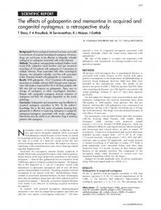
Fig. 1. Preoperative MR angiography imaging of anterior tibial artery pseudoaneurysm.25)
17,22,23)
injuries by lots of authors. Compartment syndrome of the muscles can progress after late revascularization, which ischemic for a long period. Under these conditions, immediately decompressing fasciotomies should be applied.9,21) In our clinic, following lower extremity injuries, fasciotomies were performed to the 4 anatomic compartments within the related extremity. Fewer thrombotic events occurred with respect to some publications.5) We encountered few thrombotic events after end-to-end anastomosis method, applied to approximately 83 arterial injuries. We think that several factors are effective in this result. The first is that we particularly take care to free anastomotic ends from neighboring tissues in order to achieve a good anastomotic line and this feature leads to a loose anastomosis. Another factor is that we perform longitudinal incisions on vessel walls on anastomotic line to maintain a wider diameter on anastomosis. Using heparin with a prophylactic dose (500 IU/ h), leads to greater patency rates only on the vessels with a medium and large diameter. Although etiological factors of vascular injuries differ between publications, penetrating injuries are the most commonly encountered reasons.3,7) Firearm injuries, compared to stab injuries, cause greater damage and more serious tissue loss directly proportional to its kinetic energy.3) When cases within our series are inspected, one can see that the first rank of the etiological factors belongs to firearm injuries, and this effects the treatment
Ann Thorac Cardiovasc Surg Vol. 10, No. 6 (2004)
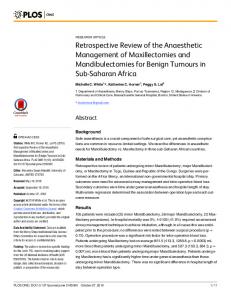
Fig. 2. Postoperative peripheral angiography (saphenous vein interposition to anterior tibial artery).25)
method applied, and thus saphenous vein graft interposition is used more frequently during both artery or vein injuries encountered. Actually the most popular conduits; PTFE or Dacron grafts for arteries with a diameter larger than 6-8 mm and autogenous saphenous vein for venous reconstruction or arteries with a diameter of less than 5 mm, are strongly recommended.1) The most interesting point within our series is the excessive frequency of pseudoaneurysms. The first factor causing this increase is iatrogenic reasons. These pseudoaneurysms, progressing after diagnostic or treatment aimed angiographies,3,24) were among the most important reason within our study. Besides, there is another patient group, in whom vascular injury diagnosis is not performed with simple investigation methods in surrounding hospitals. These patients, in whom even a Doppler ultrasound is not carried out, with suspected penetrative and non-penetrative vascular injuries came to our hospital with a pseudoaneurysm at late period. One anterior tibial artery pseudoaneurysm case, presented with pain and peripheral circulation complaints 18 months after firearm injury, was one of the best examples (Fig. 1 and 2).25) We believe that advanced imaging methods should be applied to all patients especially with firearm injury, stab injury and particularly to patients with blunt trauma in
377
Iriz et al.
whom pathological findings with physical inspection and hand Doppler were undetermined. In conclusion, early diagnosis and treatment during vascular injuries has an importance for saving the extremity and life of the patient. Vascular injuries require immediate surgical intervention, regardless of and localization. We think that mortality and morbidity rates of patients will highly decrease with suitable surgical technique, in case of requirement, liberal application of fasciotomy, aggressive debridement of necrotic tissues and suitable cure of other accompanying pathologies as well as postoperative suitable wound care and medical support.
References 1. Mattox KL, Hirshberg A. Vascular trauma. In: Haimovici H, Ascer E, Hollier LH, Strandness DE, Towne JB eds.; Haimovici’s Vascular Surgery, 4th ed. USA: Blackwell Science, 1996; pp 480–96. 2. Yaycıoglu A, Arıbal D, Tatlıcıoglu E. Cerrahi Damar Hastalıkları, 2. Baskı, Ankara-Türkiye Klinikleri Yayınevi, 1978. 3. Weaver FA, Hood DB, Yellin AE. Vascular injuries of the extremities. In: Rutherford RB, ed.; Vascular Surgery, 5th ed. W.B. Saunders Company, 2000; pp 862– 71. 4. Feliciano DV, Bitondo CG, Mattox KL, et al. Civilian trauma in the 1980s. A 1-year experience with 456 vascular and cardiac injuries. Ann Surg 1984; 199: 717– 24. 5. Razmadze A. Vascular injuries of the limbs: a fifteenyear Georgian experience. Eur J Vasc Endovasc Surg 1999; 18: 235–9. 6. Andrikopoulos V, Antonıou I, Panoussis P. Arterial injuries associated with lower-extremity fractures. Cardiovasc Surg 1995; 3: 15–8. 7. Cihan HB, Gülcan O, Hazar A, Türköz R. Periferik damar yaralanmaları. Ulus Travma Derg 2001; 7: 113– 6. 8. Johansen K, Lynch K, Paun M, Copass M. Non-invasive vascular tests reliably exclude occult arterial trauma in injured extremities. J Trauma 1991; 31: 515– 22. 9. Hood DB, Yellin AE, Weaver F. Vascular trauma. In: Dean RH, ed.; Current diagnosis & Treatment in Vascular surgery. Connecticut: Lange, 1996; pp 405–28. 10. Velinovic MM, Davidovic BL, Lotina IS, et al. Complications of operative treatment of injuries of peripheral arteries. Cardiovasc Surg 2000; 8: 256–64.
378
11. Khoury G, Sfeir R, Nabbout G, Jabbour-Khoury S, Fahl M. Traumatic arteriovenous fistulae: ‘the Lebanese war experience’. Eur J Vasc Surg 1994; 8: 171–3. 12. Klein SR, Bongard FS, White RA. Neurovascular injuries of the thoracic outlet and axilla. Am J Surg 1988; 156: 115–8. 13. Weaver FA, Rosenthal RE, Waterhouse G, Adkins RB. Combined skeletal and vascular injuries of the lower extremities. Am Surg 1984; 50: 189–97. 14. Keçeligil HT, Arıkan A, Kolbakır F, Keyik T, Erk MK. Periferik vasküler yaralanmalar: 221 olgunun degerlendirilmesi. Damar Cerrahisi Dergisi 1995; 4: 27–33. 15. Yılmaz AT, Arslan M, Demirkılıç U, et al. Missed arterial injuries in military patients. Am J Surg 1997; 173: 110–4. 16. Solak H, Yeniterzi M, Yüksek T, Eren N, Ceran S, Göktogan T. Injuries of the peripheral arteries and their surgical treatment. Thorac Cardiovasc Surg 1990; 38: 96–8. 17. Anderson RJ, Hobson RW 2nd, Lee BC, et al. Reduced dependency on arteriography for penetrating extremity trauma: influence of wound location and non-invasive vascular studies. J Trauma 1990; 30: 1059–65. 18. Peck JJ, Eastman AB, Bergan JJ, Sedwitz MM, Hoyt DB, McReynolds DG. Popliteal vascular trauma. A community experience. Arch Surg 1990; 125: 1339– 44. 19. Gahtan V, Bramson RT, Norman J. The role of emergent arteriography in penetrating limb trauma. Am Surg 1994; 60: 123–7. 20. Padberg FT, Rubelowsky JJ, Hernandez-Maldonado JJ, et al. Infrapopliteal arterial injury: prompt revascularization affords optimal limb salvage. J Vasc Surg 1992; 16: 877–86. 21. Flint LM, Richardson JD. Arterial injuries with lower extremity fracture. Surgery 1983; 93: 5–8. 22. Menzoian JO, Doyle JE, Cantelmo NL, LoGerfo FW, Hirsch E. A comprehensive approach to extremity vascular trauma. Arch Surg 1985; 120: 801–5. 23. Katsamouris AN, Steriopoulos K, Katonis P, et al. Limb arterial injuries associated with limb fractures: clinical presentation, assessment and management. Eur J Vasc Endovasc Surg 1995; 9: 64–70. 24. Nehler MR, Lawrence WA, Whitehill TA, Charette SD, Jones DN, Krupski WC. Iatrogenic vascular injuries from percutaneous vascular suturing devices. J Vasc Surg 2001; 33: 943–7 25. Akar H, Saraç A, Konuralp C, Kolbakır F, Keçeligil HT. Posttraumatic pseudoaneurysm of the anterior tibial artery. Eur Journal of Trauma 2002; 28: 44–6.
Ann Thorac Cardiovasc Surg Vol. 10, No. 6 (2004)