Hindawi Publishing Corporation BioMed Research International Volume 2013, Article ID 374395, 22 pages http://dx.doi.org/10.1155/2013/374395
Review Article Actin Cytoskeleton Manipulation by Effector Proteins Secreted by Diarrheagenic Escherichia coli Pathotypes Fernando Navarro-Garcia, Antonio Serapio-Palacios, Paul Ugalde-Silva, Gabriela Tapia-Pastrana, and Lucia Chavez-Dueñas Department of Cell Biology, Centro de Investigación y de Estudios Avanzados del IPN (CINVESTAV-IPN), Apartado Postal 14-740, 07000 México, DF, Mexico Correspondence should be addressed to Fernando Navarro-Garcia;
[email protected] Received 24 August 2012; Accepted 22 October 2012 Academic Editor: Gad Frankel Copyright © 2013 Fernando Navarro-Garcia et al. is is an open access article distributed under the Creative Commons Attribution License, which permits unrestricted use, distribution, and reproduction in any medium, provided the original work is properly cited. e actin cytoskeleton is a dynamic structure necessary for cell and tissue organization, including the maintenance of epithelial barriers. Disruption of the epithelial barrier coincides with alterations of the actin cytoskeleton in several disease states. ese disruptions primarily affect the paracellular space, which is normally regulated by tight junctions. ereby, the actin cytoskeleton is a common and recurring target of bacterial virulence factors. In order to manipulate the actin cytoskeleton, bacteria secrete and inject toxins and effectors to hijack the host cell machinery, which interferes with host-cell pathways and with a number of actin binding proteins. An interesting model to study actin manipulation by bacterial effectors is Escherichia coli since due to its genome plasticity it has acquired diverse genetic mobile elements, which allow having different E. coli varieties in one bacterial species. ese E. coli pathotypes, including intracellular and extracellular bacteria, interact with epithelial cells, and their interactions depend on a speci�c combination of virulence factors. In this paper we focus on E. coli effectors that mimic host cell proteins to manipulate the actin cytoskeleton. e study of bacterial effector-cytoskeleton interaction will contribute not only to the comprehension of the molecular causes of infectious diseases but also to increase our knowledge of cell biology.
1. Introduction Epithelial layers are essential to keep normal organ operation by creating boundaries to the movement of ions and molecules. is event allows the formation of different tissue compartments and ion gradients that drive transport across the epithelium. e tissue compartments include kidney tubules, ducts within the liver, and the lining of the gastrointestinal tract and lungs. In many of these tissues, the epithelium is also a barrier between the organ tissue and the external environment, representing the �rst layer of defense against pathogens. Epithelial cells form sheets by binding to each other through apically located adherent junctions (AJs) and more basally located desmosomes [1]. Polarized epithelial cells are characterized by the separation of distinct apical and basolateral domains that sort, traffic, and localize unique subsets of plasma membrane proteins. Epithelial cells undergo polarization through crucial interactions with
the actin cytoskeleton and associated signaling molecules, resulting in the formation of junctional complexes. e apical plasma membrane surfaces of polarized epithelial cells are deeply associated with the actin cytoskeletal network [2]. In the case of absorptive intestinal epithelia, the apical cell surface (the intestinal lumen) is composed of �nger-like projections called microvilli. Microvilli are comprised of parallel actin bundles that anchor to the subapical actin network through direct interactions with several actin bundling proteins. e ezrin, radixin, and myosin (ERM) family of actin-binding proteins provides stability to the microvilli and these proteins are important components of epithelial cell architecture as they provide a link between the cortical membrane and the actin cytoskeleton [3]. e most commonly described cellular target of pathogens is the cytoskeleton. Various intracellular microorganisms harness cytoskeletal components to gain entry to and to propel themselves within host cells [4]. e cytoskeleton of
2 eukaryotic cells is composed of actin �laments, microtubules, and intermediate �laments. In terms of bacterial pathogenesis, the most extensively studied of these are actin �laments [5].
2. Actin Cytoskeleton e shape and movement of cells, as well as phagocytosis, intercellular communication and the distribution of organelles depend on actin [6]. Actin persists in the cell as two different forms: monomeric globular actin (G-actin) and polymeric �lamentous actin (F-actin). Actin is one of the most abundant proteins in eukaryotic cells and is composed of 375 amino acids forming a single chain of 42 kDa. Its atomic structure was �rst solved for its complex with deoxyribonuclease I [7]. G-actin is a �at molecule of about 50 × 50 × 35 Å. Under physiological salt conditions puri�ed monomeric or G-actin polymerizes to its �lamentous form, F-actin. G-actin contains �rmly bound one molecule of ATP that is hydrolyzed to ADP and Pi aer incorporation into a growing F-actin �lament. e ADP remains attached to the actin subunit, whereas the Pi dissociates slowly from the �lament generating two �lament ends with actin subunits differing in their bound nucleotide: either ATP or ADP [8]. roughout polymerization, ATPbound G-actin preferentially associates to the end containing ATP-actin subunits, the fast growing end, which has also been termed the plus or barbed end. Aer reaching equilibrium, actin monomers associate to the barbed end and an identical number dissociates preferentially from the opposite end, which has also been termed the minus or pointed end. us, under these conditions and in the presence of ATP actin subunits constantly associate to the barbed end and travel through the whole �lament until they dissociate from the pointed end [9]. is event has been termed treadmilling or actin cycling and represents the sole basis for force generation for a number of motile processes [10]. In eukaryotic cells, monomeric G-actin is in dynamic equilibrium with polymerized F-actin. Polymerization is regulated by capping proteins (such as gelsolin) at the plus ends, through the binding to these ends with high affinity and prevents elongation. Rapid signal-induced actin polymerization, including that which occurs during host-pathogen interactions, can be triggered by different mechanisms, including de novo nucleation that can involve the Arp2/3 (actin-related protein 2/3) complex [11]. Actin nucleation by the Arp2/3 complex, which is essential for phagocytosis, cell polarity and migration, forms an orthogonal Y-branched network of F-actin [12]. e Arp2/3 complex itself consists of seven proteins; it binds ATP and is activated by nucleationpromoting factors from WASP (Wiskott-Aldrich syndrome protein) family including WASP, neural (N-)WASP and three SCAR/WAVE (WASP family verprolin homologous protein) isoforms, as well as other factors such as cortactin and the actin-binding protein Abp1p [13]. WASP family proteins possess a C-terminal Arp2/3-binding and activation domain (WCA domain). At the N terminus, WASP and NWASP proteins contain a WASP homology 1 (WH1) domain, which is related to a domain typical of the ENA/VASP
BioMed Research International (Ena/vasodilator stimulated phosphoprotein-like protein) protein family (also known as the EVH1 domain), and a Cdc42-interactive GTPase-binding domain (GBD) motif, which preferentially binds Cdc42. In addition, these proteins possess a phosphatidylinositol bisphosphate (PtdIns(4,5)P2)binding region (B) adjacent to the GBD domain. e WCA domain increases actin nucleation by Arp2/3 by about 80fold. WASP family proteins are autoinhibited by binding between the central region of the WCA domain and the B/GBD region, which is also near to the CRIB (Cdc42/Rac interactive binding) domain. is autoinhibition can be released by binding of GTP-bound Cdc42 to the GBD domain [14]. Cdc42 can thereby directly activate actin nucleation [11]. Filament formation is promoted and stabilized through the action of proteins such as pro�lin and cortactin, and the �lament is depolymerized through the action of proteins such as co�lin or gelsolin [15]. Actin �laments (also called micro�laments) also bundle with other actin-interacting proteins, including fascins [16], forming more substantial structures. Alternatively, the �laments can be cross-linked by branching, which is initiated by actin-nucleating proteins [17] to form a meshwork such as cortical actin. F-actin �bers form the micro�lament network inside the cell, varying from myosin-containing contractile stress �bers to the cortical actin network that resides beneath the plasma membrane and around intracellular organelles. Actin �bers are also used to make sheet-like extensions, such as lamellipodia, membrane ru�es and blebs; �nger-like protrusions, such as microvilli and �lopodia; or dot-like podosomes. ese structures are modi�ed by the action of several actin-binding and signaling proteins [18]. e actin cytoskeleton is highly dynamic and is mainly manipulated by members of the Rho-family GTPases that control signal transduction pathways linking membrane receptors to the cytoskeleton. Rho-family GTPases regulate several cellular processes, including F-actin polymerization, assembly of intercellular junctions, cell polarity, cell migration, and membrane trafficking [19]. e Rho GTPases belong to the superfamily of Ras proteins. To date, more than 20 different mammalian Rho GTPases have been identi�ed, which are ∼50% homologous to each other and ∼30% similar to Ras [20]. Rho proteins function as molecular switches in many eukaryotic signal transduction pathways and, most importantly, regulate the actin cytoskeleton. e switch function of Rho proteins depends on their regulation by a GTPase cycle. Rho GTPases are inactive when bound to GDP and are associated with a guanine nucleotide dissociation inhibitor (GDI). ey are activated by the GDP/GTP exchange caused by guanine nucleotide-exchange factors (GEFs; ∼60 have been recognized to date), which themselves are controlled by extracellular-induced signaling. GTP-bound Rho proteins induce many downstream effects through interactions with an array of effector proteins, including protein kinases, lipid kinases, phospholipases, and various adaptor proteins. e active state of Rho GTPases is terminated by hydrolysis of the bound GTP, a process that is facilitated by GTPase-activating proteins (GAPs; ∼70 have been identi�ed to date) [11]. RhoA, Rac1, and Cdc42 are the most extensively characterized members of the Rho protein family. RhoA regulates the
BioMed Research International formation of contractile actin�myosin stress �bers and the organization of focal contacts [21], Rac is involved in the formation of lamellipodia and focal complexes [21], and Cdc42 governs the formation of �lopodia or microspikes as well as focal contacts [22]. Accordingly, these GTPases have crucial roles in several cellular processes, including morphogenesis, migration, cytokinesis, phagocytosis, and cell-matrix contacts [23]. However, it is the role of Rho GTPases in regulating the actin cytoskeleton that is particularly important during a bacterial infection process [11]. Bacterial pathogens do not usually interact directly with actin �laments themselves. But, they subvert and control the polymerization of actin �laments by modulating cellular regulators of this process [5]. Actin interacts with a variety of proteins. Around 150 different known speci�c actin-binding proteins (ABPs) are found both at extracellular and intracellular (mainly) localizations that modify particular properties or its supramolecular organization [6]. e ABPs can be grouped into at least eight classes: (a) proteins that stabilize or sequester the monomeric actin; (b) proteins that bind along �-actin �laments (such as tropomyosin); (c) motor proteins that generate the force for the sliding of �-actin �laments; (d) proteins that nucleate actin polymerization; (e) proteins that bundle �-actin �laments; (f) proteins that stabilize �lament networks; (g) proteins that cut �-actin �laments; (h) proteins that attach �laments to specialized membrane areas [24]. Even if they have different functions many of these proteins attach to a few target zones on the actin surface such as the hydrophobic region. Maybe because of these multiple interactions, the sequence and three-dimensional structure of actin has been very conserved during the billions of years of evolution. Many ABPs are at the end of signaling cascades and are regulated by phospholipid interaction, Ca2+ ion concentration, phosphorylation, or small GTPases [25]. ese signals either deactivate or activate the supramolecular organization of actin during cell migration, exocytosis or endocytosis, or cytokinesis [8]. us, the actin cytoskeleton is a dynamic structure necessary for cell and tissue organization, including the maintenance of epithelial barriers. e epithelial barrier regulates the movement of ions, macromolecules, immune cells, and pathogens, and it is thus essential for normal organ function. Disruption in the epithelial barrier has been shown to coincide with alterations of the actin cytoskeleton in several disease states. ese disruptions primarily manifest as increased movement through the paracellular space, which is normally regulated by tight junctions (TJ) [26]. e TJs restrict the movement of pathogens and large macromolecules through the space between two cells [27]. Disruption of the junctions, and the barrier they establish, is a common feature of disease states and is associated with the establishment of infections, increased in�ammation, and malabsorption [28]. e actin cytoskeleton is directly connected to cell junctions and plays an important role in the assembly and maintenance of these structures [29]. e main barrier function of the epithelium is thought to depend on tight junctions, which are connected with the actin cytoskeleton. e TJ is closely associated with the actin
3 cytoskeleton and even subtle modulation of the cortical actin cytoskeleton can induce structural changes to the TJ. is strong association is attributed to the direct contacts between several TJ-localized protein components and actin [29]. ree types of transmembrane proteins are part of tight junctions: occludin, claudins, and junctional adhesion molecules (JAMs), and they are connected to adaptor proteins such as zonula occludens 1 (ZO-1), ZO-2, and ZO-3 [11]. Claudins are necessary for the barrier formation, while occludin and JAM family proteins seem to be required for the regulation of barrier permeability and signaling [30]. Many additional proteins are also essential, including PAR6, atypic Ca2+ - and diacylglycerol-independent protein kinase C (aPKC), and PAR3. is proteins complex, which is important for cell polarity, is regulated by Cdc42, as is the CRUMBS3-PALS1PATJ-complex, which is essential for tight junction assembly [31]. e precise role of RhoA and Rac in the tight junction regulation has still to be elucidated. Recently, it was suggested that RhoA-dependent phosphorylation of occludin is crucial for the tight junction function [32]. us, the �-actin �laments are highly dynamic structures, whose supramolecular organization is constantly modi�ed according to cellular needs. eir dynamic behavior is regulated by a large number of binding proteins that are oen the effectors of intracellular and extracellular signaling pathways. It is therefore not surprising that the actin cytoskeleton is one of the main targets of bacterial proteins, and thus of major importance for the host-pathogen interaction [8]. Bacteria have developed numerous toxins and effectors to target the actin cytoskeleton. To induce cytoskeletal changes, pathogenic microbes must ensure delivery of effector molecules onto or into host cells. Bacterial effectors are typically proteins that interface with and in�uence hostcell pathways and can facilitate the disease. Bacteria use several methods to deliver effector proteins to the host cell. Some effectors, such as toxins, are secreted by bacteria in the vicinity of the host cell, where they bind speci�c receptors and are taken up by endocytosis [33]. Other effector proteins can facilitate their own uptake by pore-forming subunits or autotransporter domains. Some gram-negative pathogenic bacteria have acquired sophisticated “molecular syringes,” such as type III or type IV secretion systems, which are multisubunit molecular machines that span the bacterial and host membranes and translocate effectors directly into host cells [34, 35]. As mentioned before, the common and recurring target of pathogens is the host cell cytoskeleton, which is utilized by these microbes for purposes that include attachment, entry into cells, movement within and between cells, vacuole formation and remodeling, and avoidance of phagocytosis. A relevant example is Escherichia coli, which is a bacterial species genetically versatile that comprises a huge group of nonpathogenic and pathogenic bacteria. E. coli is an important member of the normal intestinal micro�ora of humans and other mammals. Due to its genome plasticity through horizontal transfer, E. coli has been widely exploited as a cloning host in recombinant DNA technology. E. coli, beside being a laboratory workhorse or harmless intestinal
4 inhabitant, is a highly versatile and frequently deadly pathogen. Several different E. coli strains cause diverse intestinal and extraintestinal diseases by means of virulence factors that affect a wide range of cellular processes.
3. Diarrheagenic Escherichia coli ere are several highly adapted E. coli clones that have acquired speci�c virulence attributes, which confer an increased ability to adapt to new niches and allow them to cause a broad spectrum of disease. e virulence factors are oen encoded on genetic elements, that can be mobilized into different strains to generate novel combinations of virulence factors. Only the most successful combinations of virulence factors have persisted to become speci�c E. coli pathotypes that are capable of causing disease in healthy individuals [36]. Among the intestinal pathogens, there are six categories of pathotypes: enteropathogenic E. coli (EPEC), enterohemorrhagic E. coli (EHEC), enterotoxigenic E. coli (ETEC), enteroaggregative E. coli (EAEC), enteroinvasive E. coli (EIEC), and diffusely adherent E. coli (DAEC) [36]. Most of the pathogenic E. coli strains remain extracellular, but EIEC is a true intracellular pathogen that is capable of invading and replicating within epithelial cells and macrophages. Other E. coli strains might be internalized by epithelial cells at low levels, but do not appear to replicate intracellularly [34]. us, E. coli pathotypes interact with epithelial cells and these interactions depend on the speci�c combination of virulence factors that de�ne their life style and their capability to cause a speci�c pathology (pathotype). Except for ETEC, which produce the heat-labile (LT) and heat-stable (ST) enterotoxins that are their main virulence factors [36], the other �ve pathotypes subvert and control the polymerization of actin �laments by modulating cellular regulators of this process. EIEC manipulates the cytoskeleton for invasion of a host cell and/or to gain motility in the cell. Aer invasion and escape from membrane-enclosed vesicles into the cytosol, EIEC also manipulate actin-�lament dynamics to move within the infected host cell [4]. ey do so by recruiting actin in one of their poles, through bacterialprotein-mediated nucleation of actin. e hijacking of actinassociated cytoskeletal components can also occur during the infection with extracellular pathogens, such as EPEC and EHEC, which share similar features, and DAEC [5]. Finally, virulence factors secreted by EAEC and DAEC, which cause proteolysis of proteins that stabilize actin �lament networks.
4. Enteroinvasive E. coli Enteroinvasive Escherichia coli (EIEC) are closely related to Shigella spp., showing remarkable phenotypic and genotypic similarities. However, the disease induced by EIEC is generally less severe than that induced by Shigella spp. [37]. is difference in severity may be associated with a lower expression level of virulence factors by EIEC in the presence of various host cells, such as macrophages and intestinal epithelial cells. Phylogenetic studies have suggested that Shigella and EIEC form a single pathovar of E. coli. EIEC strains are regarded as precursors of full-blown Shigella evolved from E.
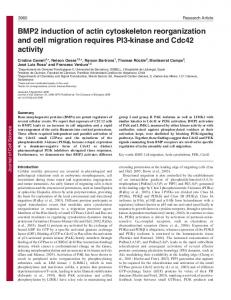
BioMed Research International coli [38]. Shigella �e��eri and EIEC are pathogenic microorganisms known to cause disease in humans by very similar mechanisms of pathogenicity. Once ingested, bacteria reach the colonic mucosa and invade various cell types, including M cells, macrophages, and epithelial cells. Invasion of the intestinal mucosa is followed by intracellular bacterial multiplication, spread of the infection to adjacent cells, induction of severe in�ammation of the colon, and destruction of the mucosa [39]. e virulence of these strains depends on the presence of both chromosomal genes and a large virulence plasmid (pINV). pINVs represent a family of highly related plasmids, and they are functionally interchangeable between Shigella and EIEC strains. Several virulence traits (Figure 1) are encoded by pINV genes, including (i) the ability to invade and multiply within epithelial cells [40], and (ii) the ability to spread infection intracellularly and to adjacent cells [41]. e uptake of S. �e��eri/EIEC depends on complex rearrangements of membranes and the actin cytoskeleton, which are mediated by the controlled temporal and spatial actions of a multitude of bacterial and host cell factors [42]. e bacteria directly target intracellular host cell signaling pathways by transporting effector proteins across the bacterial inner membrane (IM) and outer membrane (OM) as well as the host plasma membrane. e S. �e��eri/EIEC Mxi-Spa T3SS is a representative of sophisticated molecular export machineries known as injectisomes, that translocate proteins into the host cell cytoplasm in one step [43]. e initial contact between Shigella/EIEC and host cells takes place at lipid ra membrane domains and is mediated by the receptors CD44 and 𝛼𝛼5𝛽𝛽1 integrin. Receptor binding induces early actin cytoskeleton rearrangements and primes the cell for uptake, but the efficient and complete engulfment of the bacteria is triggered by the translocation of at least six T3SS effector proteins. IpaB, IpaC, and IpaD elicit actin rearrangements at the site of bacterial attachment that are required for entry, whereas IpaA promotes the reorganization of these actin-rich structures [44]. IpaB binds to CD44, a member of the immunoglobulin superfamily that binds hyaluronan. is interaction is required for Shigella-induced cytoskeletal rearrangements that lead to bacterial entry, presumably by determining early events of the internalization process [45]. e reorganization of the eukaryotic cytoskeleton is governed predominantly by small Rho GTPases and tyrosine kinases, which renders these enzymes targets for manipulation by pathogenic bacteria [46]. S. �e��eri, as well as many other bacterial pathogens, requires and manipulates these Rho GTPase pathways to promote its uptake [46]. S. �e��eri induces massive actin polymerization, leading to the formation of large membrane protrusions at the entry site, which form a macropinocytic pocket enclosing the bacteria. is process requires the activation of the small GTPases Rac1 and Cdc42, that recruit the actin-nucleating complex Arp2/3 [47]. Several bacterial effectors have been implicated in the induction of actin polymerization. e translocator protein IpaC is able to initiate actin nucleation and the formation of �lopodial and lamellipodial membrane extensions, but it is unclear how IpaC stimulates Rac1 and Cdc42 [47]. IpaC invasion function requires its immediate
BioMed Research International
5
M cell
Actin tail
VirA
IcsB,P IscA
N-WASP
EIEC
Microtubule degradation
Phagosome escape Dock18 ELMO
PIP2
IpgB1 VirA IpgB2
Rac1 Cdc42 IpaC,B T3SS IpaA Arp2/3
Macrophage Raft (cholesterol)
Receptor recognition Cytoskeleton reorganization
Bacterial propulsion
PIP Endoplasmic reticulum
Enterocyte
Nucleous
Monocyte
Apoptotic macrophage
F 1: A model for Shigella/EIEC infection of colonic epithelial cells. Shigella/EIEC invade the epithelium from the intestinal lumen through M-cells. Aer reaching the epithelium they invade epithelial cells and are phagocytosed by resident macrophages. Shigella/EIEC escape the phagosome of both cells but while Shigella/EIEC replicate within epithelial cells, they induce apoptosis in macrophages. Bacteria are released and can invade the epithelial cells from the basolateral side, move into the cytoplasm by triggering actin polymerization, and spread to adjacent cells (for full details, see main text).
C-terminus and this general region may be involved in its ability to trigger actin nucleation. A pivotal role for the T3SS effector IpgB1 in triggering invasion has been suggested [48]. IpgB1 mimics the active small GTPase RhoG and elicits Rac1 activation and membrane ruffling through the ELMODock180 pathway [49]. Rac1 is further activated in response to the destabilization of the microtubule network by the S. �e��e�i VirA, a cysteine protease [50]. Mxi-Spa effectors, which induce actin polymerization at the entry site, might also include IpgB2. IpgB2 (a IpgB1 homologue) was shown to mimic the GTP-bound form of RhoA, and its expression in eukaryotic cells triggered the formation of actin stress �bers and membrane ruffling [51]. However, the speci�c contribution of IpgB2 to the signaling induced in the course of an infection of epithelial cells with S. �e��e�i remains to be established. Two additional T3SS effector proteins are necessary for the full invasiveness of S. �e��e�i. e phosphoinositide 4-phosphatase IpgD hydrolyzes phosphatidylinositol-4,5-biphosphate [PtdIns(4,5)P2] to yield phosphatidylinositol-5-phosphate [PtdIns(5)P] [52]. Hydrolysis of [PtdIns(4,5)P2] leads to the dissociation of the actin cytoskeleton from the plasma membrane, which facilitates the remodeling of membranes and actin. Finally, the translocated effector IpaA binds vinculin and enhances its association to actin �laments, thus mediating localized actin depolymerization and a reduction of the adhesion between cells and the extracellular matrix. is process might promote the closure of the phagocytic cup around the bacteria [53]. Aer triggering its uptake into host cells, S. �e��e�i/EIEC is entrapped in the phagosome. Aer that, S. �e��e�i lyses the
surrounding membranes and escapes into the cytoplasm in less than 10 min [54]. Membrane lysis does not depend on the acidi�cation of the phagosome and is mediated by the MxiSpa T3SS and the translocator proteins IpaB, IpaC, and IpaD [55]. Although all three translocator proteins are needed for the escape from the phagosome, there is evidence that IpaC is the decisive factor mediating membrane lysis. e cytoplasm of epithelial cells is the main replicative niche for S. �e��e�i/EIEC. e intracellular survival and intercellular spread of S. �e��e�i are linked to the machinery allowing the bacteria to move by directed actin polymerization. e molecular mechanisms mediating the actinbased motility of S. �e��e�i and other intracellular pathogens have been studied extensively [4]. IcsA (intracellular spread) protein, which localizes to one pole of the bacterium, is the central bacterial mediator of actin polymerization [56, 57]. Surface-bound IcsA recruits and activates host cell factors, including N-WASP and the Arp2/3 complex [58]. e complex thus formed serves as an actin nucleator and catalyzes the directed elongation of an actin tail, which propels S. �e��e�i/EIEC through the cytoplasm. In addition to the actin-nucleating complex, the T3SS-secreted cysteine protease VirA was recently identi�ed to be pivotal for e�cient intracellular movement and subsequent intercellular spread [59]. VirA degrades 𝛼𝛼-tubulin, thus creating a “tunnel” in the dense intracellular microtubule network. However, a large body of evidence, including structural studies, has now shown that VirA is not a cysteine protease and does not directly destabilize microtubules [60, 61]. Intracellular motility, replication, and survival further depend on the T3SS
6 substrate IcsB, that protects the bacteria from being recognized and entrapped by the host cell autophagy machinery [62]. Autophagy is a host cell recycling and defense system, which engulfs and sequesters cytoplasmic components in double-membrane-bound compartments [63]. Interestingly, IcsA contains an autophagy-inducing recognition site, which has to be masked by IcsB to prevent engulfment by autophagic vacuoles and to ensure the intracellular survival of S. �e�neri [62]. Due to propulsion through the host cell cytoplasm by actin polymerization, moving S. �e�neri eventually impinges on the plasma membrane at the tight junctions of adjacent epithelial cells. e arising protrusions can be endocytosed by the neighboring cell in a process requiring host cell factors like myosin light-chain kinase [64]. Aer lysis of the surrounding double membrane, which depends on the T3SS and the translocator proteins IpaB, IpaC, and IpaD, S. �e�neri is free in the cytoplasm, and a new cycle of replication and cell-to-cell spread can start [55, 65].
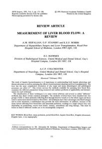
5. Enteropathogenic and Enterohemorrhagic E. coli Enterohemorrhagic E. coli (EHEC) and enteropathogenic E. coli (EPEC) cause serious diarrhea in humans that can result in death [36]. EPEC, EHEC, and a number of animal pathogens such as the mouse-speci�c pathogen Citrobacter rodentium and rabbit-speci�c enteropathogenic E. coli (REPEC) [66–68] belong to a group of mainly extracellular diarrheagenic pathogens that colonize the gut epithelium by attaching and effacing (A/E) lesions [69]. e A/E histopathology is characterized by effacement of the brush border microvilli, intimate bacterial adherence to the enterocyte apical plasma membrane, and the accumulation of polymerized actin beneath the attached bacteria [70]. All A/E pathogens carry a pathogenicity island, the locus of enterocyte effacement (LEE) [71] that encodes gene regulators [72, 73], the adhesin called intimin [74], a type III secretion system (T3SS) [71], chaperones [75, 76], and several secreted proteins, including the translocated intimin receptor called Tir [76, 77]. EPEC is a cause of gastroenteritis in infants less than 2 years of age, while EHEC causes bloody diarrhea in children and the elderly. EHEC is distinguished from EPEC by the production of Shiga toxins that can cause severe kidney damage leading to the hemolytic uremic syndrome, a form of acute renal failure [78]. Upon contact with epithelial cells, EHEC/EPEC inject a variety of effectors into the cells to modulate cellular functions involved in the host defense response, the dynamics of cytoskeleton and the maintenance of tight junctions (Figure 2). A major target of virulence factors is the cellular signaling cascade involved in the construction and modulation of the cytoskeleton and micro�laments. �hile the bacteria remain mostly extracellular in the lumen of the gut, the T3SS effectors of A/E pathogens access and manipulate the intracellular environment of host cells. e effectors subvert various host cell processes, which enable the bacteria to colonize, multiply and cause the disease. A group of 21 core effectors, including all those encoded within LEE
BioMed Research International and a number of non-LEE effectors, are shared by all A/E pathogens [79, 80]. e T3SS is an injection device (injectisome) that can transfer bacterial virulence proteins directly into host cells. e apparatus is made up of a basal body that spans both bacterial membranes and an extracellular needle that possesses a channel that is thought to act as a conduit for protein secretion [81]. e needle projects from the basal body and is assembled by the polymerization of a single, small needle subunit [82]. Contact with a host-cell membrane triggers the insertion of a pore into the target membrane, and effectors are translocated through this pore into the host cell. e T3SS exports two distinct categories of proteins: the translocators that form a pore in the target membrane and the effectors that traffic through this pore into the host cell. LEE encodes the translocator proteins EspA, EspD, and EspB, which are part of the T3SS and are required for the translocation of effectors into the host cell. Other factors encodes by LEE include the adhesin intimin, which binds to Tir and is essential for the intimate attachment of the bacteria with the cytoplasmic membrane of the host cell. Other effector proteins such as EspF, EspG, EspZ, EspH, Map, and Tir, some chaperone proteins such as CesD for EspB and EspD, CesF for EspF, and CesT for Tir are also encoded within LEE [83]. 5.1. Tir. One of the most important effectors studied that manipulates the actin cytoskeleton by EHEC-EPEC is translocated intimin receptor (Tir). EHEC-EPEC Tir mainly helps the bacteria to bind intimately to the epithelial cell surface. is intimate union requires intimin in the outer membrane of the bacterium and Tir embedded into the epithelial cell membrane. In sites of contact with epithelial cells, EPEC Tir is translocated via T3SS and is inserted into the plasma membrane of epithelial cells, where it is tyrosine phosphorylated and acts as a receptor for intimin [84]. is effector is considered the most important in the process of manipulating the actin cytoskeleton and the formation of actin-rich structures known as pedestals. Among the Tir Cterminal sequence, there are regions necessary to carry out the processes of controlling the actin machinery [85, 86]. Although EPEC Tir (TirEPEC ) and EHEC Tir (TirEHEC ) are structurally identical (60–67% identity) [84], the signaling pathways to manipulate the actin cytoskeleton within the epithelial cell are very different. In the case of TirEPEC , Tir has two transmembrane domains and six tyrosine residues, all located in the Cterminus. A loop model has been proposed for Tir when is inserted into the host membrane, where it uses its two transmembrane domains to traverse the membrane of the enterocyte, with their N- and C-terminal regions located inside the host cell; the region between the two transmembrane domains form a extracellular hairpin. Tirintimin interaction guides the intimate attachment of EPEC to epithelial cells, leading to the pedestal formation and actin polymerization [77]. TirEHEC and TirEPEC show a high degree of amino acid identity, particularly in their N-termini but are most divergent in their N-terminal domains. is region of the protein contains tyrosine residues that in TirEPEC
BioMed Research International
7
BFP EPEC
P Nck
IQGAP1
IQGAP1
N-WASP Arp2/3
Ca
Increase in Ca2+ CaCaCa CaCa concentration Disassembly of tight junction
Ibe
Intimin Tir
Nucleolin IQGAP1 Calmodulin
EspC
ZO1 Claudin
Pedestal maduration
ZO1
EspH IRTKS/ WIP IRSp53 N-WASP EspFu N-WASP Arp2/3
Occludin
Map
Enterocyte
Tight junctions
EspM
EspM EspF
Mitochondria
EspL Annexin 2 Actin stress fibers
Cif
EspG
Pedestal formation COP1 vesicle
NleA COP1
Microtubule disruption
Endoplasmic reticulum
EspC
Nucleous
Ebp50 Map Ezrin
Frodrin Cdc42 polarization
Cdc42
EspT EspV
Lamellipodia
c-Fyn, Abl, Arg and Etk
EHEC
Pe de sta l
Intimin Tir
Dendritic-like structure Filopodia
F 2: Models of actin cytoskeleton manipulation by enteropathogenic E. coli (EPEC) and enterohemorrhagic E. coli (EHEC). EPEC and EHEC are attaching and effacing (A/E) pathogens that destroy the microvilli and subvert host cell actin to form pedestals beneath the attachment site. EPEC attaches to the small bowel through the bundle-forming pilus (BFP), forming localized adhesions. Using the type III secretion system, a large repertoire of effector proteins is injected into the host cell. Intimate attachment is mediated by the interaction between the translocated intimin receptor (Tir) and intimin, an adhesin. Tir is phosphorylated by host tyrosine kinases, and phosphorylated Tir recruits Nck, which activates neural Wiskott-Aldrich syndrome protein (N-WASP) and the actin-related protein 2/3 (Arp2/3) complex to mediate actin rearrangements and pedestal formation. Whereas, the mechanism of pedestal formation by EHEC is slightly different from that used by EPEC. Tir is not phosphorylated, and pedestal formation is Nck-independent, but it is mediated by Tir cytoskeleton-coupling protein (EspFu ; also known as TccP), which is linked to Tir through the host protein insulin receptor tyrosine kinase substrate (IRTKS) and interacts with N-WASP to activate the Arp2/3 complex. EHEC injects many of the same effectors as EPEC into the host cell to manipulate host processes (for full details, see main text).
are potential substrates for phosphorylation. TirEHEC , which is not tyrosine phosphorylated, lacks one of these residues [87]. ereby, pedestal formation by EHEC and EPEC differs in the requirements for Tir tyrosine phosphorylation and additional bacterial factors delivered to the host cell. TirEPEC is phosphorylated by the host cell kinase, c-Fyn, just aer it is translocated to the epithelial cell membrane. Subsequently, Abl, Arg, and Etk through their SH3 domains recognize the proline-rich motif of Tir at the N-terminus then maintain the tyrosine phosphorylation persistent and turning the signaling pathway on to polymerize actin under attached bacteria, on the pedestal [88, 89]. In TirEPEC , a 12residue peptide encompassing phosphorylated Y474 binds the SH2 domains of the Nck adaptor proteins. WIP-like proteins may be involved in the subsequent recruitment of N-WASP to a complex of Tir and Nck, but the three tandem SH3 domains of Nck1 and Nck2 likely activate NWASP by directly binding to its PRD [90]. Multimerization of N-WASP can further enhance Arp2/3-mediated actin nucleation and pedestal assembly, which causes membrane
protrusion beneath the bacterium [91]. It worthy to note that actin pedestal formation observed in vitro is different to A/E lesion formation in vivo, as both tyrosine residues are not required for A/E lesion formation, demonstrating that in vitro phenotypes do not always correlate with that of in vivo observations [92]. ere are other proteins that bind to Tir and modulate the actin cytoskeleton, such as IQGAP1. IQGAP1 is involved in diverse cellular functions, since GTPase signaling to control cell proliferation and motility. IQGAP1 binds directly to actin, promoting the polymerization and placing it onto actin lamellipodia. It has been determined that certain middle region of the IQGAP1 C-terminal binds to the CRIB domain of N-WASP, removing autoinhibition of the latter and promoting actin polymerization, such as Cdc42 does [93]. IQGAP1 binds to Tir, causing IQGAP1 accumulation at sites of adherent bacterial. Since the Ca2+ signaling is required for remodeling and handling polymerization actin machinery in the cell surface, an increase in the Ca2+ concentration is required for EHEC-EPEC pedestal formation. e increase in
8 Ca2+ intracellular concentration promotes the association of IQGAP1 with calmodulin, recruiting the last to the pedestal. is interaction reduces the interaction of IQGAP1 with Rac1 and Cdc42. e recruitment of IQGAP1 to the pedestal increases the activation of N-WASP and therefore the actin polymerization and formation of pedestal in EHEC-EPEC adherent sites [94]. Another T3SS effector, termed IQGAP1-binding effector protein (Ibe) was recently discovered to be associated with LEE. is effector was shown to regulate Tir phosphorylation and actin pedestal formation [94]. Like Tir, IQGAP1 binds Ibe and colocalizes with Ibe at actin pedestals. However, the mechanism underlying the contribution of Ibe to EPEC pathogenesis remains to be de�ned [95]; it should also be noted that Ibe is not conserved and thus not a core LEE efector. EHEC and EPEC have evolved somewhat different mechanisms to activate the actin polymerization machinery. EHEC also generates pedestals in a Tir dependent manner without recruitment of Nck to the sites of adherent bacteria, but in the absence of tyrosine phosphorylation, EHEC translocates a second effector protein, EspFu [96]. Although EHEC does not recruit Nck, N-WASP is recruited and necessary for the pedestals formation by EHEC [97]. e expression of EspFu in EHEC allows the same adaptor to function independently of tyrosine phosphorylation. All the information necessary to manipulate the actin polymerization signaling pathway lay in a 12 amino acids region at the C-terminal (452-463) of TirEHEC [98]. is region contains a tyrosine (Y458), which is not phosphorylated, but is very important in the formation of a motif known as NPY458 . is motif is also crucial for the process of actin polymerization by EPEC independently of Nck, which uses the NPY454 motif in the C-terminal of TirEPEC [99]; although the T3SS effector translocated by EPEC could be related to NPY454 the signaling pathway has not yet been discovered. e NPY458 motif at the C-terminal of TirEHEC is detected by two homologous eukaryotic proteins that are members of the I-BAR (Bin-Amphiphysin-Rvs167) family, IRTKS, and IRSp53. ese proteins belong to the I-BAR family because its amino acid sequence laids I-BAR domains that bind to the eukaryotic membrane deforming it and producing bumps. ese proteins also have SH3 domains, which help them to recognize proline-rich motifs [100]. IRTKS and IRSp53 contain an IMD (IRSp53/MIM-homology) domain within its Nterminal sequence, which normally deforms the eukaryotic membrane and binds to eukaryotic small G proteins [100]. rough its IMD domain, IRSp53 and IRTKS, recognizes the NPY458 motif of TirEHEC , while IRTKS and IRSp53 use the SH3 domain to recognize the repetitive proline-rich protein EspFu, which is also translocated by EHEC T3SS. en EspFu activates N-WASP, allowing a potent activation of actin polymerization mediated by N-WASP/Arp2/3 [86]. Recently, it was found that spectrin is a key protein involved in the signaling required for actin polymerization during EHEC pedestal formation. A colocalization of spectrin cytoskeletal proteins with IRSp53 and IRTKS, which are required for EHEC pedestal formation, has also been reported [101].
BioMed Research International EspFu, also known as TccP (Tir cytoskeleton-coupling Protein) is an effector protein that is translocated by the EHEC T3SS. is effector is encoded on a cryptic phage known as 933U, outside the pathogenicity island LEE. EspFu comprises a translocation signal in the N-terminus recognized by the T3SS, followed by �ve and a half 47 amino acid repeats each, which are rich in prolines. Within each of these repeats, the �rst 17 amino acids are important for activation of N-WASP, which is normally given by other molecules such as the small GTPases Cdc42, the adaptor protein Nck and phosphoinositides such as PtdIns(4,5)P2 [102]. EspFu controls actin by activating members of the WASP family. EspFu binds to the autoinhibitory GTPase binding domain (GBD) in WASP proteins displacing it from the activity-bearing VCA domain (for verprolin homology, central hydrophobic, and acidic regions). is interaction potently activates WASP and neural (N)-WASP in vitro and induces localized actin assembly in cells. EspFu forms an amphipathic helix that binds the GBD, mimicking interactions of the VCA domain in autoinhibited WASP. us, EspFu activates WASP by competing directly for the VCA binding site on the GBD [103]. e 𝛼𝛼-helix of 17 amino acid binds to the autoinhibitory domain GBD (GTPase-binding domain) of N-WASP, releasing the catalytic domain WCA (WASP homology C-terminal acidic domain 2 connector), which activates the Arp2/3 complex [103, 104]. us, EspFu repeats cooperate to promote synergistic activation of NWASP and Arp2/3-mediated actin assembly in EHEC adherent sites [102]. 5.2. Other LEE Encoded Effectors. EspF is one of many effector proteins exclusive to the A/E pathogen that includes EPEC and EHEC. EspF is one of the most known multifunctional effector proteins, with roles in several host cellular processes, including disruption of the epithelial barrier, antiphagocytosis, microvillus effacement, host membrane remodeling, modulation of the cytoskeleton, targeting and disruption of the nucleolus, intermediate �lament disruption, cell invasion, mitochondrial dysfunction, apoptosis, and inhibition of several important epithelial transporters. For the purpose of this paper, we will only highlight those functions related or associated to the actin cytoskeleton. EspF exhibits a modular architecture composed of several distinct functional domains including a well-conserved Nterminal region (residues 1 to 20) containing the bacterial secretion signal that is sufficient for EspF secretion and translocation into host cells [105]. In the same N-terminal region are two organelle-targeting domains; one superimposed with the secretion signal that directs EspF to the host mitochondria (residues 1 to 24) and another to the nucleolus (residues 21 to 74) [81, 106]. e remaining C-terminal region of EspF (residue 74 and onward in all variants) comprises the eukaryotic-like proline-rich repeats (PRR), which are almost identical in size and sequence among the EspF variants [107, 108]. Each PRR comprises two putative overlapping Src homology 3 (SH3) binding domains with the consensus PxxP motif that is reported to mediate the binding to a large number of eukaryotic signaling proteins containing the well-characterized SH3 domain [109]. e
BioMed Research International PRR modules also contain a functional N-WASP binding motif [107] toward the C-terminal end of each repeat. EspF has been found in all A/E pathogens, although its size varies, being dictated by the number of PRR modules, with the rabbit-speci�c EPEC strain RDEC-1 possessing two, prototypical EPEC three, EHEC four, and C. rodentium �ve modules [107, 108]. Some A/E pathogens carry additional EspF homologues, such as Tccp/EspF(U), which displays partial similarity to EspF and possesses the PRR regions [85, 110]. However, Tccp/EspFu effectors, as mentioned before, play a clear role in EHEC manipulation of the cytoskeleton. EspF has been found to bind several host proteins including actin, pro�lin, Arp2, N-WASP, sorting nexin 9 (SNX9), Abcf2, and the intermediate �lament protein cytokeratin 18 [107, 108, 111, 112]. EPEC EspF has six putative PxxP SH3 binding motifs within its three PRR domains, and two studies have revealed that it speci�cally binds to the SH3 domain of the host protein SNX9. EspF interacts with SNX9 in the cytosol during the infection to induce the formation of membrane tubules [107, 113]. e preferred EspF binding site of SNX9 is RxAPxxP, a repeat sequence found at the N-terminal end of each PRR module [107]. In the host cell, SNX9 is normally involved in host membrane cytoskeleton processes, and the binding of EspF to SNX9 appears to regulate host membrane alterations [107]. However, the role of membrane remodeling during bacterial infection has not been determined and it is unlinked to the disruption of tight junctions (TJ), NHE3 inhibition (see below), or antiphagocytosis [114]. e putative N-WASP binding motif of each PRR has also been shown to be functional, mediating the direct interaction of EspF with the Cdc42/Rac-interactive binding (CRIB) domain of N-WASP [107]. All EspF variants possess the N-WASP binding sites, suggesting some level of functional similarity between these effectors. In vitro biochemical studies have revealed that EspF binds and activates N-WASP, which subsequently induces actin polymerization [107]. Interestingly, a recent study has shown a direct interaction between actin itself and EspF from rabbit EPEC strain E22 [108]. In the same study, EspF was shown to contribute to the extent and size of actinbased pedestals in EPEC-infected cells, presumably through its direct modulation of the cytoskeleton, although no role for N-WASP was demonstrated [108]. EspF modulation of actin may be linked to several prominent host cellular changes, including microvillus elongation [115], microvillus effacement [116], TJ disruption [117], and antiphagocytosis [118]. erefore, the ability of EspF to bind actin and signal through N-WASP may have important implications in the disease [108]. Additional to its role on the actin cytoskeleton, EspF also binds cytokeratin 18, a protein that forms part of the intermediate �lament network in epithelial cells, and this interaction was suggested to facilitate the collapse of this important cellular structure [112]. 5.3. Map. Mitochondrial associated protein (Map) was �rst described as an EPEC effector protein that is targeted to the mitochondria [119] via a small peptide signal corresponding to the N-terminal 44 amino acids. Map alters mitochondrial morphology and membrane potential in vitro and in vivo
9 [120]. In addition, Map triggers transient formation of �lopodia in cultured human cell lines [51, 121]. Map activates Cdc42 [121], but it has also been suggested that Map can bypass Cdc42, triggering �lopodia via a Cdc42 molecular mimicry mechanism [51]. Moreover, Map was shown to bind, via its C-terminal PDZ ligand motif TRL, PDZ1 of the scaffold protein sodium/hydrogen exchanger regulatory factor-1 (NHERF1) [51]. However, structural studies of Map have demonstrated Map mimics the activation mechanism of RhoGEFs and is not a Cdc42 mimic as previous postulated [122]. Berger et al. [123] have suggested a model in which EPEC infection triggers signal transduction pathways that while individually lead to either �lopodia or pedestal formation, when crossed over �ne-tune modulation of actin dynamic for the bene�t of the adherent bacteria. According to this model, Map activates Cdc42, either directly or via the interaction with as yet unidenti�ed GEF, leading to the �lopodia formation. Before clearing the cytosol and targeting the mitochondria, Map (alone or in complex with the GEF) transitorily interacts via its PDZ ligand motif with PDZ1 of NHERF1 [51, 124]. is leads to recruitment of activated ezrin [125]. Ezrin can then interact with GEF Dbl [126] or with Rho guanine nucleotide dissociation inhibitors [127], leading to activation of RhoA [127] and the RhoA-ROCK pathway, which stabilizes the actin micro�laments within the �lopodia via phosphorylation of co�lin [128]. Activation of Tir signal transduction pathway (see above), probably in conjunction with Map mitochondria targeting, signals �lopodia withdrawal. Filopodia downregulation could be due to the fact that Nck sequesters N-WASP from the Cdc42-GTP pathway, since Nck has a higher affinity to N-WASP [129]. Alternatively, Tir-bound Nck might trigger local activation of GC-GAPs [130], which were reported to speci�cally inactivate Rac-1 and Cdc42. It has been suggested that �lopodia withdrawal from infected HeLa cells is due to a GAP “GXLR” motif localized at the C-terminus of Tir [121]. Recently, Orchard et al. [131] assess the effects of Map on F-actin dynamics and Cdc42 activation and found that, in the absence of external cues, Map polarized Cdc42 on the cell surface, resulting in the formation of spatially restricted clusters of actin-rich membrane protrusions. In addition to its GEF domain, Map contains a C-terminal PDZ domainbinding motif that interacts with EBP50, and both domains are required to polarize Cdc42 activity on the cell membrane. Analysis of the Map-Cdc42 signaling cascade showed that EBP50 does not target Map to cell membrane receptors. In its place, EBP50 formed a scaffolding complex with another host cell protein, ezrin, which linked Map to the actin cytoskeleton. In vitro analysis, combined with structural and mathematical modeling of the minimal MapABD (fusion of the actin-binding domain (ABD) of ezrin directly to Map) signaling network, revealed a key role for actin dynamics in the localization of Map and Cdc42. e models suggested that Cdc42 activation occurs as a result of the stochastic cycling of Map and F-actin between the cytosol and the plasma membrane, and that actin nucleation locally ampli�es Cdc42 signal transduction through a Map-dependent positive feedback loop [132].
10 5.4. EspG. EspG and EspG2, conserved proteins of A/E pathogens, including EPEC, EHEC and C. rodentium [115], induce the disruption of microtubule networks beneath adherent bacteria by the direct association of EspG and EspG2 with tubulins [115]. EspG2 is a distant, redundant homolog of LEE-encoded EspG that is not LEE-encoded [115]. Both EspG and EspG2 effectors share homology with the Shigella VirA effector. Although it has been demonstrated that Shigella VirA promotes the destabilization of host microtubules and Rac1 activation [50], the contributions of EspG/EspG2 effectors to EPEC virulence remain unclear. Moreover, EspG and EspG2 effectors are involved in the induction of actin stress �ber formation in a Tir-intimin interaction-independent manner [133]. Interestingly, GEFH1 (a guanine nucleotide exchange factor) is exploited by EspG/EspG2 effectors to induce actin stress �ber formation during EPEC infection. GEF-H1 switches to the active form as a result of its dissociation from microtubule networks. Activated GEF-H1 promotes the binding of GTP to RhoA, resulting in the activation of RhoA. Finally, the activation of ROCK, which is located downstream of the RhoA signal, induces actin stress �ber assembly and probably exerts an in�uence on paracellular permeability in EPEC-infected cells [134]. Yeast expressing EspG were unable to control cortical actin polarity [133]. Furthermore, while the deletion of espG alone had no effect on the decrease in TER by EPEC, deletion of both espG and espG2 signi�cantly delayed TJ disruption of T84 monolayers as measured by TER [135]. Recently, it has been found that EspG binds directly to the N-terminal inhibitory domain of human p21 activated kinase (PAK), which binds GTP-bound Rac or Cdc42. EspG directly binds to the inhibitory domain of PAK, mimicking a small GTPase, implying that the primary role of EspG during pathogenesis is to promote actin remodeling [136]. EspG binds to the Rac/Cdc42-binding site of PAK1 leading to the conclusion that by imitating a small GTPase, EspG permits EPEC to bypass host cell GTPases and enable PAK-dependent actin remodeling regardless of the status of native Rac/Cdc42 [137]. 5.5. EspH. e effector EspH is efficiently translocated into the infected cells by the T3SS and is localized beneath the EPEC microcolonies. Interestingly, EspH represses the formation of �lopodia and enhances the formation of actin pedestals. Moreover, overexpression of EspH by EHEC induces a marked elongation of the typically �at pedestals. e repression of �lopodium formation by EspH is independent of Tir. EspH transiently expressed by COS cells was localized to the membrane and disrupted the actin cytoskeletal structure indicating that EspH is a modulator of the host actin cytoskeleton structure [138]. Furthermore, EspH markedly disrupts actin cytoskeleton structure and induces cell rounding up when ectopically expressed or delivered into HeLa cells by the bacterial T3SS. EspH inactivates host Rho GTPase signaling pathway at the level of RhoGEF. Since EspH directly binds to the tandem Dblhomology and pleckstrin-homology (DH-PH) domain in multiple RhoGEFs, which prevents their binding to Rho and
BioMed Research International thereby inhibits nucleotide exchange-mediated Rho activation and actin cytoskeleton dynamics [139]. Interestingly, bacterial RhoGEFs are resistant to the DH-PH mammalian RhoGEF inhibitor EspH. erefore, EPEC and EHEC neutralize mammalian RhoGEFs while translocating their own bacterial RhoGEFs to hijack Rho GTPase signaling for bene�ting the pathogen. Moreover, EspH progressively induces disassembly of focal adhesions concomitantly with actin disruption [140]. Recently, it has been found that EspH promotes actin polymerization at the bacterial attachment sites independently of the Tir tyrosine residues Y474 and Y454, which are implicated in binding Nck and IRSp53/IRTKS, respectively. Furthermore, EspH promotes recruitment of NWASP and the Arp2/3 complex to the bacterial attachment site, in a mechanism involving the C-terminus of Tir and the WH1 domain of N-WASP. Tir and EspH recruit WIP in an N-WASP dependent manner. WIP in EPEC mediates actin polymerization and pedestal elongation and represents the �rst instance whereby N-WASP is efficiently recruited to the EPEC attachment sites independently of the Tir:Nck and Tir:IRTKS/IRSp53 pathways. ese events reveal the intricacies of Tir and EspH-mediated actin signaling pathways that comprise distinct, convergent, and synergistic signaling cascades [141]. 5.6. EspM and EspT. A number of known T3SS from Shigella (IpgB1 and IpgB2), Salmonella (SifA and SifB), and EPEC and EHEC (Map) have been grouped together as WxxxE effectors, based on a conserved motif comprising an invariant tryptophan (W) and a glutamic acid (E) separated by three variable amino acids [51]. Recently, new WxxxE effectors encoded by EPEC and EHEC, EspM [142] and EspT [143] were identi�ed. Both proteins are non-LEE encoded effectors. Ectopic expression of Map leads to �lopodia formation [51], IpgB2 and EspM trigger stress �bers [51, 144] and IpgB1 and EspT induce membrane ruffles and lamellipodia [51, 145]. ese phenotypes are typically associated with activated Cdc42, RhoA and Rac1 [146], respectively. Alto et al. [51] suggested that the WxxxE effectors, which play important roles in cell invasion (IpgB proteins) and intracellular survival (SifA), mimic the function of Rho GTPases. Handa et al. [49] subsequently demonstrated that IpgB1 stimulates formation of membrane ruffles by activating Rac1 through recruitment of the Rac1-speci�c ELMO-Dock180 GEF complex. Moreover, the structure of SifA in complex with the PH domain of SKIP has shown that its C-terminus domain, which includes the WxxxE motif, adopts a fold similar to SopE [147]. However, neither direct binding to the Rho GTPases nor GEF activity was detected in this study. Furthermore, it has been reported that Map [123], EspM [144], and EspT [145] activate the Rho GTPases Cdc42, RhoA, and Rac1. Recently, Simovitch et al. [148] con�rmed that EHEC EspM1 and EspM2 activate the RhoA signaling pathway and induce the formation of stress �bers upon infection of host cells. In addition, they found that EspM inhibits the formation of actin pedestals, and that the translocation of EspM into polarized epithelial cells induces dramatic changes in the TJ localization and in the morphology and architecture of infected polarized monolayers. Surprisingly, despite the
BioMed Research International dramatic changes in their architecture, cells remain alive and the epithelial monolayer maintains a normal barrier function. All these results show that the EspM effectors inhibit pedestal formation and induce TJ mislocalization, as well as dramatic changes in the architecture of the polarized monolayer [148]. Recently, it has also been shown that EspM2 is a RhoA guanine nucleotide exchange factor. A direct interaction between EspM2 or SifA and nucleotide-free RhoA was identi�ed. EspM2 has a similar fold to SifA and the guanine nucleotide exchange factor (GEF) effector SopE. EspM2 induced nucleotide exchange in RhoA but not in Rac1 or H-Ras, while SifA induced nucleotide exchange in none of them. Substitution of Q124, located within the catalytic loop of EspM2, by alanine, greatly attenuated the RhoA GEF activity in vitro and the ability of EspM2 to induce stress �bers upon ectopic expression [149]. us, Map and EspM exhibit GEF activity towards Cdc42 [150] and RhoA [149], respectively, thereby triggering formation of �lopodia (Map) and stress �bres (EspM) [51, 143, 144]. EspT activates both Cdc42 and Rac1, which trigger membrane ruffles and lamellipodia and EPEC-induced invasion [143]. EspT exploits the “trigger” mechanism to promote EPEC invasion into nonphagocytic cells via Rac1dependent membrane ruffle formation [143]. Intriguingly, internalized EPEC mobilizes Tir to the vacuolar membrane, forming intracellular actin rich-pedestals that contribute to intracellular bacterial survival [143]. EspT thus de�nes an invasive group of EPEC through its ability to manipulate Rho GTPase signalling pathways. 5.7. EspL. EspL is well conserved among EHEC strains [151]. EspL is encoded by the espL2 gene on Sakai prophage-like element 3 (SpLE3) [142], and thereby it is non-LEE encoded effector. EspL is secreted and delivered into host cells in a T3SS-dependent manner, whereas espL1 seems to be a nonfunctional gene because it does not produce any detectable product or transcript [152]. Furthermore, an espL homologue was found in other A/E pathogens, such as EPEC strains, B171 and E2348/69, and C. rodentium. It was found that EspL protein is accumulated inside the cells and colocalizes with F-actin near the plasma membrane. Moreover, EspL directly binds F-actin-aggregating annexin 2, increasing its activity. EspL stimulates annexin 2 activity for cross-linking F-actin, while having no apparent direct effect on the F-actin itself [153]. EspL induces F-actin accumulation in the absence of Tir, but this effect is enhanced when combined with the Tir activity. Interestingly, the EspL-induced pseudopodlike protrusion in the host plasma membrane supports colonization by the bacteria, independent of Tir-mediated actin polymerization. EspL supports efficient colonization by increasing annexin 2 ability to aggregate Tir-induced Factin and by modifying the morphology of the host cell membrane. us, EspL directly interacts with annexin 2 at the cytosolic surface of the plasma membrane close to the sites of adherent bacteria and increases annexin 2 Factin-bundling activity, resulting in the aggregation of Factin beneath bacterial microcolonies and the formation of pseudopod-like structures [153].
11 5.8. EspV. EspV is the latest T3SS effector reported that modulates actin dynamics [154]. espV is not a LEE-encoded effector and is present in ∼16% of EPEC and EHEC strains and the expression of EspV in mammalian cells leads to drastic and unique morphological alterations, which are characterized by nuclear condensation, cell rounding, and formation of actin-rich, dendrite-like projections [154]. EspV appears to modulate the actin cytoskeleton by a unique mechanism that is still unde�ned and expands the activities of cytoskeleton-targeting T3SS effectors beyond Rho GTPase signaling and pedestal formation. 5.9. Other Non-LEE Encoded Effectors: Cif. Cif (cycle inhibiting factor) was the �rst identi�ed effector molecule not encoded on the LEE but on a lambdoid phage. Cif is a type III effector molecule translocated into host cells. Cif mediates a G2 cell cycle arrest characterized by inactive phosphorylated Cdk1, resulting in the accumulation of cells with 4N DNA content [155]. Moreover, Cif induces the formation of stress �bers through the recruitment and accumulation of focal adhesions with nuclei and cell enlargement [155]. It has been demonstrated that Cif is sufficient to induce stress �ber formation and G2 arrest [156]. Recently, it has been found that Cif has a catalytic triad strictly conserved and was shown to be crucial for cell cycle arrest, cytoskeleton reorganization and cyclin-dependent kinase inhibitor accumulation [157]. 5.10. NleA. NleA, a Non-LEE encoded type III translocated bacterial effector protein (also known as EspI), is necessary for the disruption of intestinal TJs by EPEC, in addition to EspF and Map [158]. NleA binds and inhibits COPII, a protein complex involved in trafficking of integral membrane proteins, proteins destined for secretion, or localization to “post-Golgi” compartments such as lysosomes. e effect of NleA on TJ integrity is related to its inhibition of host cell protein trafficking through COPII dependent pathways [159]. is suggests that EspF and Map induce the disassembly of TJs, whereas NleA disruption of COPII function blocks the delivery of new TJ proteins, which would otherwise repair the disassembled tight junctions. us, the concerted actions of NleA, EspF, and Map (and perhaps others) are required to cause a stable disruption of tight junctions and the epithelial barrier integrity [158]. NleA has a predicted PDZ-binding domain at its C-terminus, since several PDZdomain-containing proteins localize into TJs. However, the putative PDZ-binding domain of NleA is not critical in the alteration of epithelial barrier integrity or tight junction disruption [160].
6. Diffusely Adherent E. coli Diffusely adherent Escherichia coli (DAEC) strains are a heterogeneous group of isolates, all of which exhibit diffuse adherence (DA) to epithelial cells in the classical laboratory assay of adherence to HEp-2 or HeLa cells [161]. e implication of DAEC strains in diarrhea remains controversial [161–163]. Although several cell signaling events have been reported that occur aer epithelial cells have been infected by Afa/Dr DAEC (DAEC producing the Afa/Dr adhesin family),
12 the pathophysiological processes that allow intestinal and extraintestinal infections to develop are not fully understood. Data accumulated from several studies have suggested that interaction of Afa/Dr DAEC strains with fully differentiated polarized epithelial cells is associated with: (i) brush border lesions resulting from dramatic rearrangements in apical cytoskeleton proteins, (ii) changes in the distribution of tight junction-associated proteins that lead to an increase in paracellular permeability, (iii) secretion of toxin Sat, which induces marked �uid accumulation in the intestine, (iv) induction of mucosal in�ammation, and additionally, and (v) the interaction of Afa/Dr DAEC strains with undifferentiated cells show internalization of bacteria by a mechanism involving lipid ras and microtubules [164, 165] (Figure 3). 6.1. Afa/Dr Family of Adhesins and Receptors. e DA pattern of DAEC isolates is due to the production of adhesins encoded by a family of afa, dra, and daa related operons [164], which express the Afa/Dr adhesin complex on the surface of the bacteria. Afa/Dr adhesins (�mbrial and a�mbrial) have similar genetic organizations and are expressed by E. coli strains of human origin and have been demonstrated to recognize as a receptor the Cromer blood group antigen Dr(a) on the human decay-accelerating factor (DAF, also known as CD55) [164], a characteristic that enables the bacteria to promote agglutination of human erythrocytes even in the presence of mannose. Bacterial adherence to host cells is the initial step in infections caused by uropathogens or enteropathogens such as E. coli. Afa/Dr adhesins mediate bacterial attachment onto target cells by binding to the complement regulatory glycosylphophatidylinositol-(GPI-)anchored protein DAF [166]. DAF, a 70 kDa membrane glycoprotein involved in protecting cells against lysis by homologous complement, is widely distributed in hematopoietic cells, intestinal and urinary epithelia, and endothelial cells [165]. Afa/Dr DAEC strains recognize the short consensus repeat 3 (SCR3) domain of DAF, which plays a pivotal role in the regulatory function of DAF. Afa/Dr adhesins binding results in a dense accumulation of DAF molecules beneath adherent bacteria that could be detected by �uorescent DAF-staining test [167, 168]. DAF is known to have signal transduction capacity; one characteristic feature of GPI-anchored proteins is their lateral membrane mobility, which facilitates coupling to signaling molecules [169]. GPI-anchored molecules associate with tyrosine kinase proteins, which are important regulators of the signal transduction [170]. Furthermore, some carcinoembryonic, antigen-related molecules (CEACAMs) including CEACAM1, CEA, and CEACAM6, also act as receptors for a subfamily of Afa/Dr adhesins (designated as Afa/DrCEA) [171, 172]. is interaction, as well as the binding to DAF [171, 173], induces the recruitment of CEACAM molecules around adhering bacteria [171, 172] in detergent-insoluble microdomains, which is consistent with a role of lipid ras in the pathogenicity of Afa/Dr strains [174, 175]. Recognition of DAF, CEA, and CEACAM6, but not of CEACAM1, is accompanied by the induction of microvilli extensions at the cell surface, promoting the tight attachment
BioMed Research International of bacteria. Signaling controls the induced microvilli extensions. 6.2. Brush Border Microvillus Injury. DAEC strains promote structural and functional injuries in the intestinal cells, including the recognition of DAF in infected epithelial cells by Dr and F1846 adhesins and which is followed by microvillus injury. Brush border lesions result from dramatic rearrangements in apical cytoskeleton proteins, such as Factin, villin, 𝛼𝛼-actinin, ezrin and occasionally tropomyosin, proteins that play a pivotal role in the organization and maintenance of brush border integrity [163, 173, 176]. At the same time, it has also been observed an alteration in the distribution of functional brush border-associated proteins controlling the absorption/secretion function. In cultured intestinal cells forming a monolayer mimicking an epithelial intestinal barrier, Afa/Dr DAEC infection is followed by brush border lesions characterized by an injury of microvilli, evidenced by dramatic changes in the architecture of the microvilli limited to the point of bacterial contact, showing disruption of the tip of the microvilli and then nucleation [163]. e brush border lesion is the consequence of the disassembly of two major cytoskeleton proteins, F-actin, and villin as a result of the activation of Ca2+ -dependent signaling. is phenomenon involves the activation of a cascade of signaling coupled to the glycosylphosphatidylinositol-anchored receptor of the adhesin, without bacterial cell entry [176, 177]. is signal pathway involves protein tyrosine kinase, phospholipase C𝛾𝛾, phosphatidylinositol 3-kinase, protein kinase C, and Ca2+ [177]. In turn, the loss of brush border results from defective expression of brush border-associated functional intestinal proteins, including sucrose-isomaltase (SI), dipeptidylpeptidase IV (DPPIV), glucose transporter SGLT1, and fructose transporter GLUT5 [176]. 6.3. Lesions in Tight Junctions. Afa/Dr DAEC infection also leads to an increase in paracellular permeability and dramatic rearrangement of the distribution of the tight junction (TJ)associated proteins, ZO-1 (which, as mentioned before, is linked to the cytoskeleton and plays a pivotal role in the TJ architecture) and occludins (which are important in the sealing of the TJ), without affecting the transepithelial electrical resistance of the cell monolayer [178]. However, the distribution of the zonula adherens-associated E-cadherin is not affected. Functional alterations in TJs are independent of the C1845-induced apical cytoskeleton rearrangements, indicating that pathogenic factor(s) other than F1845 adhesin may be operanting in Afa/Dr DAEC C1845 infection [179]. Additionally, there is evidence that the secreted autotransporter toxin (Sat) belonging to the subfamily of serine protease autotransporters of Enterobacteriaceae (SPATEs) acts as a virulence factor in Afa/Dr DAEC. is matter will be discussed in the next section. 6.4. Internalization of Afa/Dr DAEC. Several reports have indicated that various Afa/Dr-positive strains are able to enter epithelial cells in vitro [174, 180–182]. Afa/Dr DAEC enter epithelial cells by a zipper-like mechanism [175, 180], but to
BioMed Research International
13
Sat Disassembly of villin, α-actinin and ezrin
Sat
DAEC
EAEC
DAF
Brush border microvillus injury
Disassembly of tight junctions
Ca Ca
Afa/Dr adhesins DAF CEACAMs
Ca
PTK PLCγ PI3K PKC
Ca
Ca
Ca Ca
Sat
DAF accumulation Microtubules
Ca
Ca Ca Ca CaCa+2 influx Ca Ca Ca
Sat
Ca
Pet
Ca Ca
Ca
Ca
Disassembly of tight junctions
Endocytosis Fodrin
Actin Vesicular traffic
Ca
Sat Fodrin
Sat
Lipid raft Internalization
Pet
Sat
Golgi apparatus
Endosome
Endoplasmic reticulum Nucleous
Integrin
Sat
ER translocation
F 3: Models of actin cytoskeleton manipulation by enteroaggregative E. coli (EAEC) and diffusely adherent E. coli (DAEC). EAEC secrete to the serine protease autotransporte of Enterobacteriaceae known as Plasmid-encoded toxin (Pet). Pet enters the eukaryotic cell and that trafficking through the vesicular system is required for the induction of cytopathic effects. us, aer clathrin-mediated endocytosis, Pet undergoes a retrograde trafficking from the Golgi apparatus to the endoplasmic reticulum to be translocated into the cytosol. Where, Pet reaches one of its intracellular target, 𝛼𝛼-fodrin (𝛼𝛼II spectrin). Pet cleaves epithelial fodrin (between M1198 and V1199 residues), causing fodrin redistribution within the cells; fodrin is a structural link between the plasma membrane and the cortical actin cytoskeleton. On the other hand, Afa/Dr DAEC interact with membrane-bound receptors including the recognition of DAF by Afa/Dr adhesins and of CEACAMs. is receptor recognition develops loss of microvilli, which results from a signaling pathway involving protein tyrosine kinase (PKT), phospholipase C𝛾𝛾, phosphatidylinositol 3-kinase (PLC𝛾𝛾), protein kinase C (PKC), and an increase in [Ca2+ ]𝑖𝑖 . ese events control the rearrangements of brush border-associated F-actin and villin cytoskeletal proteins. e elongation of the microvilli also involves the activation of Rho GTPase Cdc42. Secreted autotransporter toxin (Sat) plays an important role in the increased paracellular permeability, which is associated with the reorganization of TJ-associated proteins, ZO-1 and occludin, but does not affect the TER. Internalization occurs in non-polarized epithelial cells via a mechanism involving lipid ras and dynamic microtubules. Internalized Afa/Dr DAEC bacteria survive within a large, late vacuole (for full details, see main text).
a lesser extent than that achieved by invasive bacteria such as Salmonella; thus, they are not true invasive pathogens, since only a small percentage of bacteria adhering to the cells are internalized. To enter undifferentiated epithelial cells, adhering Afa/Dr DAEC cells promote plasma membrane projections to form a zipper-like structure that engulfs the adhering bacteria [175]. ese events resemble that of the uptake of Neisseria, where exist distinct mechanisms of internalization of Neisseria gonorrhoeae by members of the GPI-anchored CEACAM receptor family involving Rac1and Cdc42-dependent and -independent pathways [183]. e bacterial factors involved in Afa/Dr DAEC internalization have not been clearly identi�ed, but DraE or AfaE adhesin subunits are necessary and sufficient to promote the receptor-mediated bacterial internalization [184]. Internalization of Dr-positive bacteria occurs by a zipper-like mechanism which is independent of the Dr-induced mobilization of the micro�lament cytoskeleton and of the signaling molecules that control the Dr-induced F-actin rearrangements. e lipid ra-dependent internalization of Dr-positive bacteria occurs in both cells expressing or no expressing the marker VIP21/caveolin of caveolae, but cholesterol is critical for bacterial internalizatio [175, 184].
7. Actin Cytoskeleton Disruption by Serine Protease Autotransporters Sat from DAEC, Pet from EAEC, and EspC from EPEC belong to the SPATE subfamily of autotransporters [185]. SPATEs and other autotransporters use a type V secretion system for export to the extracellular space [185, 186]. e autotransporters contain all of the information necessary for passage through the inner membrane as well as the outer membrane. To mediate its own secretion, an autotransporter contains three functional domains: an N-terminal signal sequence, an extracellular passenger domain, and a Cterminal 𝛽𝛽-barrel domain. e signal sequence initiates Secdependent transport across the inner membrane and is proteolytically removed in the periplasmic space. e C-terminal domain forms a 𝛽𝛽-barrel pore in the outer membrane, which facilitates delivery of the passenger domain to the extracellular space. e passenger domains of some autotransporters remain anchored to the extracellular face of the outer membrane, but SPATEs are released from the bacterial cell by proteolytic nicking of a site between the 𝛽𝛽-barrel pore and the passenger domain. e mature, secreted SPATEs are 104–110 kDa toxins that contain a typical N-terminal serine
14 protease catalytic domain followed by a highly-conserved 𝛽𝛽helix motif, which is present in nearly all autotransporters [185–187]. Although the general process of SPATE secretion is understood, the details of many events in SPATE biogenesis (chaperone function in the periplasm, mechanism of 𝛽𝛽-barrel insertion into the outer membrane, translocation pathway across the outer membrane, proteolytic release of the mature protein from the outer membrane, etc.) remain unresolved [185]. It has been proposed that SPATEs can be divided phylogenetically into two distinct classes, designated 1 and 2 [188]. Class 1 SPATEs are cytotoxic in vitro and induce mucosal damage on intestinal explants. Although the actions of class 1 SPATEs are not fully understood, several have been shown to enter eukaryotic cells and to cleave cytoskeletal proteins [189–191]. While the class 2 SPATEs induces mucus release, cleaves mucin, and confers a subtle competitive advantage in mucosal colonization [192–194]. e class 1 SPATEs secreted by E. coli pathotypes include Sat, Pet, and EspC (Figures 3 and 2). Interestingly, despite their high level of homology, the SPATE proteins demonstrate distinct mechanisms of internalization and interaction with the cytoskeletal proteins. For instance, EAEC and EPEC use distinct mechanisms of toxin delivery (Pet and EspC, resp.) to the host cell. Pet is internalized by receptor-mediated endocytosis, followed by a retrograde transport to reach the cytosol [195, 196], whereas EspC is internalized by a mechanism that requires EPEC contact with the host cell and the production of the type III secretion system [197, 198]. Aer gaining access to the epithelial cytosol, both Pet and EspC target the actin-binding protein fodrin (also known as spectrin) [190, 199]. However, Pet and EspC modify fodrin by different mechanisms. Fodrin is an elongated heterodimer consisting of a 280 kDa 𝛼𝛼 subunit and a 246 kDa 𝛽𝛽 subunit [200, 201]. Two heterodimers associate in a headto-head orientation to form a functional tetramer. Both fodrin subunits contain multiple copies of a 106 amino acid motif termed the spectrin repeat as well as a src homology 3 domain, a pleckstrin homology domain, a calmodulin binding domain, and an actin-binding domain. rough these domains, the fodrin tetramer anchors membrane lipids and transmembrane proteins to the cortical actin cytoskeleton, which lies beneath the plasma membrane. e interaction between fodrin and �lamentous actin provides a degree of structural organization to the actin cytoskeleton, which helps the cell withstand mechanical stress. Fodrin is also involved in epithelial morphogenesis [202] and apoptotic cell death, when it is cleaved in the 11th repetitive unit by calpain or caspase-3, [203, 204]. Pet displays affinity for 𝛼𝛼-fodrin in vitro and cleaves epithelial fodrin in vivo [190, 205]. Two breakdown products of 37 kDa and 72 kDa were generated from in vitro Pet activity against a recombinant GST-tagged construct that contained a 109 kDa fragment of 𝛼𝛼-fodrin representing the 8th to the 14th spectrin repeats (codons 809–1529) [190]. is was the �rst report showing the cleavage of 𝛼𝛼-fodrin by a bacterial protease. e cleavage occurs between residues M1198 and V1199 within the calmodulin-binding domain of fodrin 11th repetitive unit. Site-directed mutagenesis of
BioMed Research International these amino acids prevented GST-fodrin degradation by Pet. An inactivating S260I mutation in the Pet serine protease motif also prevented the proteolysis of GST-fodrin. Pet also cleaves epithelial fodrin in cultured cells [190, 205]. In vivo proteolysis of fodrin did not occur in the presence of PMSF (a serine protease inhibitor) or with the Pet S260I mutant toxin. Pet activity against fodrin generated a 120 kDa breakdown product, which was found in intracellular aggregates as membrane blebs. Loss of fodrin disrupts the structural link between the plasma membrane and the cortical actin cytoskeleton. Contraction of the actin cytoskeleton, loss of actin stress �bers, cell rounding, and eventual detachment from the substratum resulted from the loss of proper actin architecture. In intestinal epithelial cells, fodrin proteolysis may also lead to disassembly of the microvilli; the lower part of the actin �lament bundle in the microvilli core is anchored and stabilized by a specialized structure that contains a dense core of fodrin [206]. Cleavage of 𝛼𝛼-fodrin to a 120 kDa fragment has been detected in apoptotic cells [203, 204], then Pet activity may contribute to enterocyte death by triggering an apoptotic pathway. Like Pet, EspC cleaves GST-fodrin in a reaction that requires a functional serine protease motif [199]. However, unlike Pet, fodrin proteolysis by EspC generates four subproducts with apparent molecular masses of 72, 43, 45, and 34 kDa. e last two fragments come from further processing of an initial fodrin proteolytic fragment of around 72 kDa, suggesting the existence of two cleavage sites: fodrin’s 11th and 9th repetitive units. e recognition of separate fodrin proteolytic sites by Pet and EspC was further emphasized by competition studies using the inactive Pet S260I and EspC S256I mutant toxins [199]. ese nonfunctional toxins are able to enter epithelial cells but are unable to cleave fodrin or damage the actin cytoskeleton. An excess of EspC S256I blocked the cytoskeletal damage caused by wild-type EspC, but it did not block the cytoskeletal damage caused by Pet. Moreover, an excess of Pet S260I did not prevent the cytoskeletal damage caused by wild-type EspC. e lack of competition between Pet and EspC consequently indicated that the two toxins bind to separate regions of fodrin. ese data collectively demonstrated that Pet and EspC recognize different binding and cleavage sites in fodrin. e distinct patterns of fodrin proteolysis by Pet and EspC result in distinct cellular effects. Both toxins generate cytopathic and enterotoxic effects through disruption of the actin cytoskeleton. However, fodrin degradation by EspC is not accompanied by redistribution of the proteolytic fragments to membrane blebs [199]. Furthermore, the fodrin proteolytic fragments generated by EspC do not correspond to the 120 kDa breakdown product resulting from Pet activity against the calmodulin-binding domain of fodrin [190, 205]. EspC cleavage sites occur outside of the calmodulin-binding domain, although one cleavage site is close to this domain. Since calmodulin and calpain I coordinately regulate the interaction between fodrin and �lamentous actin to maintain the cytoskeletal integrity [207], the fact that Pet but not EspC cleaves within the calmodulin-binding domain of fodrin is perhaps related to the different cellular effects elicited by the two toxins. e cleavage of fodrin by calcium-dependent
BioMed Research International proteases has also been observed in several cellular processes, and this proteolysis can lead to various necrotic and apoptotic events [208]. In a similar manner, the differential cleavage of fodrin by Pet and EspC could trigger different biological events in addition to the shared disruption of �lamentous actin. Sat (which is also secreted by uropathogenic E. coli) enters host cells by an unknown mechanism and localizes to the cytoskeletal fraction, where it can cleave target proteins such as spectrin (fodrin) and integrin. e cytoskeletal effects mediated by Sat on urinary epithelial cells are likely associated with the degradation of fodrin (nonerythrocyte spectrin). Fodrin/spectrin is involved in stabilizing membrane structures, maintaining cell shape, and linking actin �laments with the plasma membrane [201, 202, 206]. e results of Maroncle et al. [209] affirmed that Sat is able to degrade both 𝛼𝛼 and 𝛽𝛽-spectrin chains as previously demonstrated by Dutta et al. [188] and similarly shown for Pet and EspC [199, 210]. Proteolytic attack on fodrin, thereby altering the cytoskeleton, may explain the rounding, elongation, membrane ruffling, and detachment observed when urinary cells are treated with wild-type and revertant Sat. Sat also acts as virulence factor in Afa/Dr DAEC by promoting lesions in the tight junctions of polarized epithelial Caco-2/TC7 cells by inducing rearrangements of the TJs-associated proteins ZO-1, ZO-3, occluding, and claudin-1 [211]. Guignot et al. [211] demonstrated that despite the fact that Afa/Dr DAEC expresses Sat, this toxin is not involved in the Afa/Dr DAECinduced alteration of the brush border-associated F-actin cytoskeleton in intestinal cells [211].
8. Conclusion Pathogenic microbes, such as pathogenic E. coli, subvert normal-cell processes to create a specialized niche, which enhances their survival. A common and recurring target is the cytoskeleton, mainly the actin cytoskeleton, since the F-actin �laments are highly dynamic structures, whose supramolecular organization is constantly modi�ed according to cellular needs. Actin dynamic behavior is regulated by a large number of binding proteins, which drive intracellular and extracellular signaling pathways. It is therefore not surprising that the actin cytoskeleton is one of the main targets of bacterial proteins, and a key role for the host-pathogen interaction. Microbes utilize the host cell cytoskeleton for cell attachment, entry into cells, movement within and between cells, vacuole formation and remodeling, and avoidance of phagocytosis. In order to accomplish these processes, bacteria secrete and inject toxins and effectors to hijack the cell machinery. It is clear that the biology of the different E. coli pathotypes is complex, since each pathotype has a distinct subset of genes involved in the subversion of host responses and hijacking of host cell machinery. Furthermore, in many pathotypes, the same machinery or process is target but the mechanism and outcome are different. It will be easy to imagine that the effects of some effectors on the actin dynamics cause structural changes on the cell morphology that allow to other effector proteins to better reach their molecular target. For instance, for pedestal formation Tir play
15 a relevant role as an adaptor for actin polymerization, but for pedestal maturation and growing EspF has to remove tight junction proteins, which are recruited into the pedestal. ese two later events have to take place in order to expose integrin proteins at the basolateral side of the epithelial cells, which can be used as receptors, as well as to cause luminal channel mislocalization. All these events results the �nal outcoming during a speci�c pathotype infection. A speci�c pathotype can produce many bacterial effectors, which can create an intricate interaction circuit that synergize, regulate, inhibit, or have redundant functions, and all together occur for the proprietary bene�t of the pathogen. Most of these effectors have a modular structure including different motifs that mimic the function of these large actin-binding proteins and regulatory factors of actin dynamics such as Rho GTPases, Nck, N-WASP, among others. Bacterial pathogenesis is a quick evolving and expanding �eld. Genetics, genomics, and proteomics efforts continue to identify more potential virulence factors, but our understanding of the interactions between virulence factors (i.e., effectors and toxins) and host components remain incomplete. It is a considerable challenge to integrate the numerous targets and effectors, and to integrate this knowledge into an accurate understanding of the mechanisms by which effector proteins cause diseases. Furthermore, our increased understanding of these processes in recent years, as well as in the next years will contribute to greater our comprehension of the molecular causes of infectious diseases, and also to increase our knowledge of cell biology.
Acknowledgments F. Navarro-Garcia was supported by a CONACYT Grant (128490). e authors apologize to those whose work was not speci�cally cite in this paper.
References [1] A. Hartsock and W. J. Nelson, “Adherens and tight junctions: structure, function and connections to the actin cytoskeleton,” Biochimica et Biophysica Acta, vol. 1778, no. 3, pp. 660–669, 2008. [2] R. Wedlich-Soldner and R. Li, “Closing the loops: new insights into the role and regulation of actin during cell polarization,” Experimental Cell Research, vol. 301, no. 1, pp. 8–15, 2004. [3] E. Delorme-Axford and C. B. Coyne, “e actin cytoskeleton as a barrier to virus infection of polarized epithelial cells,” Viruses, vol. 3, no. 12, pp. 2462–2477, 2011. [4] J. M. Stevens, E. E. Galyov, and M. P. Stevens, “Actin-dependent movement of bacterial pathogens,” Nature Reviews Microbiology, vol. 4, no. 2, pp. 91–101, 2006. [5] A. P. Bhavsar, J. A. Guttman, and B. B. Finlay, “Manipulation of host-cell pathways by bacterial pathogens,” Nature, vol. 449, no. 7164, pp. 827–834, 2007. [6] T. D. Pollard and J. A. Cooper, “Actin, a central player in cell shape and movement,” Science, vol. 326, no. 5957, pp. 1208–1212, 2009. [7] W. Kabsch, H. G. Mannherz, D. Suck, E. F. Pai, and K. C. Holmes, “Atomic structure of the actin: DNase I complex,” Nature, vol. 347, no. 6288, pp. 37–44, 1990.
16 [8] K. Aktories, A. E. Lang, C. Schwan, and H. G. Mannherz, “Actin as target for modi�cation by bacterial protein toxins,” FEBS Journal, vol. 278, no. 23, pp. 4526–4543, 2011. [9] A. Wegner, “Head to tail polymerization of actin,” Journal of Molecular Biology, vol. 108, no. 1, pp. 139–150, 1976. [10] F. P. L. Lai, M. Szczodrak, J. Block et al., “Arp2/3 complex interactions and actin network turnover in lamellipodia,” e EMBO Journal, vol. 27, no. 7, pp. 982–992, 2008. [11] K. Aktories and J. T. Barbieri, “Bacterial cytotoxins: targeting eukaryotic switches,” Nature Reviews Microbiology, vol. 3, no. 5, pp. 397–410, 2005. [12] T. H. Millard, S. J. Sharp, and L. M. Machesky, “Signalling to actin assembly via the WASP (Wiskott-Aldrich syndrome protein)-family proteins and the Arp2/3 complex,” Biochemical Journal, vol. 380, no. 1, pp. 1–17, 2004. [13] M. D. Welch and R. D. Mullins, “Cellular control of actin nucleation,” Annual Review of Cell and Developmental Biology, vol. 18, pp. 247–288, 2002. [14] R. Rohatgi, L. Ma, H. Miki et al., “e interaction between NWASP and the Arp2/3 complex links Cdc42-dependent signals to actin assembly,” Cell, vol. 97, no. 2, pp. 221–231, 1999. [15] V. DesMarias, F. Macaluso, J. Condeelis, and M. Bailly, “Synergistic interaction between the Arp2/3 complex and co�lin drives stimulated lamellipod extension,” Journal of Cell Science, vol. 117, no. 16, pp. 3499–3510, 2004. [16] F. Matsumura, S. Yamashiro Matsumura, and J. J. C. Lin, “Isolation and characterization of tropomyosin-containing micro�laments from cultured cells,” Journal of Biological Chemistry, vol. 258, no. 10, pp. 6636–6644, 1983. [17] T. Uruno, J. Liu, P. Zhang et al., “Activation of Arp2/3 complex-mediated actin polymerization by cortactin,” Nature Cell Biology, vol. 3, no. 3, pp. 259–266, 2001. [18] M. P. Taylor, O. O. Koyuncu, and L. W. Enquist, “Subversion of the actin cytoskeleton during viral infection,” Nature Reviews Microbiology, vol. 9, no. 6, pp. 427–439, 2011. [19] A. A. P. Schmitz, E. E. Govek, B. Böttner, and L. Van Aelst, “Rho GTPases: signaling, migration, and invasion,” Experimental Cell Research, vol. 261, no. 1, pp. 1–12, 2000. [20] K. Wennerberg and C. J. Der, “Rho-family GTPases: it’s not only Rac and Rho (and i like it),” Journal of Cell Science, vol. 117, no. 8, pp. 1301–1312, 2004. [21] A. J. Ridley and A. Hall, “Distinct patterns of actin organization regulated by the small GTP- binding proteins Rac and Rho,” Cold Spring Harbor Symposia on Quantitative Biology, vol. 57, pp. 661–672, 1992. [22] C. D. Nobes and A. Hall, “Rho, Rac, and Cdc42 GTPases regulate the assembly of multimolecular focal complexes associated with actin stress �bers, lamellipodia, and �lopodia,” Cell, vol. 81, no. 1, pp. 53–62, 1995. [23] M. Raopoulou and A. Hall, “Cell migration: Rho GTPases lead the way,” Developmental Biology, vol. 265, no. 1, pp. 23–32, 2004. [24] B. L. Goode and M. J. Eck, “Mechanism and function of formins in the control of actin assembly,” Annual Review of Biochemistry, vol. 76, pp. 593–627, 2007. [25] S. Etienne-Manneville and A. Hall, “Integrin-mediated activation of Cdc42 controls cell polarity in migrating astrocytes through PKC𝜁𝜁,” Cell, vol. 106, no. 4, pp. 489–498, 2001. [26] L. S. Rodgers and A. S. Fanning, “Regulation of epithelial permeability by the actin cytoskeleton,” Cytoskeleton, vol. 68, no. 12, pp. 653–660, 2011.
BioMed Research International [27] K. Shen, C. E. Tolbert, C. Guilluy et al., “e vinculin C-terminal hairpin mediates F-actin bundle formation, focal adhesion, and cell mechanical properties,” e Journal of Biological Chemistry, vol. 286, no. 52, pp. 45103–45115, 2011. [28] D. Hollander, “Intestinal permeability, leaky gut, and intestinal disorders,” Current Gastroenterology Reports, vol. 1, no. 5, pp. 410–416, 1999. [29] A. S. Fanning, T. Y. Ma, and J. M. Anderson, “Isolation and functional characterization of the actin binding region in the tight junction protein ZO-1,” e FASEB Journal, vol. 16, no. 13, pp. 1835–1837, 2002. [30] E. A. Severson and C. A. Parkos, “Structural determinants of Junctional Adhesion Molecule A (JAM-A) function and mechanisms of intracellular signaling,” Current Opinion in Cell Biology, vol. 21, no. 5, pp. 701–707, 2009. [31] T. W. Hurd, L. Gao, M. H. Roh, I. G. Macara, and B. Margolis, “Direct interaction of two polarity complexes implicated in epthelial tight junction assembly,” Nature Cell Biology, vol. 5, no. 2, pp. 137–142, 2003. [32] T. Hirase, S. Kawashima, E. Y. M. Wong et al., “Regulation of tight Junction permeability and occludin phosphorylation by RhoA-p160ROCK-dependent and -independent mechanisms,” Journal of Biological Chemistry, vol. 276, no. 13, pp. 10423–10431, 2001. [33] K. Sandvig and B. van Deurs, “Transport of protein toxins into cells: pathways used by ricin, cholera toxin and Shiga toxin,” FEBS Letters, vol. 529, no. 1, pp. 49–53, 2002. [34] S. Gruenheid and B. B. Finlay, “Microbial pathogenesis and cytoskeletal function,” Nature, vol. 422, no. 6933, pp. 775–781, 2003. [35] S. R. Shames, S. D. Auweter, and B. B. Finlay, “Co-evolution and exploitation of host cell signaling pathways by bacterial pathogens,” International Journal of Biochemistry and Cell Biology, vol. 41, no. 2, pp. 380–389, 2009. [36] J. B. Kaper, J. P. Nataro, and H. L. T. Mobley, “Pathogenic Escherichia coli,” Nature Reviews Microbiology, vol. 2, no. 2, pp. 123–140, 2004. [37] A. C. Moreno, A. F. Filho, A. Gomes Tdo et al., “Etiology of childhood diarrhea in the northeast of Brazil: signi�cant emergent diarrheal pathogens,” Diagnostic Microbiology and Infectious Disease, vol. 66, no. 1, pp. 50–57, 2010. [38] R. Lan, M. C. Alles, K. Bonohoe, M. B. Martinez, and P. R. Reeves, “Molecular evolutionary relationships of enteroinvasive Escherichia coli and Shigella spp,” Infection and Immunity, vol. 72, no. 9, pp. 5080–5088, 2004. [39] P. J. Sansonetti, “Rupture, invasion and in�ammatory destruction of the intestinal barrier by Shigella, making sense of prokaryote-eukaryote cross-talks,” FEMS Microbiology Reviews, vol. 25, no. 1, pp. 3–14, 2001. [40] P. J. Sansonetti, H. D’Hauteville, S. B. Formal, and M. Toucas, “Plasmid-mediated invasiveness of ’Shigella-like’ Escherichia coli,” Annales de Microbiologie, vol. 133, no. 3, pp. 351–355, 1982. [41] M. L. Bernardini, J. Mounier, H. D’Hauteville, M. CoquisRondon, and P. J. Sansonetti, “Identi�cation of icsA, a plasmid locus of Shigella �exneri that governs bacterial intra- and intercellular spread through interaction with F-actin,” Proceedings of the National Academy of Sciences of the United States of America, vol. 86, no. 10, pp. 3867–3871, 1989. [42] G. T. Van Nhieu, J. Enninga, P. Sansonetti, and G. Grompone, “Tyrosine kinase signaling and type III effectors orchestrating
BioMed Research International
17
Shigella invasion,” Current Opinion in Microbiology, vol. 8, no. 1, pp. 16–20, 2005. [43] G. R. Cornelis, “e type III secretion injectisome,” Nature Reviews Microbiology, vol. 4, no. 11, pp. 811–825, 2006.
[59]
[44] G. Tran Van Nhieu, A. Ben-Ze’ev, and P. J. Sansonetti, “Modulation of bacterial entry into epithelial cells by association between vinculin and the Shigella IpaA invasin,” e EMBO Journal, vol. 16, no. 10, pp. 2717–2729, 1997.
[60]
[45] A. Skoudy, J. Mounier, A. Aruffo et al., “CD44 binds to the Shigella IpaB protein and participates in bacterial invasion of epithelial cells,” Cellular Microbiology, vol. 2, no. 1, pp. 19–33, 2000.
[61]
[46] P. Boquet and E. Lemichez, “Bacterial virulence factors targeting Rho GTPases: parasitism or symbiosis?” Trends in Cell Biology, vol. 13, no. 5, pp. 238–246, 2003. [47] G. T. Van Nhieu, E. Caron, A. Hall, and P. J. Sansonetti, “IpaC induces actin polymerization and �lopodia formation during Shigella entry into epithelial cells,” e EMBO Journal, vol. 18, no. 12, pp. 3249–3262, 1999. [48] K. Ohya, Y. Handa, M. Ogawa, M. Suzuki, and C. Sasakawa, “IpgB1 is a novel Shigella effector protein involved in bacterial invasion of host cells: its activity to promote membrane ruffling via Rac1 and Cdc42 activation,” Journal of Biological Chemistry, vol. 280, no. 25, pp. 24022–24034, 2005.
[62] [63] [64]
[65]
[49] Y. Handa, M. Suzuki, K. Ohya et al., “Shigella IpgB1 promotes bacterial entry through the ELMO-Dock180 machinery,” Nature Cell Biology, vol. 9, no. 1, pp. 121–128, 2007. [50] S. Yoshida, E. Katayama, A. Kuwae, H. Mimuro, T. Suzuki, and C. Sasakawa, “Shigella deliver an effector protein to trigger host microtubule destabilization, which promotes Rac1 activity and efficient bacterial internalization,” e EMBO Journal, vol. 21, no. 12, pp. 2923–2935, 2002. [51] N. M. Alto, F. Shao, C. S. Lazar et al., “Identi�cation of a bacterial type III effector family with G protein mimicry functions,” Cell, vol. 124, no. 1, pp. 133–145, 2006. [52] H. Hilbi, “Modulation of phosphoinositide metabolism by pathogenic bacteria,” Cellular Microbiology, vol. 8, no. 11, pp. 1697–1706, 2006. [53] N. Ramarao, C. Le Clainche, T. Izard et al., “Capping of actin �laments by vinculin activated by the Shigella IpaA carboxylterminal domain,” FEBS Letters, vol. 581, no. 5, pp. 853–857, 2007. [54] K. Nothelfer, C. D. Rodrigues, A. Bobard, A. Phalipon, and J. Enninga, “Monitoring Shigella �e�neri vacuolar escape by �ow cytometry,” Virulence, vol. 2, no. 1, pp. 54–57, 2011. [55] W. L. Picking, H. Nishioka, P. D. Hearn et al., “IpaD of Shigella �e�neri is independently required for regulation of Ipa protein secretion and efficient insertion of IpaB and IpaC into host membranes,” Infection and Immunity, vol. 73, no. 3, pp. 1432–1440, 2005. [56] S. E. Girardin, I. G. Boneca, L. A. M. Carneiro et al., “Nod1 detects a unique muropeptide from gram-negative bacterial peptidoglycan,” Science, vol. 300, no. 5625, pp. 1584–1587, 2003. [57] M. B. Goldberg, O. Barzu, C. Parsot, and P. J. Sansonetti, “Unipolar localization and ATPase activity of IcsA, a Shigella �e�neri protein involved in intracellular movement,” Journal of Bacteriology, vol. 175, no. 8, pp. 2189–2196, 1993. [58] M. Hanajima-Ozawa, T. Matsuzawa, A. Fukui et al., “Enteropathogenic Escherichia coli, Shigella �e�neri, and Listeria
[66] [67]
[68]
[69]
[70]
[71]
[72]
monocytogenes recruit a junctional protein, zonula occludens1, to actin tails and pedestals,” Infection and Immunity, vol. 75, no. 2, pp. 565–573, 2007. S. Yoshida, Y. Handa, T. Suzuki et al., “Microtubule-severing activity of Shigella is pivotal for intercellular spreading,” Science, vol. 314, no. 5801, pp. 985–989, 2006. K. L. Germane, R. Ohi, M. B. Goldberg, and B. W. Spiller, “Structural and functional studies indicate that Shigella VirA is not a protease and does not directly destabilize microtubules,” Biochemistry, vol. 47, no. 39, pp. 10241–10243, 2008. J. Davis, J. Wang, J. E. Tropea et al., “Novel fold of VirA, a type III secretion system effector protein from Shigella �e�neri,” Protein Science, vol. 17, no. 12, pp. 2167–2173, 2008. M. Ogawa, T. Yoshimori, T. Suzuki, H. Sagara, N. Mizushima, and C. Sasakawa, “Escape of intracellular Shigella from autophagy,” Science, vol. 307, no. 5710, pp. 727–731, 2005. V. Deretic, “Autophagy as an immune defense mechanism,” Current Opinion in Immunology, vol. 18, no. 4, pp. 375–382, 2006. M. Rathman, P. de Lanerolle, H. Ohayon, P. Gounon, and P. Sansonetti, “Myosin light chain kinase plays an essential role in S� �e�neri dissemination,” Journal of Cell Science, vol. 113, no. 19, pp. 3375–3386, 2000. M. Rathman, N. Jouirhi, A. Allaoui, P. Sansonetti, C. Parsot, and G. T. Van Nhieu, “e development of a FACS-based strategy for the isolation of Shigella �e�neri mutants that are de�cient in intercellular spread,” Molecular Microbiology, vol. 35, no. 5, pp. 974–990, 2000. S. W. Barthold, G. L. Coleman, R. O. Jacoby, E. M. Livestone, and A. M. Jonas, “Transmissible murine colonic hyperplasia,” Veterinary Pathology, vol. 15, no. 2, pp. 223–236, 1978. D. B. Schauer and S. Falkow, “e eae gene of Citrobacter freundii biotype 4280 is necessary for colonization in transmissible murine colonic hyperplasia,” Infection and Immunity, vol. 61, no. 11, pp. 4654–4661, 1993. R. M. Robins-Browne, A. M. Tokhi, L. M. Adams et al., “Adherence characteristics of attaching and effacing strains of Escherichia coli from rabbits,” Infection and Immunity, vol. 62, no. 5, pp. 1584–1592, 1994. G. Frankel and A. D. Phillips, “Attaching effacing Escherichia coli and paradigms of Tir-triggered actin polymerization: getting off the pedestal,” Cellular Microbiology, vol. 10, no. 3, pp. 549–556, 2008. S. Knutton, D. R. Lloyd, and A. S. McNeish, “Adhesion of enteropathogenic Escherichia coli to human intestinal enterocytes and cultured human intestinal mucosa,” Infection and Immunity, vol. 55, no. 1, pp. 69–77, 1987. S. J. Elliott, L. A. Wainwright, T. K. McDaniel et al., “e complete sequence of the locus of enterocyte effacement (LEE) from enteropathogenic Escherichia coli E2348/69,” Molecular Microbiology, vol. 28, no. 1, pp. 1–4, 1998. S. J. Elliott, V. Sperandio, J. A. Giron et al., “e locus of enterocyte effacement (LEE)-encoded regulator controls expression of both LEE- and non-LEE-encoded virulence factors in enteropathogenic and enterohemorrhagic Escherichia coli,” Infection and Immunity, vol. 68, no. 11, pp. 6115–6126, 2000.
[73] W. Deng, J. L. Puente, S. Gruenheid et al., “Dissecting virulence: systematic and functional analyses of a pathogenicity island,” Proceedings of the National Academy of Sciences of the United States of America, vol. 101, no. 10, pp. 3597–3602, 2004.
18 [74] A. E. Jerse, J. Yu, B. D. Tall, and J. B. Kaper, “A genetic locus of enteropathogenic Escherichia coli necessary for the production of attaching and effacing lesions on tissue culture cells,” Proceedings of the National Academy of Sciences of the United States of America, vol. 87, no. 20, pp. 7839–7843, 1990. [75] A. Abe, M. De Grado, R. A. Pfuetzner et al., “Enteropathogenic Escherichia coli translocated intimin receptor, Tir, requires a speci�c chaperone for stable secretion,” Molecular Microbiology, vol. 33, no. 6, pp. 1162–1175, 1999. [76] S. J. Elliott, S. W. Hutcheson, M. S. Dubois et al., “Identi�cation of CesT, a chaperone for the type III secretion of Tir in enteropathogenic Escherichia coli,” Molecular Microbiology, vol. 33, no. 6, pp. 1176–1189, 1999. [77] B. Kenny, R. DeVinney, M. Stein, D. J. Reinscheid, E. A. Frey, and B. B. Finlay, “Enteropathogenic E. coli (EPEC) transfers its receptor for intimate adherence into mammalian cells,” Cell, vol. 91, no. 4, pp. 511–520, 1997. [78] R. M. Robins-Browne and E. L. Hartland, “Escherichia coli as a cause of diarrhea,” Journal of Gastroenterology and Hepatology, vol. 17, no. 4, pp. 467–475, 2002. [79] A. Iguchi, N. R. omson, Y. Ogura et al., “Complete genome sequence and comparative genome analysis of enteropathogenic Escherichia coli O127:H6 strain E2348/69,” Journal of Bacteriology, vol. 91, no. 1, pp. 347–354, 2009. [80] A. R. C. Wong, J. S. Pearson, M. D. Bright et al., “Enteropathogenic and enterohaemorrhagic Escherichia coli: even more subversive elements,” Molecular Microbiology, vol. 80, no. 6, pp. 1420–1438, 2011. [81] J. E. Deane, P. Abrusci, S. Johnson, and S. M. Lea, “Timing is everything: the regulation of type III secretion,” Cellular and Molecular Life Sciences, vol. 67, no. 7, pp. 1065–1075, 2010. [82] D. Wang, A. J. Roe, S. McAteer, M. J. Shipston, and D. L. Gally, “Hierarchal type III secretion of translocators and effectors from Escherichia coli O157:H7 requires the carboxy terminus of SepL that binds to Tir,” Molecular Microbiology, vol. 69, no. 6, pp. 1499–1512, 2008. [83] E. Mills, K. Baruch, X. Charpentier, S. Kobi, and I. Rosenshine, “Real-time analysis of effector translocation by the type III secretion aystem of Enteropathogenic Escherichia coli,” Cell Host and Microbe, vol. 3, no. 2, pp. 104–113, 2008. [84] B. Kenny, “Phosphorylation of tyrosine 474 of the enteropathogenic Escherichia coli (EPEC) Tir receptor molecule is essential for actin nucleating activity and is preceded by additional host modi�cations,” Molecular Microbiology, vol. 31, no. 4, pp. 1229–1241, 1999. [85] K. G. Campellone, S. Rankin, T. Pawson, M. W. Kirschner, D. J. Tipper, and J. M. Leong, “Clustering of Nck by a 12-residue Tir phosphopeptide is sufficient to trigger localized actin assembly,” Journal of Cell Biology, vol. 164, no. 3, pp. 407–416, 2004. [86] S. M. Weiss, M. Ladwein, D. Schmidt et al., “IRSp53 links the enterohemorrhagic E. coli effectors tir and EspFU for actin pedestal formation,” Cell Host and Microbe, vol. 5, no. 3, pp. 244–258, 2009. [87] R. DeVinney, M. Stein, D. Reinscheid, A. Abe, S. Ruschkowski, and B. Brett Finlay, “Enterohemorrhagic Escherichia coli O157:H7 produces Tir, which is translocated to the host cell membrane but is not tyrosine phosphorylated,” Infection and Immunity, vol. 67, no. 5, pp. 2389–2398, 1999.
BioMed Research International [88] B. Bommarius, D. Maxwell, A. Swimm et al., “Enteropathogenic Escherichia coli Tir is an SH2/3 ligand that recruits and activates tyrosine kinases required for pedestal formation,” Molecular Microbiology, vol. 63, no. 6, pp. 1748–1768, 2007. [89] K. G. Campellone, “Cytoskeleton-modulating effectors of enteropathogenic and enterohaemorrhagic Escherichia coli: Tir, EspFU and actin pedestal assembly,” FEBS Journal, vol. 277, no. 11, pp. 2390–2402, 2010. [90] S. Gruenheid, R. DeVinney, F. Bladt et al., “Enteropathogenic E. coli Tir binds Nck to initiate actin pedestal formation in host cells,” Nature Cell Biology, vol. 3, no. 9, pp. 856–859, 2001. [91] D. Kalman, O. D. Weiner, D. L. Goosney et al., “Enteropathogenic E. coli acts through WASP and Arp2/3 complex to form actin pedestals,” Nature Cell Biology, vol. 1, no. 6, pp. 389–391, 1999. [92] V. F. Crepin, F. Girard, S. Schüller, A. D. Phillips, A. Mousnier, and G. Frankel, “Dissecting the role of the Tir:Nck and Tir:IRTKS/IRSp53 signalling pathways in vivo,” Molecular Microbiology, vol. 75, no. 2, pp. 308–323, 2010. [93] C. Le Clainche, D. Schlaepfer, A. Ferrari et al., “IQGAP1 stimulates actin assembly through the N-wasp-Arp2/3 pathway,” Journal of Biological Chemistry, vol. 282, no. 1, pp. 426–435, 2007. [94] H. Kim, C. D. White, and D. B. Sacks, “IQGAP1 in microbial pathogenesis: targeting the actin cytoskeleton,” FEBS Letters, vol. 585, no. 5, pp. 723–729, 2011. [95] C. Buss, D. Müller, C. Rüter, G. Heusipp, and M. A. Schmidt, “Identi�cation and characterization of Ibe, a novel type III effector protein of A/E pathogens targeting human IQGAP1,” Cellular Microbiology, vol. 11, no. 4, pp. 661–677, 2009. [96] K. G. Campellone, A. Giese, D. J. Tipper, and J. M. Leong, “A tyrosine-phosphorylated 12-amino-acid sequence of enteropathogenic Escherichia coli Tir binds the host adaptor protein Nck and is required for Nck localization to actin pedestals,” Molecular Microbiology, vol. 43, no. 5, pp. 1227–1241, 2002. [97] D. L. Goosney, R. DeVinney, and B. B. Finlay, “Recruitment of cytoskeletal and signaling proteins to enteropathogenic and enterohemorrhagic Escherichia coli pedestals,” Infection and Immunity, vol. 69, no. 5, pp. 3315–3322, 2001. [98] K. G. Campellone, M. J. Brady, J. G. Alamares et al., “Enterohaemorrhagic Escherichia coli Tir requires a C-terminal 12residue peptide to initiate EspFU-mediated actin assembly and harbours N-terminal sequences that in�uence pedestal length,” Cellular Microbiology, vol. 8, no. 9, pp. 1488–1503, 2006. [99] M. J. Brady, K. G. Campellone, M. Ghildiyal, and J. M. Leong, “Enterohaemorrhagic and enteropathogenic Escherichia coli Tir proteins trigger a common Nck-independent actin assembly pathway,” Cellular Microbiology, vol. 9, no. 9, pp. 2242–2253, 2007. [100] D. Vingadassalom, A. Kazlauskas, B. Skehan et al., “Insulin receptor tyrosine kinase substrate links the E. coli O157:H7 actin assembly effectors Tir and EspFU during pedestal formation,” Proceedings of the National Academy of Sciences of the United States of America, vol. 106, no. 16, pp. 6754–6759, 2009. [101] T. J. Ruetz, A. E. Lin, and J. A. Guttman, “EnterohaemorrhagicEscherichia colirequires the spectrin cytoskeleton for efficient attachment and pedestal formation on host cells,” Microbial Pathogenesis, vol. 52, no. 3, pp. 149–156, 2012. [102] K. G. Campellone, H. C. Cheng, D. Robbins et al., “Repetitive NWASP-binding elements of the enterohemorrhagic Escherichia
BioMed Research International
[103]
[104] [105]
[106]
[107]
[108]
[109]
[110]
[111]
[112]
[113] [114]
[115]
[116]
coli effector EspFU synergistically activate actin assembly,” PLoS Pathogens, vol. 4, no. 10, Article ID e1000191, 2008. H. C. Cheng, B. M. Skehan, K. G. Campellone, J. M. Leong, and M. K. Rosen, “Structural mechanism of WASP activation by the enterohaemorrhagic E. coli effector EspFU,” Nature, vol. 454, no. 7207, pp. 1009–1013, 2008. N. A. Sallee, G. M. Rivera, J. E. Dueber et al., “e pathogen protein EspFU hijacks actin polymerization using mimicry and multivalency,” Nature, vol. 454, no. 7207, pp. 1005–1008, 2008. X. Charpentier and E. Oswald, “Identi�cation of the secretion and translocation domain of the enteropathogenic and enterohemorrhagic Escherichia coli effector Cif, using TEM-1 𝛽𝛽-lactamase as a new �uorescence-based reporter,” Journal of Bacteriology, vol. 186, no. 16, pp. 5486–5495, 2004. J. P. Nougayrède and M. S. Donnenberg, “Enteropathogenic Escherichia coli EspF is targeted to mitochondria and is required to initiate the mitochondrial death pathway,” Cellular Microbiology, vol. 6, no. 11, pp. 1097–1111, 2004. N. M. Alto, A. W. We�en, M. J. Rardin et al., “e type III effector EspF coordinates membrane trafficking by the spatiotemporal activation of two eukaryotic signaling pathways,” Journal of Cell Biology, vol. 178, no. 7, pp. 1265–1278, 2007. J. Peralta-Ramírez, M. Hernandez, R. Manning-Cela et al., “EspF interacts with nucleation-promoting factors to recruit junctional proteins into pedestals for pedestal maturation and disruption of paracellular permeability,” Infection and Immunity, vol. 76, no. 9, pp. 3854–3868, 2008. B. P. McNamara and M. S. Donnenberg, “A novel proline-rich protein, EspF, is secreted from enteropathogenic Escherichia coli via the type III export pathway,” FEMS Microbiology Letters, vol. 166, no. 1, pp. 71–78, 1998. J. Garmendia, A. D. Phillips, M. F. Carlier et al., “TccP is an enterohaemorrhagic Escherichia coli O157:H7 type III effector protein that couples Tir to the actin-cytoskeleton,” Cellular Microbiology, vol. 6, no. 12, pp. 1167–1183, 2004. J. P. Nougayrède, G. H. Foster, and M. S. Donnenberg, “Enteropathogenic Escherichia coli effector EspF interacts with host protein Abcf2,” Cellular Microbiology, vol. 9, no. 3, pp. 680–693, 2007. V. K. Viswanathan, S. Lukic, A. Koutsouris, R. Miao, M. M. Muza, and G. Hecht, “Cytokeratin 18 interacts with the enteropathogenic Escherichia coli secreted protein F (EspF) and is redistributed aer infection,” Cellular Microbiology, vol. 6, no. 10, pp. 987–997, 2004. O. Marchès, M. Batchelor, R. K. Shaw et al., “EspF of enteropathogenic Escherichia coli binds sorting nexin 9,” Journal of Bacteriology, vol. 188, no. 8, pp. 3110–3115, 2006. A. W. We�en, N. M. Alto, V. K. Viswanathan, and G. Hecht, “E. coli secreted protein F promotes EPEC invasion of intestinal epithelial cells via an SNX9-dependent mechanism,” Cellular Microbiology, vol. 12, no. 7, pp. 919–929, 2010. R. K. Shaw, K. Smollett, J. Cleary et al., “Enteropathogenic Escherichia coli type III effectors EspG and EspG2 disrupt the microtubule network of intestinal epithelial cells,” Infection and Immunity, vol. 73, no. 7, pp. 4385–4390, 2005. P. Dean, M. Maresca, S. Schüller, A. D. Phillips, and B. Kenny, “Potent diarrheagenic mechanism mediated by the cooperative action of three enteropathogenic Escherichia coli-injected effector proteins,” Proceedings of the National Academy of Sciences of the United States of America, vol. 103, no. 6, pp. 1876–1881, 2006.
19 [117] B. P. McNamara, A. Koutsouris, C. B. O’Connell, J. P. Nougayréde, M. S. Donnenberg, and G. Hecht, “Translocated EspF protein from enteropathogenic Escherichia coli disrupts host intestinal barrier function,” Journal of Clinical Investigation, vol. 107, no. 5, pp. 621–629, 2001. [118] S. Quitard, P. Dean, M. Maresca, and B. Kenny, “e enteropathogenic Escherichia coli EspF effector molecule inhibits PI-3 kinase-mediated uptake independently of mitochondrial targeting,” Cellular Microbiology, vol. 8, no. 6, pp. 972–981, 2006. [119] B. Kenny and M. Jepson, “Targeting of an enteropathogenic Escherichia coli (EPEC) effector protein to host mitochondria,” Cellular Microbiology, vol. 2, no. 6, pp. 579–590, 2000. [120] P. Papatheodorou, G. Domańska, M. Öxle et al., “e enteropathogenic Escherichia coli (EPEC) map effector is imported into the mitochondrial matrix by the TOM/Hsp70 system and alters organelle morphology,” Cellular Microbiology, vol. 8, no. 4, pp. 677–689, 2006. [121] B. Kenny, S. Ellis, A. D. Leard, J. Warawa, H. Mellor, and M. A. Jepson, “Co-ordinate regulation of distinct host cell signalling pathways by multifunctional enteropathogenic Escherichia coli effector molecules,” Molecular Microbiology, vol. 44, no. 4, pp. 1095–1107, 2002. [122] R. Bulgin, B. Raymond, J. A. Garnett et al., “Bacterial guanine nucleotide exchange factors SopE-Like and WxxxE effectors,” Infection and Immunity, vol. 78, no. 4, pp. 1417–1425, 2010. [123] C. N. Berger, V. F. Crepin, M. A. Jepson, A. Arbeloa, and G. Frankel, “e mechanisms used by enteropathogenic Escherichia coli to control �lopodia dynamics,” Cellular Microbiology, vol. 11, no. 2, pp. 309–322, 2009. [124] N. Simpson, R. Shaw, V. F. Crepin et al., “e enteropathogenic Escherichia coli type III secretion system effector Map binds EBP50/NHERF1: implication for cell signalling and diarrhoea,” Molecular Microbiology, vol. 60, no. 2, pp. 349–363, 2006. [125] I. Simonovic, M. Arpin, A. Koutsouris, H. J. Falk-Krzesinski, and G. Hecht, “Enteropathogenic Escherichia coli activates ezrin, which participates in disruption of tight junction barrier function,” Infection and Immunity, vol. 69, no. 9, pp. 5679–5688, 2001. [126] K. Takahashi, T. Sasaki, A. Mammoto et al., “Interaction of radixin with Rho small G protein GDP/GTP exchange protein Dbl,” Oncogene, vol. 16, no. 25, pp. 3279–3284, 1998. [127] K. Takahashi, T. Sasaki, A. Mammoto et al., “Direct interaction of the Rho GDP dissociation inhibitor with ezrin/radixin/moesin initiates the activation of the Rho small G protein,” Journal of Biological Chemistry, vol. 272, no. 37, pp. 23371–23375, 1997. [128] S. Arber, F. A. Barbayannis, H. Hanser et al., “Regulation of actin dynamics through phosphorylation of co�lin by LIM- kinase,” Nature, vol. 393, no. 6687, pp. 805–809, 1998. [129] N. Tomasevic, Z. Jia, A. Russell et al., “Differential regulation of WASP and N-WASP by Cdc42, Rac1, Nck, and PI(4,5)P2,” Biochemistry, vol. 46, no. 11, pp. 3494–3502, 2007. [130] C. Zhao, H. Ma, E. Bossy-Wetzel, S. A. Lipton, Z. Zhang, and G. S. Feng, “GC-GAP, a Rho family GTPase-activating protein that interacts with signaling adapters Gab1 and Gab2,” Journal of Biological Chemistry, vol. 278, no. 36, pp. 34641–34653, 2003. [131] R. C. Orchard, M. Kittisopikul, S. J. Altschuler et al., “Identi�cation of F-actin as the dynamic hub in a microbial-induced GTPase polarity circuit,” Cell, vol. 148, no. 4, pp. 803–815, 2012.
20 [132] S. Molloy, “Cellular microbiology: EPEC puts actin on the Map,” Nature Reviews Microbiology, vol. 10, no. 4, p. 236, 2011. [133] P. R. Hardwidge, W. Deng, B. A. Vallance et al., “Modulation of host cytoskeleton function by the enteropathogenic Escherichia coli and Citrobacter rodentium effector protein EspG,” Infection and Immunity, vol. 73, no. 5, pp. 2586–2594, 2005. [134] T. Matsuzawa, A. Kuwae, S. Yoshida, C. Sasakawa, and A. Abe, “Enteropathogenic Escherichia coli activates the RhoA signaling pathway via the stimulation of GEF-H1,” e EMBO Journal, vol. 23, no. 17, pp. 3570–3582, 2004. [135] F. L. Tomson, V. K. Viswanathan, K. J. Kanack et al., “Enteropathogenic Escherichia coli EspG disrupts microtubules and in conjunction with Orf3 enhances perturbation of the tight junction barrier,” Molecular Microbiology, vol. 56, no. 2, pp. 447–464, 2005. [136] K. L. Germane and B. W. Spiller, “Structural and functional studies indicate that the EPEC effector, EspG, directly binds p21-activated kinase,” Biochemistry, vol. 50, no. 6, pp. 917–919, 2011. [137] L. G. Glotfelty and G. A. Hecht, “Enteropathogenic E. coli effectors EspG1/G2 disrupt tight junctions: new roles and mechanisms,” Annals of the New York Academy of Sciences, vol. 1258, no. 1, pp. 149–158, 2012. [138] X. Tu, I. Nisan, C. Yona, E. Hanski, and I. Rosenshine, “EspH, a new cytoskeleton-modulating effector of enterohaemorrhagic and enteropathogenic Escherichia coli,” Molecular Microbiology, vol. 47, no. 3, pp. 595–606, 2003. [139] N. Dong, L. Liu, and F. Shao, “A bacterial effector targets host DH-PH domain RhoGEFs and antagonizes macrophage phagocytosis,” e EMBO Journal, vol. 29, no. 8, pp. 1363–1376, 2010. [140] A. R. Wong, A. Clements, B. Raymond et al., “e interplay between theEscherichia coliRho guanine nucleotide exchange factor effectors and the mammalian RhoGEF inhibitor EspH,” MBio, vol. 3, no. 1, 2012. [141] A. R. Wong, B. Raymond, J. W. Collins et al., “e enteropathogenic E. coli effector EspH promotes actin pedestal formation and elongation via WASP-interacting protein (WIP),” Cellular Microbiology, vol. 14, no. 7, pp. 1051–1070, 2012. [142] T. Tobe, S. A. Beatson, H. Taniguchi et al., “An extensive repetoire of type III secretion effectors in Escherichia coli O157 and the role of lambdoid phages in their dissemination,” Proceedings of the National Academy of Sciences of the United States of America, vol. 103, no. 40, pp. 14941–14946, 2006. [143] R. Bulgin, A. Arbeloa, D. Goulding et al., “e T3SS effector EspT de�nes a new category of invasive enteropathogenic E. coli (EPEC) which form intracellular actin pedestals,” PLoS Pathogens, vol. 5, no. 12, Article ID e1000683, 2009. [144] A. Arbeloa, R. R. Bulgin, G. Mackenzie et al., “Subversion of actin dynamics by EspM effectors of attaching and effacing bacterial pathogens,” Cellular Microbiology, vol. 10, no. 7, pp. 1429–1441, 2008. [145] R. R. Bulgin, A. Arbeloa, J. C. S. Chung, and G. Frankel, “EspT triggers formation of lamellipodia and membrane ruffles through activation of Rac-1 and Cdc42,” Cellular Microbiology, vol. 11, no. 2, pp. 217–229, 2009. [146] J. F. Hancock and A. Hall, “A novel role for RhoGDI as an inhibitor of GAP proteins,” e EMBO Journal, vol. 12, no. 5, pp. 1915–1921, 1993.
BioMed Research International [147] M. B. Ohlson, Z. Huang, N. M. Alto et al., “Structure and function of salmonella SifA indicate that its interactions with SKIP, SseJ, and RhoA family GTPases induce endosomal tubulation,” Cell Host and Microbe, vol. 4, no. 5, pp. 434–446, 2008. [148] M. Simovitch, H. Sason, S. Cohen et al., “EspM inhibits pedestal formation by enterohaemorrhagic Escherichia coli and enteropathogenic E. coli and disrupts the architecture of a polarized epithelial monolayer,” Cellular Microbiology, vol. 12, no. 4, pp. 489–505, 2010. [149] A. Arbeloa, J. Garnett, J. Lillington et al., “EspM2 is a RhoA guanine nucleotide exchange factor,” Cellular Microbiology, vol. 12, no. 5, pp. 654–664, 2010. [150] Z. Huang, S. E. Sutton, A. J. Wallenfang et al., “Structural insights into host GTPase isoform selection by a family of bacterial GEF mimics,” Nature Structural and Molecular Biology, vol. 16, no. 8, pp. 853–860, 2009. [151] Y. Ogura, T. Ooka, Asadulghani et al., “Extensive genomic diversity and selective conservation of virulence-determinants in enterohemorrhagic Escherichia coli strains of O157 and nonO157 serotypes,” Genome Biology, vol. 8, no. 7, article R138, 2007. [152] T. Tobe, “Cytoskeleton-modulating effectors of enteropathogenic and enterohemorrhagic Escherichia coli: role of EspL2 in adherence and an alternative pathway for modulating cytoskeleton through Annexin A2 function,” FEBS Journal, vol. 277, no. 11, pp. 2403–2408, 2010. [153] A. Miyahara, N. Nakanishi, T. Ooka, T. Hayashi, N. Sugimoto, and T. Tobe, “Enterohemorrhagic Escherichia coli effector EspL2 induces actin micro�lament aggregation through annexin 2 activation,” Cellular Microbiology, vol. 11, no. 2, pp. 337–350, 2009. [154] A. Arbeloa, C. V. Oates, O. Marchès, E. L. Hartland, and G. Frankel, “Enteropathogenic and enterohemorrhagic Escherichia coli type III secretion effector EspV induces radical morphological changes in eukaryotic cells,” Infection and Immunity, vol. 79, no. 3, pp. 1067–1076, 2011. [155] O. Marchès, T. N. Ledger, M. Boury et al., “Enteropathogenic and enterohaemorrhagic Escherichia coli deliver a novel effector called Cif, which blocks cell cycle G2/M transition,” Molecular Microbiology, vol. 50, no. 5, pp. 1553–1567, 2003. [156] F. Taieb, J. P. Nougayrède, C. Watrin, A. Samba-Louaka, and E. Oswald, “Escherichia coli cyclomodulin Cif induces G2 arrest of the host cell cycle without activation of the DNA-damage checkpoint-signalling pathway,” Cellular Microbiology, vol. 8, no. 12, pp. 1910–1921, 2006. [157] G. Jubelin, C. Varela Chavez, F. Taieb et al., “Cycle Inhibiting Factors (CIFs) are a growing family of functional cyclomodulins present in invertebrate and mammal bacterial pathogens,” PLoS ONE, vol. 4, no. 3, Article ID e4855, 2009. [158] A. anabalasuriar, A. Koutsouris, A. We�en, M. Mimee, G. Hecht, and S. Gruenheid, “e bacterial virulence factor NleA is required for the disruption of intestinal tight junctions by enteropathogenic Escherichia coli,” Cellular Microbiology, vol. 12, no. 1, pp. 31–41, 2010. [159] J. Kim, A. anabalasuriar, T. Chaworth-Musters et al., “e bacterial virulence factor NleA inhibits cellular protein secretion by disrupting mammalian COPII function,” Cell Host and Microbe, vol. 2, no. 3, pp. 160–171, 2007.
BioMed Research International [160] A. anabalasuriar, A. Koutsouris, G. Hecht, and S. Gruenheid, “e bacterial virulence factor NleA’s involvement in intestinal tight junction disruption during enteropathogenic E. coli infection is independent of its putative PDZ binding domain,” Gut Microbes, vol. 1, no. 2, pp. 114–118, 2010. [161] A. Cravioto, A. Tello, A. Navarro et al., “Association of Escherichia coli HEp-2 adherence patterns with type and duration of diarrhoea,” e Lancet, vol. 337, no. 8736, pp. 262–264, 1991. [162] C. Jallat, V. Livrelli, A. Darfeuille-Michaud, C. Rich, and B. Joly, “Escherichia coli strains involved in diarrhea in France: high prevalence and heterogeneity of diffusely adhering strains,” Journal of Clinical Microbiology, vol. 31, no. 8, pp. 2031–2037, 1993. [163] M. F. Bernet-Camard, M. H. Coconnier, S. Hudault, and A. L. Servin, “Pathogenicity of the diffusely adhering strain Escherichia coli C1845: F1845 adhesin-decay accelerating factor interaction, brush border microvillus injury, and actin disassembly in cultured human intestinal epithelial cells,” Infection and Immunity, vol. 64, no. 6, pp. 1918–1928, 1996. [164] C. Le Bouguénec and A. L. Servin, “Diffusely adherent Escherichia coli strains expressing Afa/Dr adhesins (Afa/Dr DAEC): hitherto unrecognized pathogens,” FEMS Microbiology Letters, vol. 256, no. 2, pp. 185–194, 2006. [165] A. L. Servin, “Pathogenesis of Afa/Dr diffusely adhering Escherichia coli,” Clinical Microbiology Reviews, vol. 18, no. 2, pp. 264–292, 2005. [166] B. Nowicki, A. Hart, K. E. Coyne, D. M. Lublin, and S. Nowicki, “Short consensus repeat-3 domain of recombinant decayaccelerating factor is recognized by Escherichia coli recombinant Dr adhesin in a model of a cell-cell interaction,” Journal of Experimental Medicine, vol. 178, no. 6, pp. 2115–2121, 1993. [167] C. Le Bouguénec, L. Lalioui, L. du Merle et al., “Characterization of AfaE adhesins produced by extraintestinal and intestinal human Escherichia coli isolates: PCR assays for detection of afa adhesins that do or do not recognize Dr blood group antigens,” Journal of Clinical Microbiology, vol. 39, no. 5, pp. 1738–1745, 2001. [168] P. Goluszko, R. Selvarangan, B. J. Nowicki et al., “Rapid receptor-clustering assay to detect uropathogenic and diarrheal Escherichia coli isolates bearing adhesins of the Dr family,” Journal of Clinical Microbiology, vol. 39, no. 6, pp. 2317–2320, 2001. [169] P. J. Robinson, “Signal transduction by GPI-anchored membrane proteins,” Cell Biology International Reports, vol. 15, no. 9, pp. 761–767, 1991. [170] I. Stefanova, V. Horejsi, I. J. Ansotegui, W. Knapp, and H. Stockinger, “GPI-anchored cell-surface molecules complexed to protein tyrosine kinases,” Science, vol. 254, no. 5034, pp. 1016–1019, 1991. [171] J. Guignot, I. Peiffer, M. F. Bernet-Camard et al., “Recruitment of CD55 and CD66e brush border-associated glycosylphosphatidylinositol-anchored proteins by members of the Afa/Dr diffusely adhering family of Escherichia coli that infect the human polarized intestinal Caco-2/TC7 cells,” Infection and Immunity, vol. 68, no. 6, pp. 3554–3563, 2000. [172] C. N. Berger, O. Billker, T. F. Meyer, A. L. Servin, and I. Kansau, “Differential recognition of members of the carcinoembryonic antigen family by Afa/Dr adhesins of diffusely adhering Escherichia coli (Afa/Dr DAEC),” Molecular Microbiology, vol. 52, no. 4, pp. 963–983, 2004.
21 [173] P. Goluszko, R. Selvarangan, V. Popov, T. Pham, J. W. Wen, and J. Singhal, “Decay-accelerating factor and cytoskeleton redistribution pattern in HeLa cells infected with recombinant Escherichia coli strains expressing Dr family of adhesins,” Infection and Immunity, vol. 67, no. 8, pp. 3989–3997, 1999. [174] J. Guignot, M. F. Bernet-Camard, C. Poüs, L. Plançon, C. Le Bouguenec, and A. L. Servin, “Polarized entry of uropathogenic Afa/Dr diffusely adhering Escherichia coli strain IH11128 into human epithelial cells: evidence for 𝛼𝛼5𝛽𝛽1 integrin recognition and subsequent internalization through a pathway involving caveolae and dynamic unstable microtubules,” Infection and Immunity, vol. 69, no. 3, pp. 1856–1868, 2001. [175] I. Kansau, C. Berger, M. Hospital et al., “Zipper-like internalization of Dr-positive Escherichia coli by epithelial cells is preceded by an adhesin-induced mobilization of ra-associated molecules in the initial step of adhesion,” Infection and Immunity, vol. 72, no. 7, pp. 3733–3742, 2004. [176] I. Peiffer, J. Guignot, A. Barbat et al., “Structural and functional lesions in brush border of human polarized intestinal Caco2/TC7 cells infected by members of the Afa/Dr diffusely adhering family of Escherichia coli,” Infection and Immunity, vol. 68, no. 10, pp. 5979–5990, 2000. [177] I. Peiffer, A. L. Servin, and M. F. Bernet-Camard, “Piracy of decay-accelerating factor (CD55) signal transduction by the diffusely adhering strain Escherichia coli C1845 promotes cytoskeletal F- actin rearrangements in cultured human intestinal INT407 cells,” Infection and Immunity, vol. 66, no. 9, pp. 4036–4042, 1998. [178] I. Peiffer, M. F. Bernet-Camard, M. Rousset, and A. L. Servin, “Impairments in enzyme activity and biosynthesis of brush border-associated hydrolases in human intestinal Caco-2/TC7 cells infected by members of the Afa/Dr family of diffusely adhering Escherichia coli,” Cellular Microbiology, vol. 3, no. 5, pp. 341–357, 2001. [179] I. Peiffer, A. B. Blanc-Potard, M. F. Bernet-Camard, J. Guignot, A. Barbat, and A. L. Servin, “Afa/Dr diffusely adhering Escherichia coli C1845 infection promotes selective injuries in the junctional domain of polarized human intestinal Caco2/TC7 cells,” Infection and Immunity, vol. 68, no. 6, pp. 3431–3442, 2000. [180] M. Jouve, M. I. Garcia, P. Courcoux, A. Labigne, P. Gounon, and C. Le Bouguénec, “Adhesion to and invasion of HeLa cells by pathogenic Escherichia coli carrying the afa-3 gene cluster are mediated by the AfaE and AfaD proteins, respectively,” Infection and Immunity, vol. 65, no. 10, pp. 4082–4089, 1997. [181] P. Goluszko, D. Niesel, B. Nowicki et al., “Dr operon-associated invasiveness of Escherichia coli from pregnant patients with pyelonephritis,” Infection and Immunity, vol. 69, no. 7, pp. 4678–4680, 2001. [182] L. Plançon, L. du Merle, S. Le Friec et al., “Recognition of the cellular 𝛽𝛽1-chain integrin by the bacterial AfaD invasin is implicated in the internalization of afa-expressing pathogenic Escherichia coli strains,” Cellular Microbiology, vol. 5, no. 10, pp. 681–693, 2003. [183] O. Billker, A. Popp, V. Brinkmann et al., “Distinct mechanisms of internalization of Neisseria gonorrhoeae by members of the CEACAM receptor family involving Rac1- and Cdc42dependent and -independent pathways,” e EMBO Journal, vol. 21, no. 4, pp. 560–571, 2002.
22 [184] J. Guignot, S. Hudault, I. Kansau, I. Chau, and A. L. Servin, “Human decay-accelerating factor and CEACAM receptormediated internalization and intracellular lifestyle of Afa/Dr diffusely adhering Escherichia coli in epithelial cells,” Infection and Immunity, vol. 77, no. 1, pp. 517–531, 2009. [185] I. R. Henderson, F. Navarro-Garcia, M. Desvaux, R. C. Fernandez, and D. Ala’Aldeen, “Type V protein secretion pathway: the autotransporter story,” Microbiology and Molecular Biology Reviews, vol. 68, no. 4, pp. 692–744, 2004. [186] N. Dautin and H. D. Bernstein, “Protein secretion in gramnegative bacteria via the autotransporter pathway,” Annual Review of Microbiology, vol. 61, pp. 89–112, 2007. [187] Y. T. Yen, M. Kostakioti, I. R. Henderson, and C. Stathopoulos, “Common themes and variations in serine protease autotransporters,” Trends in Microbiology, vol. 16, no. 8, pp. 370–379, 2008. [188] P. R. Dutta, R. Cappello, F. Navarro-García, and J. P. Nataro, “Functional comparison of serine protease autotransporters of Enterobacteriaceae,” Infection and Immunity, vol. 70, no. 12, pp. 7105–7113, 2002. [189] K. Al-Hasani, F. Navarro-Garcia, J. Huerta, H. Sakellaris, and B. Adler, “e immunogenic SigA enterotoxin of shigella �exneri 2a binds to HEp-2 cells and induces fodrin redistribution in intoxicated epithelial cells,” PLoS ONE, vol. 4, no. 12, Article ID e8223, 2009. [190] A. Canizalez-Roman and F. Navarro-García, “Fodrin CaMbinding domain cleavage by Pet from enteroaggregative Escherichia coli leads to actin cytoskeletal disruption,” Molecular Microbiology, vol. 48, no. 4, pp. 947–958, 2003. [191] F. Navarro-García, A. Canizalez-Roman, J. Luna, C. Sears, and J. P. Nataro, “Plasmid-encoded toxin of enteroaggregative Escherichia coli is internalized by epithelial cells,” Infection and Immunity, vol. 69, no. 2, pp. 1053–1060, 2001. [192] I. R. Henderson, J. Czeczulin, C. Eslava, F. Noriega, and J. P. Nataro, “Characterization of Pic, a secreted protease of Shigella �exneri and enteroaggregative Escherichia coli,” Infection and Immunity, vol. 67, no. 11, pp. 5587–5596, 1999. [193] S. M. Harrington, J. Sheikh, I. R. Henderson, F. Ruiz-Perez, P. S. Cohen, and J. P. Nataro, “e pic protease of enteroaggregative Escherichia coli promotes intestinal colonization and growth in the presence of mucin,” Infection and Immunity, vol. 77, no. 6, pp. 2465–2473, 2009. [194] F. Navarro-Garcia, J. Gutierrez-Jimenez, C. Garcia-Tovar, L. A. Castro, H. Salazar-Gonzalez, and V. Cordova, “Pic, an autotransporter protein secreted by different pathogens in the Enterobacteriaceae family, is a potent mucus secretagogue,” Infection and Immunity, vol. 78, no. 10, pp. 4101–4109, 2010. [195] F. Navarro-García, A. Canizalez-Roman, J. E. Vidal, and M. I. Salazar, “Intoxication of epithelial cells by plasmid-encoded toxin requires clathrin-mediated endocytosis,” Microbiology, vol. 153, no. 9, pp. 2828–2838, 2007. [196] F. Navarro-García, A. Canizalez-Roman, K. E. Burlingame, K. Teter, and J. E. Vidal, “Pet, a non-AB toxin, is transported and translocated into epithelial cells by a retrograde trafficking pathway,” Infection and Immunity, vol. 75, no. 5, pp. 2101–2109, 2007. [197] J. E. Vidal and F. Navarro-García, “Efficient translocation of EspC into epithelial cells depends on enteropathogenic Escherichia coli and host cell contact,” Infection and Immunity, vol. 74, no. 4, pp. 2293–2303, 2006.
BioMed Research International [198] J. E. Vidal and F. Navarro-García, “EspC translocation into epithelial cells by enteropathogenic Escherichia coli requires a concerted participation of type V and III secretion systems,” Cellular Microbiology, vol. 10, no. 10, pp. 1975–1986, 2008. [199] F. Navarro-García, A. Canizalez-Roman, B. Q. Sui, J. P. Nataro, and Y. Azamar, “e serine protease motif of EspC from enteropathogenic Escherichia coli produces epithelial damage by a mechanism different from that of Pet toxin from enteroaggregative E. coli,” Infection and Immunity, vol. 72, no. 6, pp. 3609–3621, 2004. [200] A. Chakrabarti, D. A. Kelkar, and A. Chattopadhyay, “Spectrin organization and dynamics: new insights,” Bioscience Reports, vol. 26, no. 6, pp. 369–386, 2006. [201] A. J. Baines, “Evolution of spectrin function in cytoskeletal and membrane networks,” Biochemical Society Transactions, vol. 37, no. 4, pp. 796–803, 2009. [202] K. Kizhatil, W. Yoon, P. J. Mohler, L. H. Davis, J. A. Hoffman, and V. Bennett, “Ankyrin-G and 𝛽𝛽2-spectrin collaborate in biogenesis of lateral membrane of human bronchial epithelial cells,” Journal of Biological Chemistry, vol. 282, no. 3, pp. 2029–2037, 2007. [203] R. Nath, K. J. Raser, D. Stafford et al., “Non-erythroid 𝛼𝛼-spectrin breakdown by calpain and interleukin 1𝛽𝛽-converting-enzymelike protease(s) in apoptotic cells: contributory roles of both protease families in neuronal apoptosis,” Biochemical Journal, vol. 319, no. 3, pp. 683–690, 1996. [204] K. K. W. Wang, R. Posmantur, R. Nath et al., “Simultaneous degradation of 𝛼𝛼II- and 𝛽𝛽II-spectrin by caspase 3 (CPP32) in apoptotic cells,” Journal of Biological Chemistry, vol. 273, no. 35, pp. 22490–22497, 1998. [205] J. M. Villaseca, F. Navarro-Garcia, G. Mendoza-Hernandez, J. P. Nataro, A. Cravioto, and C. Eslava, “Pet toxin from enteroaggregative Escherichia coli produces cellular damage associated with fodrin disruption,” Infection and Immunity, vol. 68, no. 10, pp. 5920–5927, 2000. [206] K. Djabali, “Cytoskeletal proteins connecting intermediate �laments to cytoplasmic and nuclear periphery,” Histology and Histopathology, vol. 14, no. 2, pp. 501–509, 1999. [207] A. S. Harris and J. S. Morrow, “Calmodulin and calciumdependent protease I coordinately regulate the interaction of fodrin with actin,” Proceedings of the National Academy of Sciences of the United States of America, vol. 87, no. 8, pp. 3009–3013, 1990. [208] K. K. W. Wang, “Calpain and caspase: can you tell the difference?” Trends in Neurosciences, vol. 23, no. 1, pp. 20–26, 2000. [209] N. M. Maroncle, K. E. Sivick, R. Brady, F. E. Stokes, and H. L. T. Mobley, “Protease activity, secretion, cell entry, cytotoxicity, and cellular targets of secreted autotransporter toxin of uropathogenic Escherichia coli,” Infection and Immunity, vol. 74, no. 11, pp. 6124–6134, 2006. [210] F. Navarro-Garcia, M. Sonnested, and K. Teter, “Host-toxin interactions involving EspC and Pet, Two serine protease autotransporters of the Enterobacteriaceae,” Toxins, vol. 2, no. 5, pp. 1134–1147, 2010. [211] J. Guignot, C. Chaplais, M. H. Coconnier-Polter, and A. L. Servin, “e secreted autotransporter toxin, Sat, functions as a virulence factor in Afa/Dr diffusely adhering Escherichia coli by promoting lesions in tight junction of polarized epithelial cells,” Cellular Microbiology, vol. 9, no. 1, pp. 204–221, 2007.