Am J Clin Exp Urol 2014;2(1):XXX-XXX www.ajceu.us /ISSN:2330-1910/AJCEU0000193
Review Article Wnt signaling in castration-resistant prostate cancer: implications for therapy
To Author: Please provide the author of affiliation 4 (Chao Family Comprehensive Cancer Center, University of California, Irvine, Orange, CA 92868, USA).
Noriko N Yokoyama1, Shujuan Shao1, Bang H Hoang2, Dan Mercola4, 5,6, Xiaolin Zi1,2,3, 4 Departments of 1Urology, 2Pharmaceutical Sciences, 3Pharmacology, 4Chao Family Comprehensive Cancer Center, University of California, Irvine, Orange, CA 92868, USA; 5Department of Othopeadic Surgery, University of California, Irvine, Orange, CA 92868, USA; 6Department of Pathology and Laboratory Medicine, University of California, Irvine, Orange, CA 92868, USA Received March 9, 2014; Accepted March 26, 2014; Epub XXX, 2014; Published XXX, 2014 Abstract: Increasing evidence has indicated that Wnt signaling plays complex roles in castration resistant prostate cancer (CRPC). Although not all data were consistent, β-catenin nuclear localization and its co-localization with androgen receptor (AR) were more frequently observed in CRPC compared to hormone naïve prostate cancer. This direct interaction between AR and β-catenin seemed to elicit a specific expression of a set of target genes in low androgen conditions in CRPC. Paracrine Wnt signaling also was shown to aid resistance to chemotherapy and androgen deprivation therapy. Results from the next generation sequencing studies (i.e. RNA-seq and whole exosome sequcing) of CRPC specimens have identified the Wnt pathway as one of the top signaling pathways with significant genomic alterations in CRPC, whereas, Wnt pathway alterations were virtually absent in hormone naïve primary prostate cancer. Furthermore, Wnt signaling has been suggested to play an important role in cancer stem cell functions in prostate cancer recurrence and resistance to androgen deprivation therapy. Therefore, in this review we have summarized existing knowledge regarding potential roles of Wnt signaling in CRPC and underline Wnt signaling as a potential therapeutic target for CRPC. Further understanding of Wnt signaling in castration resistance may eventually contribute new insights into possible treatment options for this incurable disease. Keywords: Wnt signaling, castration-resistant prostate cancer, targeted therapy
Introduction Prostate cancer is a biologically diverse disease. A significant proportion of elderly men with this disease are asymptomatic [1, 2]. Most of these asymptomatic cases are prostate cancers which may be managed by watchful waiting and active surveillance. Prostate cancer patients who have clinically localized disease at diagnosis often receive curative treatments through radical prostatectomy or radiotherapy. However, between 20% and 40% of these patients eventually experience biochemical recurrence [3, 4]. Patients with biochemically recurrent disease, metastatic prostate cancer, and locally advanced prostate cancer are then mainly treated with androgen deprivation therapy (ADT). Since the 1940s, ADT consists of surgical or medical castration method to reduce circulating androgens [5]. Unfortunately, all patients eventually develop resistance to ADT
called castration-resistant prostate cancer (CRPC) [6]. Recently, two new FDA approved drugs (i.e. Abiraterone Acetate and Enzalutamide) more effectively block androgen synthesis and to prevent activation of androgen receptor (AR) [7, 8]. Although these AR targeting agents extend life of CRPC patients by a few months, resistance to these treatments remains common and currently there is no cure for CRPC [9-18]. Therefore, understanding the molecular mechanisms leading to CRPC and identifying alternative targets are important in developing more effective treatment for CRPC. Much new evidence summarized here indicates that the Wnt signaling pathway is one of the major pathways that are involved in developing CRPC. The Wnt pathway plays a central role in the development of many tissues and organs [19]. Aberrant activation of the Wnt/β-catenin pathway contributes to the progression of sev-
Wnt signaling and castration-resistant prostate cancer eral major human cancers, including colorectal, liver and prostate cancer [19]. Evidence has accumulated that the Wnt/β-catenin pathway plays an important role in CRPC by interacting with AR signaling [20-33]. Recent studies using next-generation sequencing of CRPC tumors have revealed significant genomic alterations in multiple components of the Wnt pathway, whereas, alterations of the Wnt pathway in hormone treatment naïve prostate cancer were viturally undetectable [34, 35]. Moreover, it has been reported that abberant paracrine Wnt signaling from prostatic stroma can also contribute to the resistance to ADT before or after chemotherapy [36, 37]. Hence, the main goal of this review is to summarize the important role of the Wnt/β-catenin in progression to CRPC and discuss potential therapeutic approaches for targeting the Wnt pathway for treatment of CRPC. Proposed mechanisms for elevated Wnt signaling in CRPC are summarized in Table 1. Nuclear β-catenin expression in CRPC The Wnt (wingless-type) signaling transduction pathway has three different pathways, the canonical Wnt/β-catenin, non-canonical planar cell polarity, and the non-canonical Wnt/ Calcium pathway; essentially separating but interacting pathways (Figure 1) [19, 38, 39]. The best-studied Wnt signaling pathway is the canonical Wnt/β-catenin pathway in which Wnt ligands form a complex with Frizzled (FZD) receptor and coreceptors such as the low-density lipoprotein receptor-related protein 5 (LRP5) or LRP6. Upon receptor activation, the “destruction complex” which includes adenomatous polyposis coli (APC) protein and Axin is inhibited thereby blocking the phosphorylation of β-catenin by both casein kinase Iα and glycogen synthase kinase-3β (GSK-3β). Phosphorylation of β-catenin normally targets β-catenin for degradation. This inhibition results in cytoplasmic β-catenin stabilization and accummulation which facilitates its translocation into the nucleus. Nuclear β-catenin acts as a transcriptional coactivator and interacts with transcriptions factors such as T-cell factor (TCF) and lymphoid enhancer factor (LEF) and leads to increased transcription of target genes, such as MMP7, c-Myc, cyclin D1, c-Jun, Fra and other members of the c-Fos family. In addition, Wnt signaling can also regulate other noncononical pathways such as the JNK pathway independent of β-catenin leading to changes in cell polarity, movement, and survival [19, 38, 39].
2
β-Catenin is a dual function protein and it can regulate cell-cell adhesion and gene activation [33]. A hallmark of canonical Wnt signaling is the stabilization and nuclear localization of β-catenin [19]. Therefore, various groups have studied β-catenin localization in CRPC specimens compared to hormone-naïve prostate cancer (Table 2). Chesire et al. [40] reported that about 24% (5/21) of metastatic tumors from various anatomical sites of autopsy samples from CRPC patients were positive for β-catenin nuclear localization. de la Taille et al. [41] evaluated β-catenin expression by immunohistochemistry staining in 212 prostate cancer specimens, including 122 localized prostate cancer from prostectomy specimens and 90 from CRPC specimens from transurethral resections of the prostate due to bladder obstruction. Abnormal β-catenin expression was defined as cytoplasmic and/or nuclear staining. In this study, about 23% of radical prostatectomy specimens exhibited abnormal β-catenin expression compared to 38.8% of metastatic CRPC cases with statistically significant result of p = 0.042. Likewise, Patriarca et al. [42] found that β-catenin levels were also elevated in 20 acinar prostatic adenocarcinomas after anti-androgen therapy in prostatectomy specimens compared to that of pretreatment biopsies of the same patient group and high Gleason grade, matched and untreated controls. Wan et al. [32] also found nuclear localization of β-catenin in 11 of 27 (40.7%) CRPC bone metastases. Eight (29.6%) of 27 specimens exhibited both β-catenin and androgren receptor positive staining in the nuclei compared to only 3 (11.1%) of the 27 specimens exhibited β-catenin nuclear staining positive when AR was undetectable indicating that more nuclear β-catenin was present in AR-positive nuclei in CRPC tissues. Rajan et al. [35] detected β-catenin protein over-expression in 16 CRPC of 29 matched pairs of hormone-naïve PCa (HNPC) and CRPC; they also found a statistically significant correlation between β-catenin and nuclear AR protein expression in CRPC but not in HNPC. Chen et al. [43] observed 55% (34 out of 62) of primary prostate and 85% (20 out of 23) of prostate carcinoma metastases to lymph nodes and bone specimens with cytoplasmic and nuclear β-catenin expression, respectively. Additionally, Aaltomaa et al. [44] showed that only 18% (39
Am J Clin Exp Urol 2014;2(1):XXX-XXX
Wnt signaling and castration-resistant prostate cancer
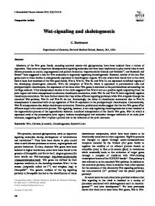
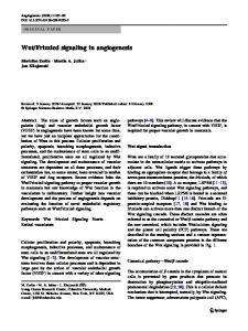
Figure 1. Overview of the Wnt signaling pathway. In the “Wnt-Off” state, Wnt/receptor interactions are interrupted by secreted Wnt antagonists, including sFRPs, DKKs and WIF1; and β-catenin is degraded by the formation of the “destruction complex” consisting of APC protein, Axin and others. In the “Wnt-On” state, Wnts are are lipid modified by the acyl transferase porcupine in the endoplasmic reticulum, and act in an autocrine and paracrine fashion. The Wnts form a complex with Frizzled receptor and coreceptors LRP5/LRP6. Upon receptor activation, the “destruction complex” which includes APC, Axin and others is inhibited thereby blocking β-catenin phosphorylation for degradation. This inhibition results in cytoplasmic β-catenin stabilization and accummulation which facilitates its translocation into the nucleus. Nuclear β-catenin acts as a transcriptional co-activator for LEF1/TCF, leading gene transcription of Wnt target genes, such as JUN, Cyclin-D1, and MMP7. In addition, Wnts bind to tyrosine-protein kinase transmembrane receptors ROR2 and RYK to activate other non-canonical planar cell polarity, the Wnt/JNK and the Wnt/Calcium pathways.
out of 186) of prostate cance specimens from radical prostatectomy were positive nuclear β-catenin. Finally, Jung et al. [45] reported that ADT-treated patients who exhibited short times to PSA progression expressed higher levels of MMP-7. The expression of cytoplasmic β-catenin, MMP-7, and AR was positively correlated. In the above section, we summarized studies that reported increased expression of nuclear β-catenin in CRPC; however, there were other groups that observed an opposite trend. Assikis et al. [46] examined β-catenin expression on a tissue microarray from 16 patients who underwent salvage surgery for symptomatic, locally aggressive androgen-independent prostate
3
cancer and found no nuclear β-catenin expression in these specimens. Whitaker et al. [47] also examined for nuclear β-catenin staining in 17 specimens from patients before hormone therapy and 13 from patients after hormone therapy, and also found no significant differences in the nuclear β-catenin staining between hormone sensitive and hormone relapsed tissues. In prostate cancer specimens that were most likely from hormone naïve patients, Bismar et al. did not detect any nuclear β-catenin staining in 101 prostatic adenocarcinomas, including 72 acinar and 29 ductal, and 16 cases of high-grade prostatic intraepithelial neoplasia (HPIN) using identical immunostaining procedures [48]. Membranous β-catenin act primarily as a cell adhesion molecule. The Am J Clin Exp Urol 2014;2(1):XXX-XXX
Wnt signaling and castration-resistant prostate cancer loss of membranous β-catenin in a small fraction of prostatic adenocarcinomas with higher Gleason scores was found in this study compared to normal prostatic epithelium. This result suggests a differential mechanism of β-catenin from its role as a cofactor for TCF/ LEF or AR. Jaggi et al. [49] also examined 17 samples of prostate cancer specimens and found a significant down-regulation of membranous β-catenin expression in prostate cancer compared to benign prostatic glands and an associatation with increasing Gleason grade (p = 0.025). In a prostate cancer prognosis study Horvath et al. [50] showed 64% (149 out of 232) of prostate cancer specimens with more than 10% of cells expressing nuclear β‑catenin. However, those patients who had less than 10% of cells expressing β-catenin in the nucleus had decreased biochemical relapse-free survival times. In this study only 17.7% (41 cases) of these prostate cancer specimens were from patients with androgen deprivation therapy, therefore, this study did not separate hormone treatment naïve specimens from CRPC specimens. In summary, the evidence suggests that there may be a role for nuclear localized β-catenin in CRPC specimens. However, the results remain conflicting. Perhaps the sample sizes and variations in the specimen processing and immunohistochemical staining methods may be contributing to the variations in the reported results with respect to β-catenin nuclear localization in CRPC. β-catenin and androgen receptor interaction β-Catenin contains 12 armadillo repeats in a highly conserved central region of the protein [33]. These armadillo repeats not only form a single structural unit to provide the interaction sites with APC, E-cadherin, and TCF/LEFs, but also can adopt an α-helical conformation for nuclear receptor binding proteins [33]. Using a yeast two-hybrid system, Yang et al. [26] demonstrated that β-catenin preferentially and directly bound to the ligand binding domain of AR in the presence of dyhydrotestosterone (DHT) over several other steroid hormone receptors which included estrogen receptor a, progesterone receptor β, and glucocorticoid receptor. This study indicated that the NH(2) terminus and the first 6 armadillo repeats of β-catenin were required components for the AR 4
interaction. The interaction between β-catenin and AR was further confirmed by several other studies showing that β-catenin bound to the activation function 2 region of the AR ligand binding domain and modulated the transcriptional effects of the transcriptional intermediary factor 2 (TIF2) and the AR N-terminal domain. Importantly, a single AR lysine (K720) has been shown to be necessary for the AR/βcatenin and TIF2 interactions [25, 30, 33]. The interactions between β-catenin and AR can be modulated by other cofactors through different signaling pathways in prostate cancer cells as well. β-Catenin interacts with AR in close proximity to the binding groove for p160 coactivators such as TIF2/glucocorticoid receptor interacting protein-1 (GRIP1) [51-54]. TIF2/ GRIP1 is one of the three p160 primary coactivator proteins, which serves as a scaffold to recruit a variety of secondary coactivators, including the protein acetyltransferases p300, CBP, and coactivator-associated arginine methyltransferase (CARM1). p300/CBP and CARM1 are reruited by the p160 complex to remodel chromatin through acetylation and methylation of histones and then functions in synergy with β-catenin as coactivators for AR and TCF/LEFs [53, 54]. The methyltransferase activity of CARM1 has also been shown to be necessary for its synergistic coactivator function with β-catenin to activate AR mediated transcription [53]. ICAT (β-catenin-interacting protein 1), an inhibitor of β-catenin and TCF, can inhibit the canonical Wnt/β-catenin signaling pathway by binding to β-catenin [27]. Expression of ICAT was observed in human prostate cancer tissues and found to be elevated in xenograft tumors in castrated mice [27]. Zhou et al. [27] showed that ICAT and AR can form a ternary complex with β-catenin and stabilize the β-catenin-AR complex, which resulted in enhanced AR-mediated transcription and cell growth. The DEAD box RNA helicase p68 (Ddx5) is often over expressed in prostate cancer tissues compared with benign tissue and studies have shown that Ddx5 is also a transcriptional co-activator of AR [55]. Interestingly, Clark et al. [56] demonstrated that the interaction between Ddx5 and β-catenin required the presence of androgens in androgen-sensitive LNCaP cells as wells as other cell lines such as LNCap AI (a CRPC derivative of LNCaP cell line) when the cells are Am J Clin Exp Urol 2014;2(1):XXX-XXX
Wnt signaling and castration-resistant prostate cancer grown in the absence of androgen. Therefore, the function of Ddx5 was shown to be required for recruitment of AR and β-catenin to the promoter regions of androgen responsive genes for AR mediated transcription. An AR variant [N-terminal truncated isoform of AR (AR45)], with an altered N-terminal domain with a replacement by a unique, short, seven amino-acid-long stretch, has been identified [57]. Overexpression of AR45 was shown to interact with the full-length AR and inhibit AR transcriptional activity and inhibit the growth of LNCaP cells [58]. However, under the conditions of β-catenin overexpression, AR45 increased dihydrotestosterone mediated AR promoter activity [58]. This result suggested that AR splicling variants may have differential effect on prostate cancer cell growth under β-catenin overexpression or overactivity. Recent success in clinical trials of second generation of the anti-androgen drugs Abiraterone and Enzalutamide strongly support that the aberrant activation of the AR pathway in the absence of high circulating levels of androgen plays a critical role in CRPC [7, 8]. It has been suggested that AR signaling in CRPC is sustained by development of AR amplication, mutation, alternate spilcing, and several alternative molecular mechanisms [6, 10]. When prostate cancer cells have been adapted to the low androgen environment, β-catenin has been shown to act as a coactivator of AR to enhance AR transcriptional activity not only in the presence of DHT, but also in the presence of androstenedione, a weaker adrenal androgen remaining present in CRPC patients [26, 30, 33]. β-Catenin is also one of the three AR coactivators (other two AR specific coactivators are ARA70 and ARA55) that can enhance AR transcriptional activity in LNCaP cells when treated with 17β-estradiol [26, 30, 33]. In addition, β-catenin can function as a coactivator with altered ARs with mutations W741C and T877A in prostate cancer cell lines [33]. These AR mutations were detected in CRPC patients that have been treated with bicalutamide leading to the W741C mutation and also in CRPC patients with lymph node metastatic lesions containing the T877A mutation [33]. In rodent studies, Chesire et al. [22] reported that castrated mice receiving androgen treatment exhibited nuclear co-localization of AR and β-catenin in normal prostatic epithelium. Nuclear β-catenin local5
ization was found to occur concomitantly with androgen-induced regrowth of normal rat prostate from androgen deprivation induced regression. Furthermore, Wang et al. [20] observed increased expression and nuclear colocalization of AR and β-catenin as well as the interaction between endogenous AR and β-catenin in CRPC from castrated mice. However, they found no interaction or colocalization of AR and β-catenin in xenografts from noncastrated mice. Mutations of β-catenin are uncommon in prostate cancer (< 5%) [59, 60]. Taken together, these results suggested that β-catenin plays an integral role in formation of the androgen-receptor transcriptional complex in CRPC. Based on available information, we proposed a simiplied relationship between Wnt and AR signaling during prostate cancr development and progression as summarized in Figure 2. AR and Wnt signaling may reinforce each other to elicit specific target genes for promoting androgen-independent growth and progression. As such, β-catenin/AR interactions could have distinct clinical relevance and be a potential therapeutic target for treatment of CRPC, especially working best at low androgens. The cross-talk between β-catenin and multiple pathways in prostate cancer Nuclear β-catenin can also arise through other mechanisms besides alterations in the canonical Wnt signaling pathway. In this section we consider examples of the accumulation of β-catenin through other mechanisms. Constitutive protein kinase B (also known as AKT) activation in prostate cancer due to loss of PTEN can inhibit GSK-3β activity leading to stabilization and nuclear accumulation of β-catenin [61]. Liu et al [62] demonstrated that addition of H2-relaxin to LNCaP cells resulted in increased phosphorylation of protein kinase B (Akt) and phosphorylation of glycogen synthase kinase-3β (GSK-3β) with subsequent cytoplasmic accumulation of β-catenin. This is followed by nuclear translocation, formation of the β-catenin/AR complex and increased AR transcriptional activity in LNCaP cells. Paradoxically, in neuronal cells, Pawlowski et al [63] demonstrated that ligand-bound AR promoted the accumulation of β-catenin in the nucleus, The nuclear co-localization of AR and β‑catenin was independent of the GSK-3β, p42/44 ERK mitogen-activated protein kinase, and phosphatiAm J Clin Exp Urol 2014;2(1):XXX-XXX
Wnt signaling and castration-resistant prostate cancer
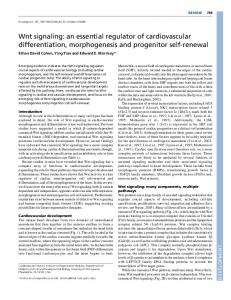
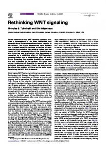
Figure 2. A simiplified and hypothetical relationship between Wnt and AR signaling during prostate cancer development and progression. In the normal prostate, Wnt signaling maintains prostate progenitor cells through regulation of Wnt target gene transcription, whereas AR signaling only functions in secretory luminal epithelial cells. In hormone treatment naïve prostate cancer cells, Wnt signaling promotes transcription of AR target genes, while androgen signaling inhibits the transcription of Wnt target genes. In CRPC, AR and Wnt signaling reinforces each other to elicit specific target genes for promoting androgen-independent growth and progression.
dylinositol 3-kinase pathways. Other groups have shown that GSK-3β can phosphorylate AR and suppress its ability to activate transcription under certain conditions. Wnt pathway activation can stimulate Akt activity which promotes an MDM-2-mediated degradation process that reduces AR protein levels in Wnt-stimulated prostate cancer cells [64, 65].
shown to be selectively expressed in apoptosisresistant and hormone-resistant human prostate cancer cells and tissues [70]. Expression of cytoplasmic protocadherin-PC can induce expression of Wnt target genes through interacting and stabilizing β-catenin [70]. These mechanisms illustrate different means of directing gene regulation involving β-catenin.
Adenomatous polyposis coli (APC) is a major regulator of β-catenin protein level through phosphorylation of β-catenin which signals the degradation bof β-catenin. Inactivation of APC gene by hypermethylation was detected in prostate cancer but not in normal prostate tissues [66, 67] thereby favoring the accumulation of β-catenin. However, Mulholland et al. [68] reported that AR can promote β-catenin nuclear translocation independently of APC. Pin1 is a peptidyl-prolyl cis/trans isomerase that stabilizes beta-catenin by inhibiting its binding to the APC gene product and subsequent GSK-3β-dependent degradation. The expression of Pin1in radical prostatectomy specimens is strongly correlated with the incidence of recurrence and metastasis [69]. Pin1 stabilizes β-catenin and abrogates the β-catenin and AR interaction, leading to increased β-catenin/TCF-4 signaling and increased expression of the WNT target genes c-Myc and TCF-4 istself [69]. The result is exclusive to PTEN-deficient LNCaP cells. Protocadherin-PC, localized on the human Y chromosome, was
Hypoxia-inducible factor-1α (HIF-1α) is a transcription factor that plays an essential role in cellular reponse to hypoxia. HIF-1α is known to enhance β-catenin activated AR transactivation in hypoxia [71]. During cellular hypoxia, increased expression of complexes composed of HIF-1α, AR and β-catenin in the nucleus were observed and activated androgen responsive genes. Knockdown of HIF-1α attenuated the recruitment of AR and β-catenin to the androgen response elements (AREs) and deactivated activation of androgen-responsive genes [71]. β‑Catenin can also interact with FOXO transcriptional factors in response to oxidative stress to promote cells exit from the cell cycle and entry into cell quiescence [72]. FOXA1 was shown to be overexpressed in advanced prostate cancer and metastases, and over expression of FOXA1 led to enrichment of the Wnt signaling pathway [73]. Heterogeneous nuclear ribonucleoprotein K (HnRNP K) was also found to be overexpressed in prostate cancer tissues and overexpression of HnRNP K positively associated with high Gleason score and poor
6
Am J Clin Exp Urol 2014;2(1):XXX-XXX
Wnt signaling and castration-resistant prostate cancer AR activator of 55 kDa (ARA55), also named hydrogen Mechanisms References peroxide-induced clone 5 Nuclear β-catenin localization [32, 41] (HIC-5), belongs to the paxillin AR/β-catenin interaction [26, 32, 35, 68] family of LIM proteins and is LEF1 overexpression [83] also a component of the focal Crosstalk between β-catenin and AKT, HIF-1α and others [69-75] adhesion complex [80, 81]. Overexpression of Wnt ligands and receptors [23, 94-106] ARA-55 can bind to AR resultParacrine Wnt signaling [36, 37, 91-93] ing in an increase of AR transcriptional activity and in turn Loss of secreted Wnt antagonists [136] alter ligand specificity of AR Epithelial to mesenchymal transition [123, 124, 137] [80, 81]. The LIM domainCancer stem cells [134] containing C-terminal half of To Author: Please provide Table 1 tag in main text. ARA-55 binds to a conserved prognosis [74, 75]. HnRNP K can bind to the alternatively spliced exon in LEF/TCF transcripβ-catenin/TCF-4 complex for regulation of pretion factor and functions as negative regulator mRNA splicing and some types of alternative of a subset of LEF/TCF family members [80, splicing have been suggested to promote pros81]. Possible function significance of these diftate cancer progression [74, 75]. Finally, ferent LEF/TCF transcription complexes requiβ-catenin can also influence the metastatic res further investigation. potential of prostate cancer cells by suppresThe Lymphoid Enhancer Factor 1 (LEF-1) is sion of transcription of a metastasis suppresanother member of TCF/LEF family and also a sor, KAI1, through formation of a β-cateninWnt target gene [82]. Li and Lee et al reported reptin chromatin remodeling complex [76]. that LEF1 mRNA levels are up 100 fold higher in LNCaP AI cells (a CRPC derivative of LNCaP TCF family memebers in prostate cancer cells) compared to the parental LNCaP cells as determined by a microarray gene expression Despite extensive research, the crosstalks profiling analysis [82]. LEF-1 expression was between AR and Wnt/β-catenin signaling pathassociated with increased cell proliferation, ways remain complex and conflicting. The migration, and invasion as well as AR exprescanonical Wnt pathway is mediated by TCF/ sion [82, 83]. LEF‑1 is usually expressed in the LEF-1 transcription factor family members basal epithelial layer of the urogenital sinus in which include TCF-1, TCF-3, TCF-4 and LEF-1 the human fetal prostate and in the urogenital [38]. The human AR gene itself has been shown mesenchyme and the basal epithelial layer of to be a Wnt target gene [65]. Activation of Wnt/ the urogenital sinus in mouse prostate developβ-catenin signaling by Wnt stimulation can ment. The survival of LEF-1-expressing basal increase AR expression via TCF/LEF-1-binding cells was not affected by treatment with the sites on the AR promoter, leading to upregulaanti-androgen bicalutamide. Moreover LEF-1 tion of AR target genes [65]. On the contrary, can repopulate the luminal compartment folAR can also compete with TCF/LEF-1 for lowing bicalutamide-indured regression of β-catenin binding and thus inhibit TCF/LEF-1 branching morphogenesis in the absence of mediated transcription [77]. Chesire et al androgen signaling [84]. This result suggested showed that anti-androgens alleviated the AR that LEF-1 may be involved in producing an mediated suppression of TCF transcriptional androgen-independent population of prostate activity and activation of TCF/LEF-1 inhibited progenitors. TMPRSS2-ERG fusion protein has the expression of AR-regulated genes [40, 78]. recently been shown to be reactivated in CRPC Other studies demonstrated that there is a [85, 86]. Wu et al. [87] provided evidence that direct interaction between the AR DNA binding LEF-1 is a critical transcriptional target of the domain and TCF-4 and that endogenous AR is ETS-family transcription factor ERG and that bound to the TCF responsive element in the LEF-1 expression is selectively upregulated in MYC promoter [79]. Ectopic expression of TCF-4 TMPRSS2-ERG fusion positive prostate cancer by transfection repressed the transcriptional as observed by microarray profiling analysis. activity of full-length AR, which was only parThe combined results suggested that LEF-1 tially attenuated by β-catenin transfection [79]. Table 1. Proposed mechanisms for elevated Wnt signaling in CRPC
7
Am J Clin Exp Urol 2014;2(1):XXX-XXX
Wnt signaling and castration-resistant prostate cancer thermore, Grasso et al. [34] performed whole-exome sequSources Percentage of positive staing References encing on 50 metaMetastatic tumors from autopsy 24% (5/21), nuclear [40] static CRPCs obtaiCRPC from TURP 38.8% (35/90), nuclear [41] ned at rapid autopsy CRPC bone metastases 40.7% (11/27), nuclear [43] (including three difCRPC matched pairs 55% (16/29), nuclear and cytoplasmic [35] ferent foci from the Localized PCa from RP 23% (49/212), nuclear [41] same patient) and 11 treatment-naïve, Localized PCa from RP 18% (39/186), nuclear [50] high-grade localized PCa: prostate cancer; RP: radical prostatectomy; CRPC: castration resistant prostate cancer; TURP: transurethral resections of the prostate. prostate cancers. They also identified To Author: Please provide Table 2 tag in main text. components of the Wnt signaling pathway to be significantly mutatand Wnt signaling may provide a good novel tared (57 somatic mutations in 38 samples) in gets for treatment of prostate cancer patients CRPC, on the other hand, Wnt pathway alterawith TMPRSS2-ERG fusion expression. tions were virtually absent in hormone naïve Expression profiling and second generation primary prostate cancer. In conclusion, these seqencing identifies activation of the Wnt results suggested that mutations in the Wnt pathway in CRPC pathway likely emerge during progression or the development of resistance after ADT in Recent gene expression profiling analyses have prostate cancer. allowed for the identification specific molecular Table 2. Nuclear β-catenin localization in CRPC and hormone naïve prostate cancer
signatures that are associated with CRPC. Wang et al. [20] performed affymetrix genechip analysis using LNCaP xenografts and hollow fiber models to identify global changes in gene expression profiling associated with CRPC. They found that the Wnt/β-catenin signaling pathway was one of the major pathways activated in CRPC. Rajan et al. [35] performed RNA sequencing (RNA-seq) profiling on tumour-rich, targeted prostatic biopsies from 7 patients with locally advanced or metastatic prostate cancer before and approximately 22 weeks after ADT. The results showed that 29 (e.g. FZD4, FZD7, JUN, and MMP7) out of 150 genes in the Wnt signalling pathway were upregulated after ADT, which was the top pathway with significantly upregulated genes. In addition, 14 of these upregulated genes were reported in previously published studies on ADT-driven gene expression changes [88, 89]. These data suggested that the Wnt signaling pathway is predominantly upregulated in CRPC. By using whole-exome sequencing technology to compare castration-resistant and androgensensitive matched pairs of prostate cancer xenografts derived from the same site of origin, Kumar et al. [90] found that 86 gene mutations were unique to CRPCs. Among them, there was a significant enrichment of mutations in the components of the Wnt pathway in CRPC tumors, including FZD6, GSK3B and WNT6. Fur8
Wnt paracrine signaling from prostatic stroma affects prostate epithelium in the settings of resistance to ADT or chemotherapy There are emerging studies indicating that Wnt paracrine signaling from neigboring prostatic stroma cells could affect prostate epithlium during prostate cancer initiation and development of resistance to ADT or chemotherapy [36, 37, 91]. Using a tissue recombination method, Zong et al. [92] showed that highmobility group AT-hook 2 (HMGA2) overexpressing urogenital sinus mesenchymal (UGSM) promoted mutifoci prostatic intraepithlial neoplasia (PIN) in the neighboring naïve epithelium. Co-overexpression of HMGA2 and AR in UGSM cells synergistically induced poorly differentiated prostate adenocarcinoma. Wnt ligands (i.e. WNT2, 4 and 9A) were shown to be the major paracrine factors from HMGA2 overexpressing UGSM. HMGA2-induced PIN formation was strongly inhibited by overexpression of the Wnt antagonists DKK1 and SFRP2, both secreted products. This study suggested that Wnt paracrine signaling may play an important stroma contribution to prostate cancer initiation and progression. In a related study, Li et al. [36] demonstrated that stroma-specific knockout mice for the TGF-β type II receptor expression (Tgfbr2fspKO) increased the expression of Wnt3a to promote PIN formation and tumorigenesis, and that systemic treatment with Wnt3a neuAm J Clin Exp Urol 2014;2(1):XXX-XXX
Wnt signaling and castration-resistant prostate cancer tralizing antibodies inhibited growth of LNCaP/ Tgfbr2fspKO xenografts. Placencio et al. [91] from the same group further demonstrated that the prostates of Tgfbr2fspKO mice had constitutively active Wnt signaling regardless of androgen status. The prostates of Tgfbr2fspKO as well as Tgfbr2fspKO prostatic stromal cells/wild-type or SV40 large T antigen expressing epithelia recombinants were resistant to androgen deprivation-mediated regression. These results suggested that the paracrine Wnt signaling from Tgfbr2fspKO prostate stroma cells not only facilitated the progression of PIN lesions to adenocarcinoma, but also confired resistance to the epithelial component to androgen deprivation. Liu et al. [93] studied the effect of dihydrotestosterone (DHT) on the interactions between preosteoblasts MC3T3 cells and bone metastasis cell line MDA-PCa-2b. They found that DHT exerted more potent growth stimulating effect on MDA-PCa-2b cells via upregulation of Wnt activity in bone cells. The effect was enhanced when the prostate cancer cells were cocultured with preosteoblasts compared to DHT treatment of MDA-PCa-2b cells alone. The enhanced growth of MDA-PCa-2b cells by DHT in this coculture experiment can be blocked by exogenous Wnt antagonists such as DKK-1 and SFRP-1 recombinant proteins. This result indicated a potential role of paracrine Wnt factors from bone cells on prostate cancer cell growth at bone metastatic sites. Sun et al. [37] showed that mitoxantrone and docetaxel therapy induced expression of stromal WNT16B and the elevated expression levels of WNT16B in prostatectomy tissue samples were associated with high risk of cancer recurrence. WNT16B expression in the stroma can also promote epithelial-to-mesenchymal transition (EMT) to increase tumor invasiveness and tumor growth. These findings provided a new mechanism of acquired resistance to chemotherapy drugs that is based on the properties of the tumor microenvironment. Wnt ligands, receptors, secreted Wnt antagonists in prostate cancer There is accumulating data showing overexpression of Wnt ligands and FZD receptors in prostate cancer potentially form autocrine or paracrine loops that support prostate cancer progression. The expression of WNT1 was detected in prostate cancer cells, tissues,
9
lymph nodes and bone metastases, and the expression positively correlated with high Gleason scores and high serum PSA levels [48]. Elevated expression of Wnt agonists: WNT5A, WNT2, WNT6, and WNT11 have also been detected in prostate cancer tissues versus normal samples [23, 94-96]. Wnt agonists like WNT5A and WNT11 can induce the non-canonical Wnt pathway (e.g. through the JNK pathway) [23]. Interestingly, WNT11 inhibited androgen-dependent but not androgen-independent prostate cancer cell growth [23]; whereas, WNT3A stimulation enhanced AR activity and prostate cancer cell growth in presence of low androgen levels [23]. WNT11 can be regulated by androgens and WNT11 can induce expression of neuroendocrine differentiation markers NSE and ASCL1 as well as promote cell invasion [97]. WNT5A can also activate Wnt/Ca2+ pathway via CaMKII [98]. Yamamoto et al. [99] showed that WNT5A overexpression enhanced cell invasion in prostate cancer cell lines (i.e. PC3 cells), which required the expression of Wnt receptors Frizzled-2 and Ror2. Abnormal expression of WNT5A was positively correlated with high Gleason scores and biochemical relapse of prostate cancer [99]. Using transgenic mouse models, Takahashi et al. [100] demonstrated that the introduction of the AR with the T877A mutation into epithelial cells of the TRAMP mice resulted in an accelerated onset of tumor formation and tumor growth, moreover, this effect of the AR T877A mutation can be blocked by crossing Wnt-5a haploinsufficient mice with the TRAMP mice. In contrast, Syed Khaja et al. [101] reported that overexpression of WNT5A protein in patients with localized prostate cancer was shown to predict a favorable outcome after surgery. Recombinant WNT5A treatment of 22Rv1 and DU145 cells resulted in a decreased invasion [101]. These results suggested that Wnts may act differentially in a context-dependent maner during prostate cancer progression. There are several Wnt receptors that are expressed in normal prostate tissues which includes FZD-1, -4, -6, and -10. Out of these receptors, expression of FZD-4 and FZD-6 were found to be increased in prostate tumors [102105]. Gupta et al. [106] reported that ERG oncogenic transcriptional factor regulated the expression of FZD-4, which mediated epithelial-to-mesenchymal transition in prostate cancer.
Am J Clin Exp Urol 2014;2(1):XXX-XXX
Wnt signaling and castration-resistant prostate cancer Secreted Wnt antagonists, which include the secreted frizzled-related protein (sFRP) family, Dickkopf (DKK) family, and Wnt inhibitory factor-1 (WIF-1), are negative modulators of Wnt signaling [107-109]. Wnts bind to FZDs via cysteine-rich domain (CRD) sequence with high affinity. Expression of CRD alone can inhibit Wnt/β-catenin signaling [107]. All sFRPs contain a CRD sequence and can inhibit Wnt signaling either by sequestering Wnt ligands or by forming nonfunctional complexes with Frizzled receptors [107]. The Dkk family proteins include DKK-1, -2, -3, and -4 in humans [108]. DKK-1 inhibits Wnt signaling by disrupting the binding of LRP6 to the Wnt/FZD ligand-receptor complex [108]. Although WIF-1 does not share any sequence similarity with the CRD sequence of FZDs and sFRPs, it can also bind to Wnts and inhibit signaling [109]. Down-regulation of sFRPs, DKKs and WIF-1 by gene deletion or promoter hypermethylation are frequently detected in many human cancers including prostate cancer [110-114], suggesting their possible role as tumor suppressors. Endogenous Dkk-3 was found to be required to limit cell proliferation both in the developing mouse prostate and in 3D cultures of human prostate epithelial cells [115]. DKK-3 was further shown to regulate the response of normal prostate epithelial cells to transforming growth factor-β (TGF-β) [116]. These studies are consistent with a model in which DKK-3 is required by normal cells to prevent the TGF-β-dependent switch from tumor suppressor to tumor promoter. In contrast, DKK-1 was shown to promote tumor growth and prostate cancer progression in part by suppression of p21 (CIP1/WAF1) through a mechanism independent of canonical Wnt signaling [117]. DKK-1 also inhibited Wnt induced osteoblastic activity [118]. DKK-1 appears to play a role in parathyroid hormone related protein (PTHrP) induced osteolytic activity and in transition from osteoblastic to osteolytic bone lesions [119]. The sFRP family member sFRP1 was also found to be down-regulated both in prostate cancer tissues and prostate cancer cell lines. SFRP1 can function as a negative regulator of the AR [120]. However, this effect of SFRP1 was not associated with Wnt inhibition [120]. Joesting et al. [121] found that sFRP1 was overexpressed in prostate cancer stromal cells and ovexpression of sFRP1 activated JNK pathway, but not the canonical Wnt pathway. Horvath et al [50] reported that sFRP4
10
overexpression can decrease cell proliferation, anchorage-independent growth, and invasiveness in PC3 cells. SFRP3/FRZB was the first identied secreted Wnt antagonist during studies of the Spemann’s organizer of Xenopus [122]. Our group has shown that expression of sFRP3/FRZB or WIF-1 in a CRPC cell line PC3 caused a reversal of epithelial-to-mesenchymal transition and inhibition of tumor growth by inhibition of canonical Wnt pathway [123, 124]. Taken together, these results suggested that the role of Wnt ligands and secreted antagonists work in context-dependent manner whether in different types of cells or by varied ligand-receptor interactions. Wnt ligands and receptors appear to be important in CRPC. Wnt signaling in disease models of CRPC In transgenic mouse models, conditionally deleted exon3 of β-catenin resulted in production of high-grade PIN (HG-PIN) and induction of Foxa2 re-expression in the adult mouse prostate through Wnt/β-catenin signaling as well as promoting prostate growth even under the conditions of androgen deprivation [125]. In mouse models with the SV40 large T-antigen, which inactivates p53 and Rb [126], or in mice expressing mutated K-ras and form invasive carcinoma [127], or in mice with loss of PTEN expression [128], β-catenin overexpression can promote highly invasive prostate cancer and squamous metaplasia, even in the absence of androgens. These findings provided strong evidence for a critical role of the Wnt/β-catenin signaling in prostate cancer development and progression. In xenograft mouse models, H2 relaxin (RLN2) was shown to facilitate castrate-resistant growth of prostate cancer cells through AKT phosphorylation-mediated activation of both the Wnt/β-cateinin and the AR pathway [129]. As mentioned before Hic-5/ARA55 is a co-factor for both TCF/LEFs and AR and can inhibit the Wnt/β-catenin pathway [36]. Overexpresssion of HIC-5/ARA55 in LNCaP cells can restore sensitivity of xenograft composed of LNCaP cells and Tgfbr2-KO fibroblasts to androgen deprivation-induced tumor regression [130]. In an orthotopic C4-2B CRPC xenograft mouse model, Placencio et al. [131] found that mesenchymal stem cells (MSCs) were recruited into tumor sites and were associated with enhanced tumor growth. The result occurs with
Am J Clin Exp Urol 2014;2(1):XXX-XXX
Wnt signaling and castration-resistant prostate cancer activation of Wnt signaling. When MSCs were used as a targeted delivery vector for the exogenously expressed Sfrp2, tumor growth was reduced and the response to androgen deprivation was restored. These animal and tissue culture studies suggested that components of the Wnt signaling pathway may be involved in prostate cancer progression to more invasive phenotype and contributed to castration resistance. Therapeutic potentials of targeting Wnt/βcatenin in CRPC and future directions Although, the results remain inconclusive, β-catenin nuclear localization as well as its co‑localization with AR has been more frequently observed in CRPC compared to hormone naïve prostate cancer. Alterations in multiple signaling pathways, including PI3K/AKT, HIF-1α, PIN1, APC gene silencing and more have been shown to cause nuclear localization of β-catenin and activation of Wnt signaling. Moreover, β-catenin acts as an AR co-factor to enhance androgen-stimulated AR transcriptional activation and increase sensitivity to low levels of androgens and to non-androgen ligands. As a result, the interaction between β-catenin and AR in CRPC may elicit specific target genes for promoting androgen-independent growth and progression. The next-generation sequencing technology (i.e. whole-exome and RNA sequencing) has revealed that the Wnt pathway is one of the top signaling pathways that were frequently mutated or genomically altered in lethal CRPC patients. Likewise, paracrine Wnt signaling also contributed to resistance to ADT after chemotherapy and androgen deprivation therapy. In addition to high expression in cancer cells, Wnt/β-catenin signaling is highly activated in cancer stem cells (CSCs) [132]. Prostate CSCs are suggested to be resistant to androgen deprivation therapy and responsible for cancer recurrence [133]. Targeting CSCs by inhibition of the Wnt pathway may have the potential to reduce the self-renewal and aggressive behavior of prostate cancer [134]. As a proof of princle, Lee et al [135] demonstrated that a novel compound that disrupts both β-catenin/TCF and β-catenin/AR protein interactions can inhibit prostate tumor growth in a xenograft model and also blocked bicalutamide-resistant sphere-forming cells. This study indicated the
11
potential of targeting the β-catenin/AR as a good treatment target for CRPC. As described in this review, the Wnt signaling pathway plays a complex role in CRPC. Given the multiple important roles of Wnt signaling in CRPC, the Wnt signaling pathway can not be ignored as a source of therapeutic targets. Inhibition of the Wnt pathway would allow therapies to target not only epithelial cells but also stromal cells, as well as, CSCs, androgen-depedent, and androgen-independent prostate cancer cells. Future therapies for CRPC would most likely benefit from combination of both antiandrogens and Wnt inhibitors. Acknowledgements We apologize for not being able to cite all of the publications in the field due to the limitations of the length of the review. NY is supported by DOD grant W81XWH-13-1-0257. XZ is supported by UCI ICTS pilot grant, DOD grant W81XWH-11-1-0312 and NIH grants R01CA122558 and R21CA152804. Disclosure of conflict of interest None. Address correspondence to: Xiaolin Zi, Department of Urology, University of California, Irvine, 101 The City Drive South, Rt. 81 Bldg. 55 Rm. 302, Orange CA 92868, USA. Tel: 714-456-8316; Fax: 714-4561786; E-mail:
[email protected]
References [1] [2]
[3]
[4]
[5]
Siegel R, Naishadham D, Jemal A. Cancer statistics, 2013. CA Cancer J Clin 2013; 63: 1130. Sakr WA, Grignon DJ, Crissman JD, Heilbrun LK, Cassin BJ, Pontes JJ, Haas GP. High grade prostatic intraepithelial neoplasia (HGPIN) and prostatic adenocarcinoma between the ages of 20-69: an autopsy study of 249 cases. In Vivo 1994; 8: 439-43. Lennernas B, Edgren M, Haggman M, Norlen BJ, Nilsson S. Postoperative radiotherapy after prostatectomy--a review. Scand J Urol Nephrol 2003; 37: 10-5. Ahlering TE, Skarecky DW. Long-term outcome of detectable PSA levels after radical prostatectomy. Prostate Cancer Prostatic Dis 2005; 8: 163-6. Kozlowski JM, Ellis WJ, Grayhack JT. Advanced prostatic carcinoma. Early versus late endo-
Am J Clin Exp Urol 2014;2(1):XXX-XXX
Wnt signaling and castration-resistant prostate cancer
[6]
[7]
[8]
[9]
[10]
[11]
[12]
[13]
[14]
12
crine therapy. Urol Clin North Am 1991; 18: 1524. Karantanos T, Corn PG, Thompson TC. Prostate cancer progression after androgen deprivation therapy: mechanisms of castrate resistance and novel therapeutic approaches. Oncogene 2013; 32: 5501-11. de Bono JS, Logothetis CJ, Molina A, Fizazi K, North S, Chu L, Chi KN, Jones RJ, Goodman OB Jr, Saad F, Staffurth JN, Mainwaring P, Harland S, Flaig TW, Hutson TE, Cheng T, Patterson H, Hainsworth JD, Ryan CJ, Sternberg CN, Ellard SL, Fléchon A, Saleh M, Scholz M, Efstathiou E, Zivi A, Bianchini D, Loriot Y, Chieffo N, Kheoh T, Haqq CM, Scher HI; COU-AA-301 Investigators. Abiraterone and increased survival in metastatic prostate cancer. N Engl J Med 2011; 364: 1995-2005. Scher HI, Fizazi K, Saad F, Taplin ME, Sternberg CN, Miller K, de Wit R, Mulders P, Chi KN, Shore ND, Armstrong AJ, Flaig TW, Fléchon A, Mainwaring P, Fleming M, Hainsworth JD, Hirmand M, Selby B, Seely L, de Bono JS; AFFIRM Investigators. Increased survival with enzalutamide in prostate cancer after chemotherapy. N Engl J Med 2012; 367: 1187-1197. Korpal M, Korn JM, Gao X, Rakiec DP, Ruddy DA, Doshi S, Yuan J, Kovats SG, Kim S, Cooke VG, Monahan JE, Stegmeier F, Roberts TM, Sellers WR, Zhou W, Zhu P. An F876L Mutation in Androgen Receptor Confers Genetic and Phenotypic Resistance to MDV3100 (Enzalutamide). Cancer Discov 2013; 3: 1030-43. Yuan X, Cai C, Chen S, Chen S, Yu Z, Balk SP. Androgen receptor functions in castration-resistant prostate cancer and mechanisms of resistance to new agents targeting the androgen axis. Oncogene 2013; [Epub ahead of print]. Nyquist MD, Li Y, Hwang TH, Manlove LS, Vessella RL, Silverstein KA, Voytas DF, Dehm SM. TALEN-engineered AR gene rearrangements reveal endocrine uncoupling of androgen receptor in prostate cancer. Proc Natl Acad Sci U S A 2013; 110: 17492-7. Nadiminty N, Tummala R, Liu C, Yang J, Lou W, Evans CP, Gao AC. NF-κB2/p52 induces resistance to enzalutamide in prostate cancer: role of androgen receptor and its variants. Mol Cancer Ther 2013; 12: 1629-37. Joseph JD, Lu N, Qian J, Sensintaffar J, Shao G, Brigham D, Moon M, Maneval EC, Chen I, Darimont B, Hager JH. A Clinically Relevant Androgen Receptor Mutation Confers Resistance to Second-Generation Antiandrogens Enzalutamide and ARN-509. Cancer Discov 2013; 3: 1020-9. Li Y, Chan SC, Brand LJ, Hwang TH, Silverstein KA, Dehm SM. Androgen receptor splice vari-
[15]
[16] [17]
[18]
[19] [20]
[21]
[22] [23]
[24]
[25]
[26]
[27]
ants mediate enzalutamide resistance in castration-resistant prostate cancer cell lines. Cancer Res 2013; 73: 483-9. Zhang X, Morrissey C, Sun S, Ketchandji M, Nelson PS, True LD, Vakar-Lopez F, Vessella RL, Plymate SR. Androgen receptor variants occur frequently in castration resistant prostate cancer metastases. PLoS One 2011; 6: e27970. Colloca G. Role of androgens in abiraterone resistance. J Clin Oncol 2012; 30: 3561-2. Li R, Evaul K, Sharma KK, Chang KH, Yoshimoto J, Liu J, Auchus RJ, Sharifi N. Abiraterone inhibits 3β-hydroxysteroid dehydrogenase: a rationale for increasing drug exposure in castration-resistant prostate cancer. Clin Cancer Res 2012; 18: 3571-9. Mostaghel EA, Marck BT, Plymate SR, Vessella RL, Balk S, Matsumoto AM, Nelson PS, Montgomery RB. Resistance to CYP17A1 inhibition with abiraterone in castration-resistant prostate cancer: induction of steroidogenesis and androgen receptor splice variants. Clin Cancer Res 2011; 17: 5913-25. Polakis P. Wnt signaling in cancer. Cold Spring Harb Perspect Biol 2012; 4: 5. Wang G, Wang J, Sadar MD. Crosstalk between the androgen receptor and beta-catenin in castrate-resistant prostate cancer. Cancer Res 2008; 68: 9918-27. Schweizer L, Rizzo CA, Spires TE, Platero JS, Wu Q, Lin TA, Gottardis MM, Attar RM. The androgen receptor can signal through Wnt/betaCatenin in prostate cancer cells as an adaptation mechanism to castration levels of androgens. BMC Cell Biol 2008; 9: 4. Chesire DR and Isaacs WB. Beta-catenin signaling in prostate cancer: an early perspective. Endocr Relat Cancer 2003; 10: 537-60. Zhu H, Mazor M, Kawano Y, Walker MM, Leung HY, Armstrong K, Waxman J, Kypta RM. Analysis of Wnt gene expression in prostate cancer: mutual inhibition by WNT11 and the androgen receptor. Cancer Res 2004; 64: 7918-26. Verras M, Brown J, Li X, Nusse R, Sun Z. Wnt3a growth factor induces androgen receptor-mediated transcription and enhances cell growth in human prostate cancer cells. Cancer Res 2004; 64: 8860-6. Truica CI, Byers S, Gelmann EP. Beta-catenin affects androgen receptor transcriptional activity and ligand specificity. Cancer Res 2000; 60: 4709-13. Yang F, Li X, Sharma M, Sasaki CY, Longo DL, Lim B, Sun Z. Linking beta-catenin to androgen-signaling pathway. J Biol Chem 2002; 277: 11336-44. Zhuo M, Zhu C, Sun J, Weis WI, Sun Z. The beta-catenin binding protein ICAT modulates an-
Am J Clin Exp Urol 2014;2(1):XXX-XXX
Wnt signaling and castration-resistant prostate cancer
[28]
[29]
[30]
[31]
[32]
[33]
[34]
[35]
[36]
[37]
13
drogen receptor activity. Mol Endocrinol 2011; 25: 1677-88. Liu S, Vinall RL, Tepper C, Shi XB, Xue LR, Ma AH, Wang LY, Fitzgerald LD, Wu Z, Gandour-Edwards R, deVere White RW, Kung HJ. Inappropriate activation of androgen receptor by relaxin via beta-catenin pathway. Oncogene 2008; 27: 499-505. Liu XH, Kirschenbaum A, Yao S, Liu G, Aaronson SA, Levine AC. Androgen-induced Wnt signaling in preosteoblasts promotes the growth of MDA-PCa-2b human prostate cancer cells. Cancer Res 2007; 67: 5747-53. Masiello D, Chen SY, Xu Y, Verhoeven MC, Choi E, Hollenberg AN, Balk SP. Recruitment of beta-catenin by wild-type or mutant androgen receptors correlates with ligand-stimulated growth of prostate cancer cells. Mol Endocrinol 2004; 18: 2388-401. Amir AL, Barua M, McKnight NC, Cheng S, Yuan X, Balk SP. A direct beta-catenin-independent interaction between androgen receptor and T cell factor 4. J Biol Chem 2003; 278: 3082834. Wan X, Liu J, Lu JF, Tzelepi V, Yang J, Starbuck MW, Diao L, Wang J, Efstathiou E, Vazquez ES, Troncoso P, Maity SN, Navone NM. Activation of β-catenin signaling in androgen receptornegative prostate cancer cells. Clin Cancer Res 2012; 18: 726-36. Song LN, Herrell R, Byers S, Shah S, Wilson EM, Gelmann EP. Beta-catenin binds to the activation function 2 region of the androgen receptor and modulates the effects of the N-terminal domain and TIF2 on ligand-dependent transcription. Mol Cell Biol 2003; 23: 1674-87. Grasso CS, Wu YM, Robinson DR, Cao X, Dhanasekaran SM, Khan AP, Quist MJ, Jing X, Lonigro RJ, Brenner JC, Asangani IA, Ateeq B, Chun SY, Siddiqui J, Sam L, Anstett M, Mehra R, Prensner JR, Palanisamy N, Ryslik GA, Vandin F, Raphael BJ, Kunju LP, Rhodes DR, Pienta KJ, Chinnaiyan AM, Tomlins SA. The mutational landscape of lethal castration-resistant prostate cancer. Nature 2012; 487: 239-43. Rajan P, Sudbery IM, Villasevil ME, Mui E, Fleming J, Davis M, Ahmad I, Edwards J, Sansom OJ, Sims D, Ponting CP, Heger A, McMenemin RM, Pedley ID, Leung HY. Next-generation Sequencing of Advanced Prostate Cancer Treated with Androgen-deprivation Therapy. Eur Urol 2013; [Epub ahead of print]. Li X, Placencio V, Iturregui JM, Uwamariya C, Sharif-Afshar AR, Koyama T, Hayward SW, Bhowmick NA. Prostate tumor progression is mediated by a paracrine TGF-beta/Wnt3a signaling axis. Oncogene 2008; 27: 7118-30. Sun Y, Campisi J, Higano C, Beer TM, Porter P, Coleman I, True L, Nelson PS. Treatment-in-
[38]
[39] [40]
[41]
[42]
[43]
[44]
[45]
[46]
[47]
[48]
duced damage to the tumor microenvironment promotes prostate cancer therapy resistance through WNT16B. Nat Med 2012; 18: 135968. Sprowl S, Waterman ML. Past visits present: TCF/LEFs partner with ATFs for β-cateninindependent activity. PLoS Genet 2013; 9: e1003745. Gómez-Orte E, Sáenz-Narciso B, Moreno S, Cabello J. Multiple functions of the noncanonical Wnt pathway. Trends Genet 2013; 29: 545-53. Chesire DR, Ewing CM, Gage WR, Isaacs WB. In vitro evidence for complex modes of nuclear beta-catenin signaling during prostate growth and tumorigenesis. Oncogene 2002; 21: 2679-94. de la Taille A, Rubin MA, Chen MW, Vacherot F, de Medina SG, Burchardt M, Buttyan R, Chopin D. Beta-catenin-related anomalies in apoptosis-resistant and hormone-refractory prostate cancer cells. Clin Cancer Res 2003; 9: 1801-7. Patriarca C, Petrella D, Campo B, Colombo P, Giunta P, Parente M, Zucchini N, Mazzucchelli R, Montironi R. Elevated E-cadherin and alpha/beta-catenin expression after androgen deprivation therapy in prostate adenocarcinoma. Pathol Res Pract 2003; 199: 659-65. Chen G, Shukeir N, Potti A, Sircar K, Aprikian A, Goltzman D, Rabbani SA. Up-regulation of Wnt1 and beta-catenin production in patients with advanced metastatic prostate carcinoma: potential pathogenetic and prognostic implications. Cancer 2004; 101: 1345-56. Aaltomaa S, Lipponen P, Kärjä V, Lundstedt S, Lappi J, Kosma VM. The expression and prognostic value of alpha-, beta- and gamma-catenins in renal cell carcinoma. Anticancer Res 2004; 24: 2407-13. Jung SJ, Oh S, Lee GT, Chung J, Min K, Yoon J, Kim W, Ryu DS, Kim IY, Kang DI. Clinical Significance of Wnt/β-Catenin Signalling and Androgen Receptor Expression in Prostate Cancer. World J Mens Health 2013; 31: 36-46. Assikis VJ, Do KA, Wen S, Wang X, Cho-Vega JH, Brisbay S, Lopez R, Logothetis CJ, Troncoso P, Papandreou CN, McDonnell TJ. Clinical and biomarker correlates of androgen-independent, locally aggressive prostate cancer with limited metastatic potential. Clin Cancer Res 2004; 10: 6770-8. Whitaker HC, Girling J, Warren AY, Leung H, Mills IG, Neal DE. Alterations in beta-catenin expression and localization in prostate cancer. Prostate 2008; 68: 1196-205. Bismar TA, HumphreyPA, Grignon DJ, Wang HL. Expression of beta-catenin in prostatic adenocarcinomas: A comparison with colorectal adenocarcinomas. Am J Clin Pathol 2004; 121: 557-563.
Am J Clin Exp Urol 2014;2(1):XXX-XXX
Wnt signaling and castration-resistant prostate cancer [49] Jaggi M, Johansson SL, Baker JJ, Smith LM, Galich A, Balaji KC. Aberrant expression of Ecadherin and beta-catenin in human prostate cancer. Urol Oncol 2005; 23: 402-6. [50] Horvath LG, Henshall SM, Lee CS, Kench JG, Golovsky D, Brenner PC, O’Neill GF, Kooner R, Stricker PD, Grygiel JJ, Sutherland RL. Lower levels of nuclear beta-catenin predict for a poorer prognosis in localized prostate cancer. Int J Cancer 2005; 113: 415-22. [51] Song LN, Gelmann EP. Interaction of betacatenin and TIF2/GRIP1 in transcriptional activation by the androgen receptor. J Biol Chem 2005; 280: 37853-67. [52] Yang CK, Kim JH, Li H, Stallcup MR. Differential use of functional domains by coiled-coil coactivator in its synergistic coactivator function with beta-catenin or GRIP1. J Biol Chem 2006; 281: 3389-97. [53] Koh SS, Li H, Lee YH, Widelitz RB, Chuong CM, Stallcup MR. Synergistic coactivator function by coactivator-associated arginine methyltransferase (CARM) 1 and beta-catenin with two different classes of DNA-binding transcriptional activators. J Biol Chem 2002; 277: 26031-5. [54] Labalette C, Renard CA, Neuveut C, Buendia MA, Wei Y. Interaction and functional cooperation between the LIM protein FHL2, CBP/p300, and beta-catenin. Mol Cell Biol 2004; 24: 10689-702. [55] Clark EL, Coulson A, Dalgliesh C, Rajan P, Nicol SM, Fleming S, Heer R, Gaughan L, Leung HY, Elliott DJ, Fuller-Pace FV, Robson CN. The RNA helicase p68 is a novel androgen receptor coactivator involved in splicing and is overexpressed in prostate cancer. Cancer Res 2008; 68: 7938-7946. [56] Clark EL, Hadjimichael C, Temperley R, Barnard A, Fuller-Pace FV, Robson CN. p68/DdX5 supports β-catenin & RNAP II during androgen receptor mediated transcription in prostate cancer. PLoS One 2013; 8: e54150. [57] Wu ZY, Chen K, Haendler B, McDonald TV, Bian JS. Stimulation of N-terminal truncated isoform of androgen receptor stabilizes human ether-ágo-go-related gene-encoded potassium channel protein via activation of extracellular signal regulated kinase 1/2. Endocrinology 2008; 149: 5061-9. [58] Ahrens-Fath I, Politz O, Geserick C, Haendler B. Androgen receptor function is modulated by the tissue-specific AR45 variant. FEBS J 2005 Jan; 272: 74-84. [59] Voeller HJ, Truica CI, Gelmann EP. Beta-catenin mutations in human prostate cancer. Cancer Res 1998; 58: 2520-3. [60] Chesire DR, Ewing CM, Sauvageot J, Bova GS, Isaacs WB. Detection and analysis of beta-
14
[61]
[62]
[63]
[64]
[65]
[66]
[67]
[68]
[69]
[70]
catenin mutations in prostate cancer. Prostate 2000; 45: 323-34. Sharma M, Chuang WW, Sun Z. Phosphatidylinositol 3-kinase/Akt stimulates androgen pathway through GSK3beta inhibition and nuclear beta-catenin accumulation. J Biol Chem 2002; 277: 30935-41. Liu S, Vinall RL, Tepper C, Shi XB, Xue LR, Ma AH, Wang LY, Fitzgerald LD, Wu Z, GandourEdwards R, deVere White RW, Kung HJ. Inappropriate activation of androgen receptor by relaxin via beta-catenin pathway. Oncogene 2008; 27: 499-505. Pawlowski JE, Ertel JR, Allen MP, Xu M, Butler C, Wilson EM, Wierman ME. Liganded androgen receptor interaction with beta-catenin: nuclear co-localization and modulation of transcriptional activity in neuronal cells. J Biol Chem 2002; 277: 20702-10. Lin HK, Wang L, Hu YC, Altuwaijri S, Chang C. Phosphorylation-dependent ubiquitylation and degradation of androgen receptor by Akt require Mdm2 E3 ligase. EMBO J 2002; 21: 4037-48. Yang X, Chen MW, Terry S, Vacherot F, Bemis DL, Capodice J, Kitajewski J, de la Taille A, Benson MC, Guo Y, Buttyan R. Complex regulation of human androgen receptor expression by Wnt signaling in prostate cancer cells. Oncogene 2006; 25: 3436-44. Henrique R, Ribeiro FR, Fonseca D, Hoque MO, Carvalho AL, Costa VL, Pinto M, Oliveira J, Teixeira MR, Sidransky D, Jerónimo C. High promoter methylation levels of APC predict poor prognosis in sextant biopsies from prostate cancer patients. Clin Cancer Res 2007; 13: 6122-9. Rosenbaum E, Hoque MO, Cohen Y, Zahurak M, Eisenberger MA, Epstein JI, Partin AW, Sidransky D. Promoter hypermethylation as an independent prognostic factor for relapse in patients with prostate cancer following radical prostatectomy. Clin Cancer Res 2005; 11: 8321-5. Mulholland DJ, Cheng H, Reid K, Rennie PS, Nelson CC. The androgen receptor can promote beta-catenin nuclear translocation independently of adenomatous polyposis coli. J Biol Chem 2002; 277: 17933-43. Chen SY, Wulf G, Zhou XZ, Rubin MA, Lu KP, Balk SP. Activation of beta-catenin signaling in prostate cancer by peptidyl-prolyl isomerase Pin1-mediated abrogation of the androgen receptor-beta-catenin interaction. Mol Cell Biol 2006; 26: 929-39. Thompson VC, Hurtado-Coll A, Turbin D, Fazli L, Lehman ML, Gleave ME, Nelson CC. Relaxin drives Wnt signaling through upregulation of
Am J Clin Exp Urol 2014;2(1):XXX-XXX
Wnt signaling and castration-resistant prostate cancer
[71]
[72]
[73]
[74]
[75]
[76]
[77]
[78]
[79]
[80]
[81]
15
PCDHY in prostate cancer. Prostate 2010; 70: 1134-45. Mitani T, Harada N, Nakano Y, Inui H, Yamaji R. Coordinated action of hypoxia-inducible factor1α and β-catenin in androgen receptor signaling. J Biol Chem 2012; 287: 33594-606. Essers MA, de Vries-Smits LM, Barker N, Polderman PE, Burgering BM, Korswagen HC. Functional interaction between beta-catenin and FOXO in oxidative stress signaling. Science 2005; 308: 1181-4. Robinson JL, Hickey TE, Warren AY, Vowler SL, Carroll T, Lamb AD, Papoutsoglou N, Neal DE, Tilley WD, Carroll JS. Elevated levels of FOXA1 facilitate androgen receptor chromatin binding resulting in a CRPC-like phenotype. Oncogene 2013; [Epub ahead of print]. Sato S, Idogawa M, Honda K, Fujii G, Kawashima H, Takekuma K, Hoshika A, Hirohashi S, Yamada T. beta-catenin interacts with the FUS proto-oncogene product and regulates premRNA splicing. Gastroenterology 2005; 129: 1225-36. Ciarlo M, Benelli R, Barbieri O, Minghelli S, Barboro P, Balbi C, Ferrari N. Regulation of neuroendocrine differentiation by AKT/hnRNPK/ AR/β-catenin signaling in prostate cancer cells. Int J Cancer 2012; 131: 582-90. Kim JH, Kim B, Cai L, Choi HJ, Ohgi KA, Tran C, Chen C, Chung CH, Huber O, Rose DW, Sawyers CL, Rosenfeld MG, Baek SH. Transcriptional regulation of a metastasis suppressor gene by Tip60 and beta-catenin complexes. Nature 2005; 434: 921-6. Mulholland DJ, Read JT, Rennie PS, Cox ME, Nelson CC. Functional localization and competition between the androgen receptor and Tcell factor for nuclear beta-catenin: a means for inhibition of the Tcf signaling axis. Oncogene 2003; 22: 5602-13. Chesire DR, Isaacs WB. Ligand-dependent inhibition of beta-catenin/TCF signaling by androgen receptor. Oncogene 2002; 21: 845369. Amir AL, Barua M, McKnight NC, Cheng S, Yuan X, Balk SP. A direct beta-catenin-independent interaction between androgen receptor and T cell factor 4. J Biol Chem 2003; 278: 3082834. Ghogomu SM, van Venrooy S, Ritthaler M, Wedlich D, Gradl D. HIC-5 is a novel repressor of lymphoid enhancer factor/T-cell factor-driven transcription. J Biol Chem 2006; 281: 1755-64. Li X, Martinez-Ferrer M, Botta V, Uwamariya C, Banerjee J, Bhowmick NA. Epithelial Hic-5/ ARA55 expression contributes to prostate tumorigenesis and castrate responsiveness. Oncogene 2011; 30: 167-77.
[82] Yang X, Chen MW, Terry S, Vacherot F, Chopin DK, Bemis DL, Kitajewski J, Benson MC, Guo Y, Buttyan R. A human- and male-specific protocadherin that acts through the wnt signaling pathway to induce neuroendocrine transdifferentiation of prostate cancer cells. Cancer Res 2005 Jun 15; 65: 5263-71. [83] Li Y, Wang L, Zhang M, Melamed J, Liu X, Reiter R, Wei J, Peng Y, Zou X, Pellicer A, Garabedian MJ, Ferrari A, Lee P. LEF1 in androgen-independent prostate cancer: regulation of androgen receptor expression, prostate cancer growth, and invasion. Cancer Res 2009; 69: 3332-8. [84] Wu X, Daniels G, Shapiro E, Xu K, Huang H, Li Y, Logan S, Greco MA, Peng Y, Monaco ME, Melamed J, Lepor H, Grishina I, Lee P. LEF1 identifies androgen-independent epithelium in the developing prostate. Mol Endocrinol 2011; 25: 1018-26. [85] Casey OM, Fang L, Hynes PG, Abou-Kheir WG, Martin PL, Tillman HS, Petrovics G, Awwad HO, Ward Y, Lake R, Zhang L, Kelly K. TMPRSS2driven ERG expression in vivo increases selfrenewal and maintains expression in a castration resistant subpopulation. PLoS One 2012; 7: e41668. [86] Qu X, Randhawa G, Friedman C, Kurland BF, Glaskova L, Coleman I, Mostaghel E, Higano CS, Porter C, Vessella R, Nelson PS, Fang M. A three-marker FISH panel detects more genetic aberrations of AR, PTEN and TMPRSS2/ERG in castration-resistant or metastatic prostate cancers than in primary prostate tumors. PLoS One 2013; 8: e74671. [87] Wu L, Zhao JC, Kim J, Jin HJ, Wang CY, Yu J. ERG is a critical regulator of Wnt/LEF1 signaling in prostate cancer. Cancer Res 2013; 73: 6068-79. [88] Holzbeierlein J, Lal P, LaTulippe E, Smith A, Satagopan J, Zhang L, Ryan C, Smith S, Scher H, Scardino P, Reuter V, Gerald WL. Gene expression analysis of human prostate carcinoma during hormonal therapy identifies androgenresponsive genes and mechanisms of therapy resistance. Am J Pathol 2004; 164: 217-227. [89] Lehmusvaara S, Erkkilä T, Urbanucci A, Waltering K, Seppälä J, Larjo A, Tuominen VJ, Isola J, Kujala P, Lähdesmäki H, Kaipia A, Tammela TLJ, Visakorpi T. Chemical castration and antiandrogens induce differential gene expression in prostate cancer. J Pathol 2012; 227: 336345. [90] Kumar A, White TA, MacKenzie AP, Clegg N, Lee C, Dumpit RF, Coleman I, Ng SB, Salipante SJ, Rieder MJ, Nickerson DA, Corey E, Lange PH, Morrissey C, Vessella RL, Nelson PS, Shendure J. Exome sequencing identifies a spectrum of mutation frequencies in advanced and
Am J Clin Exp Urol 2014;2(1):XXX-XXX
Wnt signaling and castration-resistant prostate cancer lethal prostate cancers. Proc Natl Acad Sci U S A 2011; 108: 17087-92. [91] Placencio VR, Sharif-Afshar AR, Li X, Huang H, Uwamariya C, Neilson EG, Shen MM, Matusik RJ, Hayward SW, Bhowmick NA. Stromal transforming growth factor-beta signaling mediates prostatic response to androgen ablation by paracrine Wnt activity. Cancer Res 2008; 68: 4709-18. [92] Zong Y, Huang J, Sankarasharma D, Morikawa T, Fukayama M, Epstein JI, Chada KK, Witte ON. Stromal epigenetic dysregulation is sufficient to initiate mouse prostate cancer via paracrine Wnt signaling. Proc Natl Acad Sci U S A 2012; 109: E3395-404. [93] Liu XH, Kirschenbaum A, Yao S, Liu G, Aaronson SA, Levine AC. Androgen-induced Wnt signaling in preosteoblasts promotes the growth of MDA-PCa-2b human prostate cancer cells. Cancer Res 2007; 67: 5747-53. [94] Katoh M. Frequent up-regulation of WNT2 in primary gastric cancer and colorectal cancer. Int J Oncol 2001; 19: 1003-7. [95] Iozzo RV, Eichstetter I, Danielson KG. Aberrant expression of the growth factor Wnt-5A in human malignancy. Cancer Res 1995; 55: 349599. [96] Hall CL, Bafico A, Dai J, Aaronson SA, Keller ET. Prostate cancer cells promote osteoblastic bone metastases through Wnts. Cancer Res 2005; 65: 7554-60. [97] Uysal-Onganer P, Kawano Y, Caro M, Walker MM, Diez S, Darrington RS, Waxman J, Kypta RM. Wnt-11 promotes neuroendocrine-like differentiation, survival and migration of prostate cancer cells. Mol Cancer 2010; 9: 55. [98] Wang Q, Symes AJ, Kane CA, Freeman A, Nariculam J, Munson P, Thrasivoulou C, Masters JR, Ahmed A. A novel role for Wnt/Ca2+ signaling in actin cytoskeleton remodeling and cell motility in prostate cancer. PLoS One 2010; 5: e10456. [99] Yamamoto H, Oue N, Sato A, Hasegawa Y, Yamamoto H, Matsubara A, Yasui W, Kikuchi A. Wnt5a signaling is involved in the aggressiveness of prostate cancer and expression of metalloproteinase. Oncogene 2010; 29: 203646. [100] Takahashi S, Watanabe T, Okada M, Inoue K, Ueda T, Takada I, Watabe T, Yamamoto Y, Fukuda T, Nakamura T, Akimoto C, Fujimura T, Hoshino M, Imai Y, Metzger D, Miyazono K, Minami Y, Chambon P, Kitamura T, Matsumoto T, Kato S. Noncanonical Wnt signaling mediates androgen-dependent tumor growth in a mouse model of prostate cancer. Proc Natl Acad Sci U S A 2011; 108: 4938-43. [101] Syed Khaja AS, Helczynski L, Edsjö A, Ehrnström R, Lindgren A, Ulmert D, Andersson T,
16
Bjartell A. Elevated level of Wnt5a protein in localized prostate cancer tissue is associated with better outcome. PLoS One 2011; 6: e26539. [102] Sagara N, Toda G, Hirai M, Terada M and Katoh M. Molecular cloning, differential expression, and chromosomal localization of human frizzled-1, frizzled-2, and frizzled-7. Biochem Biophys Res Commun 1998; 252: 117-122. [103] Kirikoshi H, Sagara N, Koike J, Tanaka K, Sekihara H, Hirai M, Katoh M. Molecular cloning and characterization of human Frizzled-4 on chromosome 11q14-q21. Biochem Biophys Res Commun 1999; 264: 955-961. [104] Tokuhara M, Hirai M, Atomi Y, Terada M and Katoh M. Molecular cloning of human Frizzled-6. Biochem Biophys Res Commun 1998; 243: 622-627. [105] Wissmann C, Wild PJ, Kaiser S, Roepcke S, Stoehr R, Woenckhaus M, Kristiansen G, Hsieh JC, Hofstaedter F, Hartmann A, Knuechel R, Rosenthal A, Pilarsky C. WIF1, a component of the Wnt pathway, is down-regulated in prostate, breast, lung, and bladder cancer. J Pathol 2003; 201: 204-12. [106] Gupta S, Iljin K, Sara H, Mpindi JP, Mirtti T, Vainio P, Rantala J, Alanen K, Nees M, Kallioniemi O. FZD4 as a mediator of ERG oncogeneinduced WNT signaling and epithelial-to-mesenchymal transition in human prostate cancer cells. Cancer Res 2010; 70: 6735-45. [107] Jones SE, Jomary C. Secreted Frizzled-related proteins: searching for relationships and patterns. Bioessays 2002; 24: 811-20. [108] Kawano Y, Kypta R. Secreted antagonists of the Wnt signalling pathway. J Cell Sci 2003; 116: 2627-34. [109] Hsieh JC, Kodjabachian L, Rebbert ML, Rattner A, Smallwood PM, Samos CH, Nusse R, Dawid IB, Nathans J. A new secreted protein that binds to Wnt proteins and inhibits their activities. Nature 1999; 398: 431-6. [110] Suzuki H, Watkins DN, Jair KW, Schuebel KE, Markowitz SD, Chen WD, Pretlow TP, Yang B, Akiyama Y, Van Engeland M, Toyota M, Tokino T, Hinoda Y, Imai K, Herman JG, Baylin SB. Epigenetic inactivation of SFRP genes allows constitutive WNT signaling in colorectal cancer. Nat Genet 2004; 36: 417-22. [111] Stoehr R, Wissmann C. Deletions of chromosome 8p and loss of sFRP1 expression are progression markers of papillary bladder cancer. Lab Invest 2004; 84: 465-78. [112] Rawson JB, Manno M, Mrkonjic M, Daftary D, Dicks E, Buchanan DD, Younghusband HB, Parfrey PS, Young JP, Pollett A, Green RC, Gallinger S, McLaughlin JR, Knight JA, Bapat B. Promoter methylation of Wnt antagonists DKK1 and SFRP1 is associated with opposing
Am J Clin Exp Urol 2014;2(1):XXX-XXX
Wnt signaling and castration-resistant prostate cancer tumor subtypes in two large populations of colorectal cancer patients. Carcinogenesis 2011; 32: 741-7. [113] Urakami S, Shiina H, Enokida H. Epigenetic inactivation of Wnt inhibitory factor-1 plays an important role in bladder cancer through aberrant canonical Wnt/beta-catenin signaling pathway. Clin Cancer Res 2006; 12: 383-91. [114] Lodygin D, Epanchintsev A, Menssen A, Diebold J, Hermeking H. Functional epigenomics identifies genes frequently silenced in prostate cancer. Cancer Res 2005; 65: 4218-27. [115] Kawano Y, Kitaoka M, Hamada Y, Walker MM, Waxman J, Kypta RM. Regulation of prostate cell growth and morphogenesis by Dickkopf-3. Oncogene 2006; 25: 6528-37. [116] Romero D, Kawano Y, Bengoa N, Walker MM, Maltry N, Niehrs C, Waxman J, Kypta R. Downregulation of Dickkopf-3 disrupts prostate acinar morphogenesis through TGF-β/Smad signalling. J Cell Sci 2013; 126: 1858-67. [117] Hall CL, Zhang H, Baile S, Ljungman M, Kuhstoss S, Keller ET. p21CIP-1/WAF-1 induction is required to inhibit prostate cancer growth elicited by deficient expression of the Wnt inhibitor Dickkopf-1. Cancer Res 2010; 70: 9916-26. [118] Thudi NK, Martin CK, Murahari S, Shu ST, Lanigan LG, Werbeck JL, Keller ET, McCauley LK, Pinzone JJ, Rosol TJ. Dickkopf-1 (DKK-1) stimulated prostate cancer growth and metastasis and inhibited bone formation in osteoblastic bone metastases. Prostate 2011; 71: 615-25. [119] Zhang H, Yu C, Dai J, Keller JM, Hua A, Sottnik JL, Shelley G, Hall CL, Park SI, Yao Z, Zhang J, McCauley LK, Keller ET. Parathyroid hormonerelated protein inhibits DKK1 expression through c-Jun-mediated inhibition of β-catenin activation of the DKK1 promoter in prostate cancer. Oncogene 2013; [Epub ahead of print]. [120] Kawano Y, Diez S, Uysal-Onganer P, Darrington RS, Waxman J, Kypta RM. Secreted Frizzledrelated protein-1 is a negative regulator of androgen receptor activity in prostate cancer. Br J Cancer 2009; 100: 11650-74. [121] Joesting MS, Perrin S, Elenbaas B, Fawell SE, Rubin JS, Franco OE, Hayward SW, Cunha GR, Marker PC. Identification of SFRP1 as a candidate mediator of stromal-to-epithelial signaling in prostate cancer. Cancer Res 2005; 65: 10423-30. [122] Wang S, Krinks M, Lin K, Luyten FP, Moos M Jr. Frzb, a secreted protein expressed in the Spemann organizer, binds and inhibits Wnt-8. Cell 1997; 88: 757-66. [123] Zi X, Guo Y, Simoneau AR, Hope C, Xie J, Holcombe RF, Hoang BH. Expression of Frzb/secreted Frizzled-related protein 3, a secreted Wnt antagonist, in human androgen-independent prostate cancer PC-3 cells suppresses
17
tumor growth and cellular invasiveness. Cancer Res 2005; 65: 9762-70. [124] Yee DS, Tang Y, Li X, Liu Z, Guo Y, Ghaffar S, McQueen P, Atreya D, Xie J, Simoneau AR, Hoang BH, Zi X. The Wnt inhibitory factor 1 restoration in prostate cancer cells was associated with reduced tumor growth, decreased capacity of cell migration and invasion and a reversal of epithelial to mesenchymal transition. Mol Cancer 2010; 9: 162. [125] Yu X, Wang Y, Jiang M, Bierie B, Roy-Burman P, Shen MM, Taketo MM, Wills M, Matusik RJ. Activation of beta-Catenin in mouse prostate causes HGPIN and continuous prostate growth after castration. Prostate 2009; 69: 249-262. [126] Yu X, Wang Y, DeGraff DJ, Wills ML, Matusik RJ. Wnt/beta-catenin activation promotes prostate tumor progression in a mouse model. Oncogene 2011; 30: 1868-1879. [127] Pearson HB, Phesse TJ, Clarke AR K-ras and Wnt signaling synergize to accelerate prostate tumorigenesis in the mouse. Cancer Res 2009; 69: 94-101. [128] Francis JC, Thomsen MK, Taketo MM, Swain A. β-catenin is required for prostate development and cooperates with Pten loss to drive invasive carcinoma. PLoS Genet 2013; 9: e1003180. [129] Vinall RL, Mahaffey CM, Davis RR, Luo Z, Gandour-Edwards R, Ghosh PM, Tepper CG, de Vere White RW. Dual blockade of PKA and NFκB inhibits H2 relaxin-mediated castrate-resistant growth of prostate cancer sublines and induces apoptosis. Horm Cancer 2011; 2: 224-38. [130] Li X, Martinez-Ferrer M, Botta V, Uwamariya C, Banerjee J, Bhowmick NA. Epithelial Hic-5/ ARA55 expression contributes to prostate tumorigenesis and castrate responsiveness. Oncogene 2011 Jan 13; 30: 167-77. [131] Placencio VR, Li X, Sherrill TP, Fritz G, Bhowmick NA. Bone marrow derived mesenchymal stem cells incorporate into the prostate during regrowth. PLoS One 2010; 5: e12920. [132] Bisson I, Prowse DM. WNT signaling regulates self-renewal and differentiation of prostate cancer cells with stem cell characteristics. Cell Res 2009 Jun; 19: 683-97. [133] Lawson DA, Witte ON. Stem cells in prostate cancer initiation and progression. J Clin Invest 2007; 117: 2044-50. [134] Lawson DA, Zong Y, Memarzadeh S, Xin L, Huang J, Witte ON. Basal epithelial stem cells are efficient targets for prostate cancer initiation. Proc Natl Acad Sci U S A 2010; 107: 26105. [135] Lee E, Madar A, David G, Garabedian MJ, Dasgupta R, Logan SK. Inhibition of androgen receptor and β-catenin activity in prostate can-
Am J Clin Exp Urol 2014;2(1):XXX-XXX
Wnt signaling and castration-resistant prostate cancer cer. Proc Natl Acad Sci U S A 2013; 110: 15710-5. [136] Perry AS, O’Hurley G, Raheem OA, Brennan K, Wong S, O’Grady A, Kennedy AM, Marignol L, Murphy TM, Sullivan L, Barrett C, Loftus B, Thornhill J, Hewitt SM, Lawler M, Kay E, Lynch T, Hollywood D. Gene expression and epigenetic discovery screen reveal methylation of SFRP2 in prostate cancer. Int J Cancer 2013; 132: 1771-80.
18
[137] Ju X, Casimiro MC, Gormley M, Meng H, Jiao X, Katiyar S, Crosariol M, Chen K, Wang M, Quong AA, Lisanti MP, Ertel A, Pestell RG. Identification of a cyclin D1 network in prostate cancer that antagonizes epithelial-mesenchymal restraint. Cancer Res 2014 Jan 15; 74: 508-19.
Am J Clin Exp Urol 2014;2(1):XXX-XXX