Sep 3, 2009 - from the Tertiary period (Azar et al. 1999). Approximately 48 species of Psychodidae fossils had formally been described by 1994, 6 of them ...
MORPHOLOGY, SYSTEMATICS, EVOLUTION
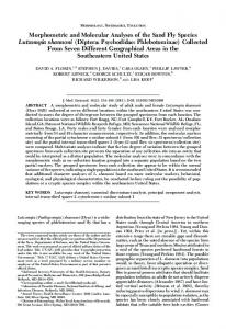
Review of American Fossil Phlebotominae (Diptera: Psychodidae) With a Description of Two New Species JOSE´ DILERMANDO ANDRADE FILHO,1 EUNICE A BIANCHI GALATI,2 AND REGINALDO PEC ¸ ANHA BRAZIL3
J. Med. Entomol. 46(5): 969Ð979 (2009)
ABSTRACT The objective of this study was to carry out a taxonomic review of fossil American phlebotomine sand ßies and describe two new species found in amber in the Dominican Republic. The gonostyle of one of these, Micropygomyia dorafeliciangeliae nov. sp., (⫽Lutzomyia dorafeliciangeliae, species group oswaldoi), has Þve spines, similar to that of Micropygomyia paterna (Quate, 1963) (⫽Lutzomyia paterna, species group oswaldoi), but they may be distinguished by the alpha/gamma ratio, which is ⬍1.0 in the new species and ⬎1 in the latter. Pintomyia dominicana nov. sp. (⫽Lutzomyia dominicana, species group verrucarum) has four spines on the gonostyle and presents a long bristle on the apex of the paramere, which distinguishes it from the other fossil species. With the description of these two new species, a total of 14 species of the American fossil phlebotomine sand ßies has been described, 10 of which belong to the genus Pintomyia. An identiÞcation key for male fossil species is presented. KEY WORDS Micropygomyia dorafeliciangeliae nov. sp., Pintomyia dominicana nov. sp., amber, fossil, Phlebotominae
The subfamily Phlebotominae constitutes a group of insects of importance to public health, because several of its species are vectors of bacteria, viruses, and especially Leishmania, protozoans implicated as etiological agents of leishmaniases throughout the world, thus motivating biological, ecological, and systematic studies of its various species. Slightly ⬎900 species or subspecies, inhabiting different ecosystems, have been described, ⬇500 of them occurring in the Neotropical region of the world. The great morphological and ecological diversity of the phlebotomines undoubtedly reßects their long evolutionary history, the group having arisen on earth 250 Ð 300 million years (myr) ago (Andrade Filho and Brazil 2003). As a consequence of this long evolutionary history, these insects present morphological variations that reßect their adaptation to diverse environments. Although members of the family Psychodidae are small insects, the fossil record of the group has been documented (Brazil and Andrade Filho 2002), specimens having been found most commonly in amber from the Tertiary period (Azar et al. 1999). Approximately 48 species of Psychodidae fossils had formally been described by 1994, 6 of them being Phlebotominae. However, with the recent description of more species (Azar et al. 1999; Brazil and Andrade 1 Corresponding author: Laborato ´ rio de Leishmanioses, Instituto Rene´ Rachou-Fiocruz, Av. Augusto de Lima 1715, CEP 30190-002 Belo Horizonte, MG, Brazil (e-mail: jandrade@cpqrr.Þocruz.br). 2 Departamento de Epidemiologia, Faculdade de Sau ´ de Pu´ blica, Universidade de Sa˜o Paulo, Sa˜o Paulo, SP, Brazil. 3 Departamento de Bioquõ´mica e Biologia Molecular, Instituto Oswaldo Cruz-Fiocruz, Rio de Janeiro, RJ, Brazil.
Filho 2002; Andrade Filho et al. 2004, 2006a, b, 2007, 2008; Poinar 2004; Pen˜ alver and Grimaldi 2005), this number has increased to 15. Phlebotomites brevifilis Hennig, 1972; Phlebotomites longifilis Hennig, 1972; Mesophlebotomites hennigi Azar, Solignac, Paicheler, and Bouchet, 1999; and Libanophlebotomus lutfallahi Azar, Solignac, Paicheler, and Bouchet, 1999 have been described from Lebanese cretaceous amber (Hennig 1972, Azar et al. 1999). Phlebotomiella tipuliforme (Meunier, 1905) has been described from Baltic tertiary amber (Meunier 1905). Another species described from the Cretaceous period is Palaeomyia burmitis Poinar, 2004, found in Burmese amber (Poinar 2004). The other species of sand ßy found in amber belonging to extant genera are Phlebotomus pungens (Loew, 1845), a subfossil of the Holocene; Sergentomyia succini Stuckenberg, 1975 of the Eocene (Loew 1845, Stuckenberg 1975), both from the Old World; Micropygomyia paterna (Quate, 1963) from Mexican Oligocene/Miocene amber; Pintomyia falcaorum Brazil and Andrade Filho, 2002; Trichopygomyia killickorum Andrade Filho, Falca˜o, and Brazil, 2004; Lutzomyia filipalpis Pen˜ alver and Grimaldi, 2005; Lutzomyia succini Pen˜ alver and Grimaldi, 2005; Lutzomyia miocena Pen˜ alver and Grimaldi, 2005; Lutzomyia paleopestis Pen˜ alver and Grimaldi, 2005; Lutzomyia schleei Pen˜ alver and Grimaldi, 2005; Pintomyia brazilorum Andrade Filho, Galati, and Falca˜o, 2006; Pintomyia paleotownsendi Andrade Filho, Falca˜o, Galati, and Brazil, 2006; Pintomyia paleotrichia Andrade Filho, Brazil, Falca˜o, and Galati, 2007; and Micropygomyia brandaoi Andrade Filho, Galati, Falca˜o, and Brazil,
0022-2585/09/0969Ð0979$04.00/0 䉷 2009 Entomological Society of America
970
JOURNAL OF MEDICAL ENTOMOLOGY
Vol. 46, no. 5
Fig. 1. General aspect of M. dorafeliciangeliae nov.sp. (holotype). (Online Þgure in color.)
2008, all of these species from Dominican Republic Miocene amber (Quate 1963; Brazil and Andrade Filho 2002; Andrade Filho et al. 2004, 2006a, b, 2007, 2008; Pen˜ alver and Grimaldi 2005). Besides these, Young and Lawyer (1987) recorded 14 fossil specimens from the Dominican Republic belonging to two species, and Antoine et al. (2006) found one species of phlebotomine sand ßy from Miocene amber from Peru, but these species were not described by the respective authors. The objective of this study was to carry out a taxonomic review of these fossils based on data from the literature and on microscopic examination of the phlebotomine fossil material. Descriptions are also provided for two more species from the Dominican Republic. Materials and Methods Review of the fossil species of American phlebotomines was based on direct observation under the microscope of specimens deposited in the Phlebotomine collection of the Instituto Rene´ Rachou. Information on the morphological characteristics of the other species was obtained from Quate (1963) and Pen˜ alver and Grimaldi (2005). One of the new species was found in a piece of amber measuring 2.5 by 1.2 by 0.3 mm, containing the male holotype and a male paratype. The other new species was found in a concave piece of amber that contained six phlebotomines (Þve males and a female). These latter phlebotomines were not well preserved, and the form of the amber also impedes more detailed study, so that few structures described in this study have been measured. One of the males was elected as holotype and the other four as paratypes. It
was not possible to associate the female with these specimens because of its imperfect state of preservation. The insects were examined under the microscope and measured using an ocular micrometer calibrated for this purpose. Drawings were made using the same microscope with the aid of a camera lucida and photographs taken with Konica digital equipment with a deÞnition of 8.0 megapixels, the photographs being taken directly through the eyepiece of the microscope. The measurements are given in micrometers. Some morphological details that help to distinguish fossil from extant species have been provided. A dichotomous key is provided to assist in the identiÞcation of the fossil species. The authors have listed species names in accordance with the classiÞcation system of Galati (1995, 2003), followed by the corresponding nomenclature of Young and Duncan (1994) in brackets when cited for the Þrst time. The new species will be deposited in the phlebotomine collection of the Instituto Rene´ Rachou, Fundac¸ a˜o Oswaldo Cruz, Belo Horizonte, Minas Gerais State, Brazil. Results Family Psychodidae Newman, 1835 Subfamily Phlebotominae Kerte´sz, 1903 Micropygomyia (Sauromyia) dorafeliciangeliae nov. sp. Andrade Filho, Galati, and Brazil, 2008 (Figs. 1Ð 6) (⫽Lutzomyia dorafeliciangeliae species group oswaldoi) Description. Male (holotype, Figs. 1Ð 6). Total length 1,298 m. The coloration of the insect not was indistinguishable.
September 2009
ANDRADE FILHO ET AL.: REVIEW OF FOSSIL PHLEBOTOMINES
971
Fig. 2. Terminalia of M. dorafeliciangeliae nov.sp. (holotype), showing the gonostyle with Þve spines. (Online Þgure in color.)
Head (Lateral View). Clypeus 83 m and labrumepipharynx 118 m long. Flagellomeres: AIII, 187 m; AIV, 94 m; AV, 83 m; AXVI longer than AXV. Interocular distance was not measured because of the lateral position of the phlebotomine heads. Ascoids visible in AVÐAX, simple, inserted at the same level, and very short, not reaching the middle of the segment. Papilla absent on AV and AXIII and present on AIII and AIV. Several papillae on AXIV, AXV, and AXVI. Palpal formula 1.4.2.3.5; palpomere lengths as follows: Þrst, 30; second, 113; third, 118; fourth, 83; Þfth, 250. NewsteadÕs spines grouped on basal third of third palpomere. Cervix. Ventro-cervical sensillae could not be observed. Thorax. Median and hind legs lost. Lengths of femur, tibia, basitarsus, and tarsomeres II ⫹ III ⫹ IV ⫹ V of the foreleg 463, 598, 339, and 463 m, respectively. Wing 1,332 m long by 294 m at maximum width. Principal alar indices in micrometers: alpha, 147; beta, 181; gamma, 226; delta, 23; R5, 847. Abdomen. Gonocoxite 154 m long by 33 m wide, without tuft of bristles. Gonostyle 81 m long, presenting Þve spines: two apical (one of them smaller and Þner than the other), one external superior, one external inferior, and one internal, the last two inserted at the same level, in the middle of the structure. Paramere length 171 m and shorter than the lateral lobe (184 m long). The ejaculatory pump, genital Þlaments, and aedeagus could not be observed. Etymology. This species was named Micropygomyia dorafeliciangeliae nov. sp. in tribute to Dr. Maria Dora Feliciangeli, Senior Lecturer at the Facultad de Ciencias de la Salud, Centro Nacional de Referencia de Flebo´ tomos y Otros Vectores, Universidad de Carabobo, Maracay, Venezuela, who has made a great contribution to our knowledge of the American phlebotomines.
Type Material. HOLOTYPE: one male, Dominican Republic, north of Santiago, specimens in Miocene amber; PARATYPE: one male, same data. Remarks. The presence of NewsteadÕs spines inserted together on the basal half of the third palpomere and the absence of the papilla on AV leads us to include the new species in the subtribe Sergentomyiina. The presence of Þve spines on the gonostyle, gonocoxite without tuft of bristles, and AIII shorter than length of head permit the inclusion of the new species within the genus Micropygomyia, subgenus Sauromyia, series oswaldoi (⫽genus Lutzomyia, species group oswaldoi). With respect to extant species, M. dorafeliciangeliae nov. sp. can be distinguished from Micropygomyia peresi (Mangabeira 1942) (⫽Lutzomyia peresi, species group oswaldoi) by the clypeus, which is shorter than the labrum-epipharynx. It can be distinguished from Micropygomyia quinquefer (Dyar, 1929) (⫽Lutzomyia quinquefer, species group oswaldoi), Micropygomyia ferreirana (Barretto, Martins, and Pellegrino, 1956) (⫽Lutzomyia ferreirana, species group oswaldoi), Micropygomyia saccai (Feliciangeli, Ramõ´rez Pe´rez, and Ramirez, 1989) (⫽Lutzomyia saccai, species group oswaldoi), Micropygomyia quechua (Martins, Llanos, and Silva, 1975) (⫽Lutzomyia quechua, species group oswaldoi), Micropygomyia rorotaensis (Floch and Abonnenc, 1944) (⫽Lutzomyia rorotaensis, species group oswaldoi), and Micropygomyia longipennis (Barretto, 1946) (⫽Lutzomyia longipennis, species group oswaldoi) by the absence of bristles on the gonocoxite. The R5/wing length ratios, 2.88 in the new species and 3.60 in Micropygomyia machupicchu (Martins, Llanos, and Silva, 1975) (⫽Lutzomyia machupicchu, species group oswaldoi), distinguish them from one
972
JOURNAL OF MEDICAL ENTOMOLOGY
Vol. 46, no. 5
Figs. 3–6. Micropygomyia dorafeliciangeliae nov.sp. (holotype). (3) Terminalia (bar ⫽ 100). (4) Third, fourth, and Þfth palpomeres (bar ⫽ 100). (5) VI antennomere (bar ⫽ 100). (6) Wing (bar ⫽ 200).
another. The species Micropygomyia pratti (Vargas and Dias-Na´ jera, 1951) (⫽Lutzomyia pratti, species group oswaldoi) has the lower external spine of the gonostyle inserted at a more apical level than the internal one, whereas in the new species they are at the same level. In Micropygomyia zikani (Barretto, 1950) (⫽Lutzomyia zikani, species group oswaldoi), there is a small projection, absent in the new species, on the ventral margin of the paramere. The terminalia of Micropygomyia villelai (Mangabeira, 1942) (⫽Lutzomyia villelai, species group oswaldoi), Micropygomyia trinidadensis (Newstead, 1922) (⫽Lutzomyia trinidadensis, species group oswaldoi), Micropygomyia oswaldoi (Mangabeira, 1942) (⫽Lutzomyia oswaldoi, species group oswaldoi), and Micropygomyia capixaba (Dias, Falca˜ o, Silva, and Martins, 1987) (⫽Lutzomyia capixaba, species group oswaldoi) are longer (⬎300 m long)
than those of Mi. dorafeliciangeliae nov. sp. (⬍250 m). The fossil species of Micropygomyia pusilla (Dias, Martins, Falca˜ o, and Silva, 1986) (⫽Lutzomyia pusilla, species group oswaldoi) may be distinguished by its smaller thorax and abdomen, which together measure ⬇1,140 m, whereas in M. pusilla, they measure 1,648 Ð1,976 m (n ⫽ 5). Regarding the fossil species, all of them present four spines on the gonostyle, except Micropygomyia paterna (Quate, 1963) (⫽Lutzomyia paterna, species group oswaldoi), which is found in Mexican amber and is morphologically very similar to the new species (Quate 1963). The drawings in the original description of this species are very rudimentary and provide little morphological information, although the alar indices are sufÞcient to differentiate them. The alpha/gamma ratio is 0.65 in M. dorafeliciangeliae nov. sp. and 1.05 in M. paterna.
September 2009
ANDRADE FILHO ET AL.: REVIEW OF FOSSIL PHLEBOTOMINES
973
Fig. 7. General aspect of Pintomyia dominicana nov.sp. (holotype). (Online Þgure in color.)
Pintomyia (Pifanomyia) dominicana nov. sp. Andrade Filho, Galati and Brazil, 2008 (Figs. 7Ð11) (⫽Lutzomyia dominicana, species group verrucarum) Description. Male (Holotype, Figs. 6Ð11). Total length 1,524 m. The color of the insect was indistinguishable. Head (Lateral View). The head is 248 m, clypeus is 79 m, and labrum-epipharynx is 192 m long. Flagellomere lengths as follows: AIII, 248 m; AIV, 112 m; AV, 112 m, AXV ⫽ AXVI. Interocular distance not measured because of position of the head. Ascoids of AVÐX
simple, inserted at same level and very short, not reaching middle of segment. Papilla absent on AVÐXIII but present on AIII and AIV. Several papillae on AXIV, AXV, and AXVI. Palpal formula 1.4.2.3.5, ßagellomere lengths as follows: Þrst, 33 m; second, 125 m; third, 135 m; fourth, 79 m; Þfth, 293 m. NewsteadÕs spines not observed. Cervix. Ventro-cervical sensillae could not be observed. Thorax. Legs without special characters. Length of femur, tibia, basitarsus, and tarsomeres II ⫹ III ⫹
Fig. 8. Terminalia of Pintomyia dominicana nov.sp. (holotype), showing the curved paramere with a bristle on its apex. (Online Þgure in color.)
974
JOURNAL OF MEDICAL ENTOMOLOGY
Vol. 46, no. 5
Figs. 9–11. Pintomyia dominicana nov.sp. (holotype) and (paratype). (9) Terminalia (bar ⫽ 100). (10) Flagellomeres (bar ⫽ 100). (11) Wing (bar ⫽ 200).
IV ⫹ V on foreleg 621, 756, 406, and 497 m, respectively. Median and hind legs lost. Wing 384 m at maximum width. Principal alar indices (m): alpha, 203; beta, 248; gamma, 260; delta, 11; R5, 982. Abdomen. Gonocoxite 350 m long, without tuft of bristles. Gonostyle could not be measured but presents four spines, of which one apical, one upper external, one lower external inserted in middle of structure and one internal implanted at same level as the last. Preapical bristle absent. Paramere curved upward, presenting three small bristles at its apex, two of which are smaller and inclined forward and downward and the third, much better developed than the others, inclined upward. Lateral lobe very long, measuring 452 m, lateral lobe/gonocoxite ratio 1.29:1. Ejaculatory pump, genital Þlaments and aedeagus could not be observed. Etymology. The name Pintomyia dominicana nov.sp. derives from that of the country in which the species was found. Type Material. HOLOTYPE: one male, Dominican Republic, north of Santiago, specimens in Miocene amber; PARATYPE: four males, same data.
Remarks. The combination of a long Þfth palpomere (longer than the sum of 3 ⫹ 4), short simple ascoids, four spines on gonostyle, gonocoxite without tuft of bristles, and long lateral lobe with pointed apex, as well as the arrangement of the spines on the gonostyle, permit the new species to be included in the genus Pintomyia, subgenus Pifanomyia, seeing that there is no longitudinal row of spines on the hind femur. We prefer not to include the new species in any of the known series at present, because its characters do not Þt any of them. The upwardly curved paramere and presence of a spiniform bristle on its apical region, clearly distinguish the new species from those of the present day. Micropygomyia (Sauromyia) paterna (Quate, 1963) (ⴝLutzomyia paterna, species group oswaldoi) Phlebotomus paternus Quate, 1963: 114, Fig. 2 Palpal formula not given and Þfth segment lost. Gonostyle with Þve spines, two being apical: one external superior close to the apex and one external inferior on the
September 2009
ANDRADE FILHO ET AL.: REVIEW OF FOSSIL PHLEBOTOMINES
middle of the structure, at the same level as the internal spine. Gonocoxite with few bristles, although these may also be deciduous (Quate 1963). Paramere long, a little shorter than the lateral lobe. The original description provides little information on this species, although there seems to be no doubts regarding its inclusion in the genus Micropygomyia, as suggested by Galati (1995). Pintomyia falcaorum Brazil and Andrade Filho, 2002 (ⴝLutzomyia falcaorum, species group verrucarum) Pintomyia falcaorum Brazil and Andrade Filho, 2002: 501, Figs. 1 and 2 Palpal formula 1.2.4.3.5, palpomere 5 approximately same size as 2 ⫹ 3 ⫹ 4. Gonostyle with four spines: one being apical, one external superior, one external inferior, and one internal. Preapical bristle absent. Gonocoxite without tuft of bristles. Paramere with spiniform bristle on basal third. Lateral lobe longer than gonocoxite. In the original description, Brazil and Andrade Filho (2002) reported the presence of a preapical bristle, which in fact seems to be the shadow of the apical spine. In the original description, the presence of longitudinal bristles along the gonocoxite is also reported; however, these are deciduous and of no taxonomic importance. Pintomyia (Pifanomyia) killickorum (Andrade Filho, Falca˜ o, and Brazil, 2004) New Combination [ⴝ Lutzomyia (Trichopygomyia) killickorum] Trichopygomyia killickorum Andrade Filho, Falca˜o, and Brazil, 2004: 71, Fig. 1 Palpal formula 1.4.2.3.5, palpomere 5 longer than 3 ⫹ 4. Gonostyle with three spines, of which one apical, one external inserted on the apical quarter and one internal, implanted in the middle of the structure. Gonocoxite without tuft of bristles. Paramere with one lobe on its dorsal margin curved downwards and covered by several bristles. In the original description, Andrade Filho et al. (2004) stated that this species bears four spines on the gonostyle, including one (the external inferior) less developed than the others. However, on re-examining this material, the latter was found to be a deciduous bristle, without taxonomic value. Thus, this species presents only three spines on the gonostyle, a conÞguration similar to that presented by some species of the series serrana. This species can be distinguished from the others by the dorsal lobe present on the middle of the paramere. Another two species of the series serrana, i.e., Pintomyia boliviana (Velasco and Trapido 1974) and Pintomyia torresi (Le Pont and Desjeux, 1991), which have branched parameres, can be distinguished from Pi. killickorum by the aspect of the dorsal branch of this latter and also by the fact that the present-day species
975
bear a group of bristles in the interlobular region (Galati 2003), absent from the fossil species. Pintomyia (Pifanomyia) filipalpis (Pen˜ alver and Grimaldi, 2005) New Combination [ ⴝ Lutzomyia (Micropygomyia) filipalpis] Lutzomyia (Micropygomyia) filipalpis Pen˜ alver and Grimaldi, 2005: 180, Figs. 2Ð 4 and 7 Palpal formula 1.4.3.2.5, palpomere 5 longer than 3 ⫹ 4. Gonostyle with four spines; gonocoxite without tuft of bristles; paramere with slight dorsal curvature in its median region and lateral lobe longer than gonocoxite. This species was described by Pen˜ alver and Grimaldi (2005), who included it within the subgenus Micropygomyia, based on the size of the palpal segments, short ascoids, simple parameres, and presence of four spines on the gonostyle; however, these features are not exclusive to this group. Based on the description and drawing (Fig. 3a), the second palpomere is slightly longer than the fourth, which would exclude Pi. filipalpis from the subtribe Sergentomyiina of Galati (2003) and consequently from the genus Micropygomyia. It is impossible to observe the point of insertion of the internal and external inferior spines, which precludes a more detailed analysis. However, the general aspect of the terminalia, principally of the paramere, indicates that this species is closer to the genus Pintomyia. In the original description, Pen˜ alver and Grimaldi (2005) stated that the hind femur has a distal spur. However, if there were a longitudinal row of spines on the hind femur, these would be between the base and median region of the appendage. Thus, the femur can be considered unarmed, and this species is included in the subgenus Pifanomyia. The presence of four well-developed spines on the gonostyle excludes it from the series pia (with Þve spines) and serrana (with three). The absence of tuft(s) on the gonocoxite excludes it from the series verrucarum and townsendi; species of the series monticola also have highly developed mouthparts, unlike this fossil species. Thus, it seems that it is most closely related to species of the series evansi. It can be distinguished from Pintomyia maranonensis (Galati, Ca´ ceres, and Le Pont, 1995) and Pintomyia ovallesi (Ortiz, 1952) by the absence of a tuft on the gonocoxite. The curvature on the dorsal margin of the paramere shown by Pintomyia filipalpis distinguishes it from the other two species of the series, Pintomyia evansi (Nun˜ ez-Tovar, 1924) and Pintomyia nevesi (Damasceno and Arouck, 1956). Pintomyia (Pifanomyia) succini (Pen˜ alver and Grimaldi, 2005) New Combination (ⴝLutzomyia succini, species group pilosa) Lutzomyia succini Pen˜ alver and Grimaldi, 2005: 181, Figs. 3 and 4
976
JOURNAL OF MEDICAL ENTOMOLOGY
Palpal formula 1.4.2.3.5, palpomere 5 longer than 3 ⫹ 4. Gonostyle with three spines: one apical, one external on the basal third, and the other internal implanted close to the external; gonocoxite without tuft of bristles; paramere simple and lateral lobe longer than the gonocoxite. Lutzomyia succini is included in the group pilosa of Young and Duncan (1994) by Pen˜ alver and Grimaldi (2005), and in their identiÞcation, description, and Fig. 4b, the authors do not mention the presence of bristles, characteristic of species belonging to the group, on the median or distal regions of the structure. However, these authors justify its inclusion in the group pilosa based on its having a gonostyle with three highly developed spines and a small median bristle, despite the length of the lateral lobeÕs being different from that presented by other species of the group. Based on our observations, the lateral lobe is incompatible with that of other species of the series pilosa, as are the entire terminalia. The gonocoxite, gonostyle, paramere, and lateral lobe of Pi. succini are 0.23, 0.10, 0.16, and 0.34 mm long, respectively (Pen˜ alver and Grimaldi 2005), whereas those of Micropygomyia mangabeirana (Martins, Falca˜o, and Silva, 1963) measure 0.18, 0.10, 0.14, and 0.18 mm (holotype number 23383) and of Micropygomyia pilosa (Damasceno and Causey, 1944) measure 0.17, 0.10, 0.17, and 0.17 mm (specimen from Venezuela). This information, principally in relation to the gonocoxite and lateral lobe, clearly excludes Pi. succini from the series pilosa of Young and Duncan (1994) and approximates it to the subtribe Lutzomyiina, genus Pintomyia (Pifanomyia), series serrana, in which some species present three spines on the gonostyle. The arrangement of these spines is different from that presented by the other present-day species, given that, on them, the external spine is inserted close to the apical one. In the fossil species, it is inserted in a median position. Pintomyia (Pifanomyia) miocena (Pen˜ alver and Grimaldi, 2005) New Combination (ⴝLutzomyia miocena, species group pilosa) Lutzomyia miocena Pen˜ alver and Grimaldi, 2005: 181, Figs. 3 and 4 Palpal formula 1.4.3.2.5, palpomere 5 equal to 3 ⫹ 4. Gonostyle with three spines: one apical, one external on the basal third, and the internal inserted proximal to the external one; gonocoxite without tuft of bristles; paramere simple and lateral lobe long. The same line of reasoning used for Pi. succini can be applied to Pi. miocena, given that the two species are closely similar and do not present a bristle on the gonostyle. According to Pen˜ alver and Grimaldi (2005), the two fossil species can be distinguished by the difference between the overall size of the two insects, palpal formula, and variation in the wing venation (gamma). However, these characteristics could be caused by intraspeciÞc variation or to a distorted orientation of the structure of the phlebotomine
Vol. 46, no. 5
within the amber affecting the measurement. The difference between the paramere of the two species may also be caused by the position in which the insect was trapped within the resin, something that occurs frequently in modern phlebotomines when mounted between slide and coverslip. We consider this species to be valid, although the material needs to be examined to settle any further doubts. Pintomyia (Pifanomyia) paleopestis (Pen˜ alver and Grimaldi, 2005) New Combination (ⴝLutzomyia paleopestis, species group verrucarum) Lutzomyia paleopestis Pen˜ alver and Grimaldi, 2005: 182, Figs. 1, 3, and 4 Palpal formula 1.4.2.3.5, palpomere 5 as long as 3 ⫹ 4; ascoids short and simple. Gonostyle with two spines, one apical and other internal, inserted on apical third; preapical bristle present. Gonocoxite with longitudinal tuft of bristles. Aedeagus straight and long. This species, placed in the group verrucarum, series serrana of Young and Duncan (1994) by Pen˜ alver and Grimaldi (2005), would correspond, in the classiÞcation of Galati (2003) to Pintomyia (Pifanomyia) paleopestis. A preapical bristle is represented in Fig. 4d, although nothing is said about it in the diagnosis of the species to justify its inclusion in the series. Pen˜ alver and Grimaldi (2005) mentioned that the aedeagus of this species is bifurcate. The phlebotomine aedeagus is a paired structure, however, and based on Fig. 4d, this bifurcation seems to be caused by misalignment of the right and left sides. Nevertheless, its long, Þne aspect may be used as a diagnostic criterion for Pi. (Pif.) paleopestis. Alternatively, the representation of the aedeagus as biÞd and atypical may be because of the points of the genital Þlaments extruding from the aedeagus, as is frequently seen in slide-mounted phlebotomines. However, without studying the species under the microscope, such deductions remain purely speculative. Psathyromyia (Forattiniella) schleei (Pen˜ alver and Grimaldi, 2005) New Combination [ⴝ Lutzomyia (Trichophoromyia?) schleei] Lutzomyia (Trichophoromyia?) schleei Pen˜ alver and Grimaldi, 2005: 183, Figs. 3Ð5 Palpal formula 1.4.5.2.3, palpomere 5 shorter than 2 or 3. Ascoid apparently simple and short. Gonostyle with four spines, of which one is apical, one external superior, one external inferior (insertion not clear from Fig. 4e), and one internal, inserted in the middle of the structure. Gonocoxite with two tufts of bristles on the apical half. The two tufts of bristles of the apical half may be deciduous, a common conÞguration in phlebotomines, or caused by an overlapping of the gonocoxites, each bearing a single tuft. However, this cannot be conÞrmed without studying the material under the microscope.
September 2009
ANDRADE FILHO ET AL.: REVIEW OF FOSSIL PHLEBOTOMINES
The authors who described this species confused the names of the subgenera Trichopygomyia Barretto, 1962 and Trichophoromyia Barretto, 1962, adopted by Young and Duncan (1994). In the original description of the species, it is named Lutzomyia (Trichopygomyia) schleei, but in the discussion, it is treated as Lutzomyia (Trichophoromyia) schleei. It seems that these authors were confused with regard to the Þgures, because morphologically the two subgenera are very different. We assume that the authors intended to include this species within the subgenus Trichophoromyia, given that the size of the Þfth palpal segment is incompatible with that of the Trichopygomyia species. This characteristic alone is sufÞcient to exclude the fossil species from the latter subgenus. The palpal formula and genitalia allow this species to be included in the subtribe Psychodopygina, although based on the description of the ascoids, it could be included in any of the extant groups, the terminalia, palpal formula, and wings being most similar to those of the genus Psathyromyia, subgenus Forattiniella. Pen˜ alver and Grimaldi (2005) described the ascoids as short and simple. Given the difÞculty of observing these structures in fossil phlebotomines, there may be a posterior prolongation that is difÞcult to see; thus, we prefer to call this species Psathyromyia (Forattiniella) schleei. Where there are two tufts of bristles on the apical half of the gonocoxite, this may be used to distinguish this species from the other present-day species of this group. The form of the paramere is another distinguishing character.
Pintomyia (Pifanomyia) brazilorum Andrade Filho, Galati, and Falca˜ o, 2006 (ⴝLutzomyia brazilorum, species group verrucarum) Pintomyia (Pifanomyia) brazilorum Andrade Filho, Galati, and Falca˜o, 2006: 141, Figs. 1 and 2 Palpal formula 1.4.2.3.5, palpomere 5 longer than the sum of 3 ⫹ 4; ascoid short and simple. Gonostyle with four spines, of which one apical, one external superior, and one external inferior implanted at a different level from the internal one, these last two being atrophied. Gonocoxite without tuft of bristles, paramere with small extension on its dorsal margin. Because of the position of the terminalia, few details can be observed, and thus it is impossible to measure the length of the lateral lobe, although it seems to be of approximately the same size as the gonocoxite (Andrade Filho et al. 2006b). The presence of four spines on the gonostyle, two of them atrophied, is sufÞcient to distinguish this species from the modern ones.
Pintomyia (Pifanomyia) paleotownsendi Andrade Filho, Falca˜ o, Galati, and Brazil, 2006 (ⴝLutzomyia paleotownsendi, species group verrucarum) Pintomyia (Pifanomyia) paleotownsendi Andrade Filho, Falca˜o, Galati, and Brazil, 2006: 57, Figs. 1Ð3
977
Palpal formula 1.4.2.3.5, segment 5 longer than 3 ⫹ 4. Gonostyle with four spines, of which one apical, one external superior implanted on the apical third, one external inferior implanted in the middle of the structure, and one internal implanted on the basal third. Gonocoxite with basal tuft of Þve bristles, paramere curved upward, with group of apical bristles and a more highly developed spiniform bristle on its basal third. The position of the terminalia and the quality of the amber permit the observation of several morphological structures of this fossil (Andrade Filho et al. 2006a), although there are doubts regarding its inclusion within the series townsendi of the subgenus Pifanomyia. Pintomyia (Pifanomyia) paleotrichia Andrade Filho, Brazil, Falca˜ o, and Galati, 2007 (ⴝLutzomyia paleotrichia, species group verrucarum) Pintomyia (Pifanomyia) paleotrichia Andrade Filho, Brazil, Falca˜o, and Galati, 2007: 901, Figs. 1Ð 6 Palpal formula 1.4.2.3.5, palpomere 5 longer than 3 ⫹ 4. Gonostyle with four spines, of which one apical, one external superior implanted on apical third, one external inferior implanted in middle of the structure, and one internal implanted on the basal third. Gonocoxite with basal tuft with ⬇13 bristles, paramere curved dorsally, with groups of setae at its apex and spiniform ones in its median region. The terminalia of this species overlap, impeding visualization of all the bristles of the gonocoxite and their point of implantation (Andrade Filho et al. 2007), although ⬇13 bristles can be seen. The apical spine of the gonostyle of this species is highly developed, as that found on the present-day species Pintomyia spinacrassa (Morales, Osorno-Mesa, Osorno, and Hoyos, 1969), both being easily distinguished (among other characters) by the paramere of the fossil species, which presents a group of setae on its apex. Micropygomyia (Micropygomyia) brandaoi Andrade Filho, Galati, Falca˜ o, and Brazil, 2008 [ⴝLutzomyia (Micropygomyia) brandaoi] Micropygomyia (Micropygomyia) brandaoi Andrade Filho, Galati, Falca˜o, and Brazil, 2008: p, Figs. 1Ð 4 Palpal formula 1.4.2.3.5, palpomere 5 longer than 3 ⫹ 4. Gonostyle with four spines, of which one apical, one external superior, one external inferior, and one internal, the last two being implanted at the same level. Terminalia small, gonocoxite without bristles. This species has been described on the basis of 56 males and is the most abundant fossil species in the Dominican Republic (Andrade Filho et al. 2008). It belongs to the cayennensis complex and can be separated from other modern species on the basis of the ␣/ ratio and by the lateral lobe, which is longer than the gonocoxite and paramere.
978
JOURNAL OF MEDICAL ENTOMOLOGY Key for the Identification of American Fossil Phlebotomines
1. Gonostyle with Þve highly developed spines . . . . . . . . . . . . . . . . . . . . . . . . 2 Gonostyle with two, three, or four highly developed spines . . . . . . . . . . . . . . . . . . 3 2. Alpha clearly shorter than gamma. . . . . . . . . . . . . . . . . . Mi. dorafeliciangeliae nov. sp. Alpha practically as long as gamma . . . . . . . . . . . . . . . . . . . . . . . . . . . . . Mi. paterna 3. Terminalia short, lateral lobe being longer than the gonocoxite and practically as long as the paramere . . . . . . . . . . . . . . Mi. brandaoi Terminalia long, lateral lobe longer than gonocoxite. Palpomere 5 longer than 3 ⫹ 4 . . . 4 4. Palpomere 5 shorter than 3 ⫹ 4 . . Ps. schleei Palpomere 5 longer than 3 ⫹ 4 . . . . . . . . 5 5. Gonostyle with two or three highly developed spines, with or without other atrophied spines . . . . . . . . . . . . . . . . . . . 6 Gonostyle with four highly developed spines . . . . . . . . . . . . . . . . . . . . . . . . 9 6. Gonostyle with two highly developed and two atrophied spines . . . . . . . . . Pi. brazilorum Gonostyle with two or three highly developed spines, without atrophied spines . . . . . . . 7 7. Gonostyle with two highly developed spines; aedeagus longer than the paramere . . . . . . . . . . . . . . . . . . . . . . . . . Pi. paleopestis Gonostyle with three highly developed spines; aedeagus shorter than the paramere . . . . 8 8. Palpal formula 1.4.2.3.5 . . . . . . . Pi. succini Palpal formula 1.4.3.2.5 . . . . . . . Pi. miocena 9. Paramere with spiniform bristle on the basal third of the dorsal region . . . . . . . . . . . 10 Paramere without spiniform bristle in this region . . . . . . . . . . . . . . . . . . . . . . . . 12 10. Gonocoxite without basal tuft of bristles. . . . . . . . . . . . . . . . . . . . . . . . . Pi. falcaorum Gonocoxite with basal tuft of bristles . . . 11 11. Gonocoxite with basal tuft of Þve bristles . . . . . . . . . . . . . . . . . . . . Pi. paleotownsendi Gonocoxite with basal tuft of ⬇13 bristles. . . . . . . . . . . . . . . . . . . . . . Pi. paleotrichia 12. Paramere elongated, simple on its dorsal margin . . . . . . . . . . . . . . . . . . . Pi. filipalpis Paramere with lobe or highly-developed bristle on dorsal margin . . . . . . . . . . . . . . 13 13. Paramere with dorsal lobe curved downward. . . . . . . . . . . . . . . . . . . . . Pi. killickorum Paramere without dorsal lobe, presenting two or three bristles on its apex. . . Pi. dominicana
Discussion Of the 14 sand ßy American fossil species, 13 are from the Dominican Republic. This is a strong indication that the local sand ßy fauna during the Miocene was highly diversiÞed, with a predominance of species belonging to the Pintomyia genus. According to Pe-
Vol. 46, no. 5
n˜ alver and Grimaldi (2005), the extinct fauna of the Dominican Republic may be as many as two to three times more than those already found, thus amounting to a total of between 24 and 36 species. However, new fossil species of sand ßies may yet be described and thus increase that estimate further. Besides the fossil adult sand ßies described by him, Poinar (2007) found two phlebotomine larvae in Burmese amber of ⬇100 million years old. They were associated with fungi that might have served as food for them. Close to these larvae were found monogenetic trypanosomatids that might have been among the ancestors of todayÕs digenetic trypanosomatids. Another fossil trypanosomatid is Paleoleishmania proterus Poinar Jr. and Poinar, 2004, found parasitizing the species Palaeomyia burmitis Poinar Jr, 2004. This species of phlebotomine may be the vector of Leishmania between primitive vertebrates of the Cretaceous period (Poinar 2004, Poinar and Poinar 2004). The Pintomyia genus presents some species responsible for the transmission of Leishmania in some countries of the Americas, and it may be that some of these fossil species were involved in the transmission of parasites. The current Psathyromyia species are commonly found in areas of the Amazonian and Atlantic forests, which would seem to conÞrm the hypothesis that the Dominican Republic was covered by dense tropical forests 20 million years ago (Allen 1956). The other genus found in the Dominican Republic, Micropygomyia, includes the only species of phlebotomine sand ßy fossil registered outside the country, having been found in amber in Chiapas, Mexico. The current species of Micropygomyia are not anthropophilic and feed on several cold-blooded animals; therefore, they have no epidemiological signiÞcance (Young and Duncan 1994, Andrade Filho et al. 1998). Several morphological structures could not be visualized because of their small size and the impossibility of their observation at an increase of ⫻400 because of the thickness of the amber. Moreover, during the process of fossilization, all the sand ßiesÕ heads remained in a lateral position, which prevented the morphological and morphometric study of some structures, such as interocular distance, length and width of the head, and interocular suture. The lack of more information makes a further comparison with existing species impossible.
Acknowledgments This study was supported by Fiocruz, Fapemig, and Faperj.
References Cited Allen, P. H. 1956. The rain forests of Golfo Dulce. University of Florida Press, Gainsville, FL. Andrade Filho, J. D., and R. P. Brazil. 2003. Relationships of new word phlebotomine sand ßies (Diptera: Psychodidae) based on fossil evidence. Mem. Inst. Oswaldo Cruz 98(Suppl 1): 145Ð149.
September 2009
ANDRADE FILHO ET AL.: REVIEW OF FOSSIL PHLEBOTOMINES
Andrade Filho, J. D., R. P. Brazil, A. L. Falca˜ o, and E.A.B. Galati. 2007. Description of Pintomyia (Pifanomyia) paleotrichia, a Miocene period new species from the Dominican Republic (Diptera: Psychodidae: Phlebotominae). Mem. Inst. Oswaldo Cruz 102: 901Ð903. Andrade Filho, J. D., A. L. Falca˜ o, and R. P. Brazil. 2004. A new phlebotomine fossil species Trichopygomyia killickorum sp. n. (Diptera: Psychodidae), found in the Dominican Republic amber. Parasite 11: 71Ð73. Andrade Filho, J. D., A. L. Falca˜ o, E.A.B. Galati, and R. P. Brazil. 2006a. Pintomyia (Pifanomyia) paleotowsendi, a new sand ßy from the Miocene amber of Dominican Republic (Diptera: Psychodidae: Phlebotominae). Mem. Inst. Oswaldo Cruz 101(Suppl II): 57Ð58. Andrade Filho, J. D., E.A.B. Galati, and A. L. Falca˜ o. 2006b. Description of Pintomyia (Pifanomyia) brazilorum sp. nov. a new fossil species from the Dominican Republic (Diptera: Psychodidae: Phlebotominae). Mem. Inst. Oswaldo Cruz 101: 141Ð142. Andrade Filho, J. D., E.A.B. Galati, A. L. Falca˜ o, and R. P. Brazil. 2008. Description of Micropygomyia brandaoi sp. nov. (Diptera: Psychodidae: Phlebotominae), a fossil phlebotomine from the Dominican Republic. Mem. Inst. Oswaldo Cruz 103: 344 Ð346. Andrade Filho, J. D., M.L.N. Lima, A. L. Falca˜ o, and R. P. Brazil. 1998. Sazonalidade dos ßebotomõ´neos (Diptera, Psychodidae) dos arredores da Gruta da Lapinha, municõ´pio de Lagoa Santa, Minas Gerais, Brasil. Rev. Bras. Entomol. 42: 93Ð95. Antoine, P. O., D. Franceschi, J. J. Flynn, A. Nel, P. Baby, M. Benammi, Y. Caldero´ n, N. Espurt, A. Goswami, and R. Salas-Gismondi. 2006. Amber from western Amazonia reveals Neotropical diversity during the middle Miocene. Proc. Natl. Acad. Sci. U.S.A. 113: 13595Ð13600. Azar, D., A. Nel, M. Solinace, J. C. Paicheler, and F. Bouchet. 1999. New genere and species of psychoid ßies from Lower Cretaceous amber of Lebanon. Paleontol 42: 1101Ð 1136. Brazil, R. P., and J. D. Andrade Filho. 2002. Description of Pintomyia (Pifanomyia) falcaorum sp. n. (Diptera: Psychodidae: Phlebotominae), a fossil sand ßy from Dominican Amber. Mem. Inst. Oswaldo Cruz. 97: 501Ð 503. Galati, E.A.B. 1995. Phylogenetics systematic of Phlebotominae (Diptera, Psychodidae) with emphasis on Amer-
979
ican groups. Bol. Dir. Malariol. Saneam. Amb. 35 (suppl. 1): 133Ð142. Galati, E.A.B. 2003. ClassiÞcac¸ a˜o de Phlebotominae, pp. 23Ð 53. In E. F. Rangel and R. Lainson (eds.), Flebotomõ´neos do Brasil, Fiocruz, Rio de Janeiro, Brazil. Hennig, W. 1972. Insektenfossilien aus der unteren Kreide. IV. Psychodidae (Phlebotominae), mit einer kritischen U¨ bersicht u¨ ber das phylogenetische System der Familie und die bisher beschriebenen Fossilien (Diptera). Stuttgarter B. Naturkunde (serie A). 241: 1Ð 69. Loew, H. 1845. Dipterologische Beitra¨ge. I K Friedrich-Wilhelms-Gymnasiums Posen, Posen. Meunier, F. 1905. Monographie deˆs Psychodidae de LÕAmbre de la Baltique. Ann. Mus. Nat. Hungarici. 3: 235Ð253. Pen˜ alver, E., and D. Grimaldi. 2005. Assemblages of mammalian hair and blood-feeding midges (Insecta: Diptera: Psychodidae: Phlebotominae) in Miocene amber. Trans. R. Soc. Edin.: Earth Sci. 96: 177Ð195. Poinar, G., Jr. 2004. Palaeomyia burmitis gen. n., sp. n. (Phlebotominidae: Diptera) a new genus of Cretaceous sandßies with evidence of blood sucking habits. Proc. Entomol. Soc. Wash. 106: 598 Ð 605. Poinar, G., Jr. 2007. Early Cretaceous trypanosomatids associated with fossil sand ßy larvae in Burmese amber. Mem. Inst. Oswaldo Cruz. 102: 635Ð 637. Poinar, G., Jr., and R. Poinar. 2004. Paleoleishmania proterus n. gen., n. sp., (Trypanosomatidae: Kinetoplastida) from cretaceous burmese amber. Protist. 155: 305Ð310. Quate, L. W. 1963. Fossil Psychodidae in Mexican amber, part 2 Diptera: Insecta J. Paleontol 37: 110 Ð118. Stuckenberg, B. R. 1975. New fossil species of Phlebotomus and Haematopoda in Baltic amber (Diptera: Psychodidae, Tabanidae). Ann. Natal. Mus. 22: 455Ð 464. Young, D. G., and P. G. Lawyer. 1987. New World vectors of the leishmaniasis, pp. 29 Ð37. In K. F. Harris (ed), Current Topics in Vector Research. Springer- Verlag, New York. Young, D. G., and M. A. Duncan. 1994. Guide to the identiÞcation and geographic distribution of Lutzomyia sand ßies in Mexico, the West Indies, Central and South America (Diptera: Psychodidae). Mem. Am. Entomol. Inst. 54: 1Ð 881. Received 3 April 2008; accepted 5 March 2009.