A soft rot of fruits caused by Rhizopus stolonifer occurred on cherry tomato in Jinju City Agricultural Products Wholesale. Market, Korea. The disease infection ...
Mycobiology 29(3): 176-178 (2001) Copyright © 2001 by The Korean Society of Mycology
Rhizopus Soft Rot on Cherry Tomato Caused by Rhizopus stolonifer in Korea Jin-Hyeuk Kwon�, Soo-Woong Kang , Jeong-Soo Kim and Chang-Seuk Park 1
1
2
Gyeongsangnam-do Agricultural Research and Extension Servies, Jinju 660-360, Korea 1 Department of Agricultural Science, Chinju National University, Jinju 660-758, Korea 2 College of Agriculture, Gyeongsang National University, Jinju 660-701, Korea A soft rot of fruits caused by Rhizopus stolonifer occurred on cherry tomato in Jinju City Agricultural Products Wholesale Market, Korea. The disease infection usually started from wounding after cracking of fruits. At first, the lesions started with water soaked and were rapidly softened and diseased lesion gradually expanded. The mycelia grew vigorously on the surface of fruits and formed stolons. Colonies on potato dextrose agar at 25oC were white cottony at first, becoming heavily speckled by the presence of sporangia and the browinish black, and spreading rapidly by means of stolons fired at various points to the substrate by rhizoids. Sporangia were 82.7×196.7 µm in size and globose or sub-globose with somewhat flattened base. The color of sporangia was white at first and then turned black with many spores, and never overhanging. Sporangiophores were 2.6~5.8×12.3~24.2 µm in width, smooth-walled, non-septate, light brown, simple, long, arising in groups of 3~5 from stolons opposite rhizoids. Sporangiospores were 8.2~18.8 µm long, irregular, round, oval, elongate, angular, and browinishblack streaked. Columella was 64.1×136.3 µm. brownish gray, and umberella-shaped when dehisced. The causal organism was identified to be R. stolonifer. This is the first report of Rhizopus soft rot on cherry tomato caused by R. stolonifer in Korea. KEYWORDS: Cherry tomato, Rhizopus soft rot, Rhizopus stolonifer
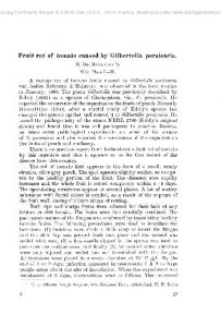
phores were carefully observed under the light microscope. The fungal colonies grown on potato dextrose agar at o 25 C were white cottony at first, and became heavily speckled with the appearance of sporangia and the browinish black, spreading rapidly by means of stolons fired at various points to the substrate by rhizoids (Table 1). Sporangia were 82.7×196.7 µm long and were globose or sub-globose with somewhat flattened base. The color of sporangia was white at first and then turned black with many spores, but never overhung. The sporangia contained thousands of spherical sporangiospores. Sporangiophores were 2.6~5.8×12.3~24.2 µm wide, smooth-walled, non-septate, light brown, simple, long, and arising in groups of 3~5 from stolons opposite rhizoids (Fig. 2A). Sporangiospores were 8.2~18.8 µm in diameter, irregular, round, oval, elongate, angular, and browinish-black streaked (Fig. 2B). Most of the sporangiospores were appeared to be readily dispersed in the air. Numerous sporangiospores were produced on the diseased fruits. Columella was 64.1×136.3 µm in size, light brownish gray, hemispheric, and umberella-shaped when dehisced (Fig. 2C). Rhizoids and stolons were hyaline to dark brown (Fig. 2D). Above characteristics are almost identical to Rhizopus stolonifer (Ehrenberg ex. Fr.) Lind (C.M.I, 1966, 1977. Gobayashi et al., 1992; Udagawa et al., 1980). The maximum and minimum temperatures for myceo o lial growth of the R. stolonifer were 33 C and 10 C, reo spectively. Optimum growth temperature was 25 C (Fig. 3). When 100 ml of the conidial suspension were sprayed to
The soft rot on the succulent tissues of vegetables, fruits and ornamentals caused by Rhizopus sp. occurs throughout the world. The disease mainly occurs during sale, transports, marketshelf and storage after harvest. Rhizopus is omnipresent as a saprophyte and sometimes as a weak parasite on stored organs of plants. When the epidermal cells are collapsed, the fungus emerges through the wounds and produces aerial sporangiophores, sporangia, stolons, and rhizoids, the latter capable of piercing the softened epidermis (Agrios, 1997). In the spring of 2001, a disease suspected as rhizopus soft rot occurred on cherry tomato (Lycopersicon esculentum) in Jinju City Agricultural Products Wholesale Market. The infection rate of the disease in some containers reached to 6.7%. Rhizopus attacked only cracks of matured fruits of cherry tomato, but not young and immatured ones (Fig. 1A, B). The infected parts of cracks on matured fruits appeared water soaked at first, then became soften. Gray hyphae grew from the sites where the fungus invaded primarily and covered the affected portions by producing tuft whiskerlike gray sporangiophores and sporangia. The infected tissues finally broke down and disintegrated in watery rot. Diseased fruits were collected from cherry tomato (cv. Pepe) in the containers and the causal organism was isolated from mycelial tips on the diseased fruits. Brownish black fungal colonies were formed on potato dextrose o agar in the darkness at 25 C. Sporangia and sporangio*Corresponding author 176
Rhizopus Soft Rot on Cherry Tomato in Korea
177
Fig. 1. Symptoms of Rhizopus soft rot on cherry tomato fruits. A: Symptoms with water-soaked lesion usually started from the cracks of fruits. B: Symptoms with mycelia, sporangia and sporangiospores. C: Symptoms in artificially inoculated fruits. Table 1. Comparison of morphological characteristics of the pathogenic fungus isolated from Rhizopus soft rot of cherry tomato with Lind's description of Rhizopus stolonifer Characteristics Colony Sporangiospores Sporangiophores Sporangia Columella
Present isolate color size size shape size size
white to brownish 8.2~18.8 µm 2.6~5.8×12.3~24.2 µm globose, sub-globose 82.7×196.7 µm 64.1×136.3 µm
a
R. stolonifer
white to brownish 10~20 µm 3~5×13~25.3 µm globose, sub-globose 85~200 µm 70~90 µm
a
Described by Lind’s (1913).
Fig. 2. Morphology of the causal organism, Rhizopus stolonifer. A: Sporangium and sporangiophore. B: Columella, C: Sporangiospores, D: Rhizoids and stolons. Scale bar: 20 µm.
cracked fruits, and the inoculated fruits were placed in a humid chamber with 100% relative humidity at 25oC for 24 hours, the typical symptoms on cherry tomato were appeared at 3 days after inoculation in fruit containers (Fig. 1C). The disease infection usually started from cracked parts of fruits. The symptoms were identical to those of naturally infected tomatoes. Morphological characteristics of conidia and mycelia of the fungi that were reisolated
from inoculated fruits were same as those of naturally infected fruits. Generally, cherry tomato is cultivated in greenhouses near urban area where high temperature and humid condition are kept. Such environments are favorable for occurring rhizopus soft rot of tomato. The soft rot disease caused by Rhizopus has been reported in pepper (Capsicum annuum L.) plants in Korea (The Korean Society of Plant Pathol-
178
Kwon et al.
References
Fig. 3. Effect of temperature on mycelial growth of Rhizopus stolonifer, the causal organism of soft rot of cherry tomato. Linear mycelial growth was measured 28 hours after inoculation on PDA. Data are means of three replications ( ).
���
ogy. 1998), but no records in tomato. Farr et al. (1995) and Abdel-Mallek, et al. (1995) have been reported the rhizopus soft rot disease in tomato. Therfore, this is the first report of Rhizopus soft rot on cherry tomato caused by R. stolonifer in Korea.
Abdel-Mallek, A. Y., Hemida. S. K. and Bagy, M. M. K. 1995. Studies on fungi associated with tomato fruits and effectivness of some commercial fungicides against three pathogens. Mycopathologia 130: 109-116: 30 ref. Agrios, G. N. 1997. Plant Pathology. Fourth edition. Academic Press. pp. 283-286. C.M.I. Descriptions of pathogenic fungi and bacteria. 1966. No. 110. C.M.I. Descriptions of pathogenic fungi and bacteria. 1977. No. 524. Farr, D. F., Bills, G. F., Chamuris, G. P. and Rossman, A. Y. 1995. Fungi on plants and plant products in the United States. APS Press. pp. 149-151. Gobayashi, T., Katumoto, K., Abiko, K., Abe, Y. and Kakishima, M. 1992. Illustrated genera of plant pathogenic fungi in Japan. The Whole Farming Educational Association. 534 pp. The Korean Society of Plant Pathology. 1998. List of plant diseases in Korea, 3rd ed. pp. 149-151 (in Korean). Udagawa, S., Tubaki, K., Horie, Y., Miura, K., Minoura, K., Yamazaki, M., Yokoyama, T. and Watanbe, S. 1980. Illustrated genera of fungi. pp. 300-301 (in Japanese).