Acta Alimentaria, Vol. 37 (1), pp. 23–39 (2008) DOI: 10.1556/AAlim.37.2008.1.3
RIPENING AND MICROSTRUCTURE OF APRICOT (PRUNUS ARMENIACA L.) E. KOVÁCSa*, P. MERÉSZb, Z. KRISTÓFc and E. NÉMETH-SZERDAHELYIa a Postharvest
Research Group of the Hungarian Academy of Sciences, Central Food Research Institute, H-1024 Budapest, Herman Ottó 15. Hungary b Department of Biochemistry and Food Technology, Budapest University of Technology and Economics, H-1111 Budapest, Műegyetem rkp 3. Hungary c Department of Plant Anatomy, Eötvös Loránd University, H-1117 Budapest, Pázmány Péter sétány 1/a. Hungary (Received: 24 April 2006; accepted: 7 July 2007)
Colour, texture, pectin autolysis, membrane permeability and microstructure (SEM, TEM), βgalactosidase and polygalacturonase were studied in apricots (cv. Magyar kajszi) harvested in mature green, straw yellow, bright orange and deep orange stages. The L* increased from mature green to straw yellow then decreased from straw yellow to deep orange state. The a* values increased with ripening. The bright and deep orange apricots were significantly softer than the mature green and straw yellow ones and the membrane permeability increased with ripening. The presence of β-galactosidase enzyme was proved by immunoblotting analysis using monoclonal anti-β-galactosidase clone GAL-13 (Sigma) in all ripening stages. The enzyme activity was very low in mature green stage and increased significantly (P>95%) with increasing ripeness and during storage. The PG activity was very low in the mature green apricot. A significant (P>95%) increase was observed in the straw yellow apricot and in the riper fruits. The mature green apricot showed a regular, the straw yellow and bright orange samples showed a moderately regular tissue structure, while the tissue of the deep orange apricot collapsed (SEM). The cell wall and the middle lamella of the green apricot (TEM) were intact. Generally, there were intact cytoplasm membranes with some damaged parts. In the straw yellow apricot, the cell wall started to loosen, the middle lamella lost pectic polysaccharides. The structure of the cytoplasm was not recognisable, the tonoplast and the cytoplasm membrane were injured. The cell wall of the bright orange apricot was similar to that of the straw yellow ones. The middle lamella dissolved and hairy, fibrillar structure of cell wall was found in the deep orange samples. Keywords: apricot, ripening, hardness, colour, β-galactosidase, polygalacturonase, membrane permeability, ultrastructure (TEM, SEM)
* To whom correspondence should be addressed (present address). Phone: (361)-482-6032; fax: (361)-482-6326; e-mail:
[email protected] 0139-3006/$ 20.00 © 2008 Akadémiai Kiadó, Budapest
24
KOVÁCS et al.: RIPENING AND MICROSTRUCTURE OF APRICOT
Fruit softening is associated with extensive solubilisation and degradation of cell wall polysaccharides. The softening involves the loss of tissue cohesion. The middle lamellae consist of pectin polysaccharides, and enzymes bounding to cell wall are responsible for loosening cell to cell adhesion. A turnover in the carbohydrate moieties in the neutral side chains of cell wall pectic polysaccharides has been detected in several types of cell wall including those of ripening fruits (GROSS & SAMS, 1984). However, it has been also suggested that loss of pectin neutral sugar side chains has no significant effect on the physico-chemical properties of the wall and there appears to be not a consistent correlation between galactose loss and extent or type of softening (REDGWELL & PERCY, 1992; REDGWELL & FISCHER, 2002). β-Galactosidase, which acts as an exo-hydrolase, removing terminal non-reducing β-D-galactosyl residues, has been well documented in many fruit species. In case of apricot, as growth occurs concomitantly with ripening, it is possible that cell wall rearrangements linked to fruit growth may play an important role in the softening process (MBÉGUIÉ-A-MBÉGUIÉ et al., 2002). Some of the macro- and microstructural changes in apricot as a function of fruit ripening and storage were investigated. The cell wall break-down, and cell wall bound enzymes such as β-galactosidase and polygalacturonase were studied. The permeability of membranes, tissue and cell wall structure were investigated by electron microscope. The aim was to determine the main factors influencing the textural changes in apricot during ripening and to propose the right ripeness stage for consumption and for storage of apricot. 1. Materials and methods 1.1. Samples (Prunus armeniaca L., cv. Magyar kajszi) The apricots were harvested at four ripening stages, mature green, straw yellow, bright orange and deep orange. The pH values varied between 2.98–3.25. Straw yellow fruits are good for storage, bright orange is good for consumption and deep orange is overripe for table consumption. Some samples were stored at 2–4 °C with humidity between 85– 90 RH% (Table 1). Table 1. Program of the harvest and storage of apricots Harvest I. harvest mature green July 12 th straw yellow July 12 th bright orange July 19 th deep orange July 19 th II. harvest III. harvest
Acta Alimentaria 37, 2008
1 st
– 11 7 5 6 6
2 nd
Storage out 3 rd Storage time (day)
4 th
5 th
31 31 12 12 11 –
34 – 14 14 13 13
39 38 19 19 18 18
41 40 21 21 20 21
KOVÁCS et al.: RIPENING AND MICROSTRUCTURE OF APRICOT
25
1.2. Methods 1.2.1. Determination of colour. Colour was determined by Hunterlab colorflex 45/0 spectrophotometer. Evaluation was carried out by UNI Universal Software (The Color Company, Reston, USA). The number of repetition was 20 for each sample. The X, Y, Z components were measured, and the CIELAB a*, b*, L* values were calculated by UNI software (MUSKOVICS et al., 2006). 1.2.2. Texture analysis. Texture was analysed by a penetrometer (Labor MIM, Hungary). The firmness was determined by penetration distance of a 50 g probe cone during 5 s. The firmness was determined for 5–5 pieces of fruits at both sides of one sample. 1.2.3. Cell wall analysis, β -galactosidase and polygalacturonase activity. The cell wall analysis was carried out according to KOVÁCS and NÉMETH-SZERDAHELYI (2002). The determination of polygalacturonase (PG) (EC.3.2.1.15) activity is based on degree of polymerisation and number of reducing end groups of pectin; the activity is in correlation with the number of reducing end groups. The activity of β-galactosidase (EC. 3.2.1.23) is defined by BARTLEY (1974). Autolysis was carried out by incubation at 30 °C for 72 h in NaOAc/NaCl solution (NaOAc solution was 50 mM; NaCl solution was 0.2 mM, pH=2.5). The free galacturonic acid was determined in the total and soluble pectin fraction (BLUMENKRANTZ & ASBOE-HANSEN, 1973). The soluble carbohydrate content of cell wall determination is based on the formation of antron complex of the carbohydrates (DREYWOOD, 1946). The protein extracts were liophylised until further use. 1.2.4. Isoelectric focusing. A gel was formed between two glass sheets (distance 0.5 mm) at 20 °C for one night. The lyophilised proteins were dissolved in a buffer solution (5.7 g 87% glycerol, 24 g urea, 250 mg ditiothreitol in 50 ml distilled water) and 10 µl of each were loaded in the gel by filtre paper stripe (10×5 mm). Filtre paper stripes dipped in anodic solution (5% phosphoric acid) or catodic solution (2% sodium hydroxide) were set to appropriate sides of the gel. The separation was carried out on Pharmacia FBE-3000 equipment (60 min, 2500 V, 150 mA, 10 °C). The proteins were fixed by agitation in 15% TCA for 15 min, dyed (0.3 g Coomassie Brilliant Blue, 0.5 g Cu-sulfate, 5 H2O, 90 cm3 MeOH, 20 cm3 glacial acetic acid, 90 cm3 distilled water) for 15 min and washed by dedyeing solution (250 cm3 MeOH+100 cm3 glacial acetic acid+650 cm3 distilled water) several times till the background became transparent (KRAUSE et al., 1982). The protein fractions were separated by IEF at pH 3.5–10.0, then they were carried through the nitrocellulose membrane by Semi-Dry Blot apparatus (Bio-Rad). The blotting time was 60 min, the current was set at 0.8 mA cm–2. After incubation period, the sample was washed 3 times for 10 min in washing solution containing antibody conjugated by biotin that was developed against β-galactosidase (monoclonal anti-β-galactosidase clone GAL-13, Sigma). This was followed by a 2-h incubation in washing solution containing avidin-peroxidase (Sigma) and repeated washing (3×10 min). The last 5 min of the incubation period was carried out in cold PBS solution (8.0 g NaCl, 0.2 g KCl, 2.86 g Na2HPO4×2H2O, 0.27 g KH2PO4 in 1000 cm3 water). Developing process was carried out with a solution containing 30 mg Acta Alimentaria 37, 2008
26
KOVÁCS et al.: RIPENING AND MICROSTRUCTURE OF APRICOT
4-chloro-naftol (Bio-Rad) solved in 10 cm3 ethanol, 25 cm3 PBS, 100 µl 37% hydrogen-peroxide. 1.2.5. Ion leakage. The cell membrane permeability was determined according to LOVÁSZ and co-workers (1998) with modifications. Twenty discs (10 mm diameter, 2 mm thickness) were cut out by a cylindrical knife from 3 pieces of every apricot sample. The discs were placed into 0.3 M mannit solution and the conductivity of the solution was measured as a function of time by a conductometer containing 100 cells (type OE420, LMIM, HU). The ion leakage is proportional to the changes of conductivity and to the rate of ions released from the fruit discs to the surrounding solution if the circumstances are fixed. The determination of conductivity at the equilibrium is necessary. To reach it in a short time and simulate the damage of cell surface, 3 discs of each sample were heat-treated at 80 °C for 20 min. The curve of ion leakage is logarithmic. The limit of ion leakage curve can be set as the current according to the estimated equilibrium conductivity. The combined ion leakage process that takes place during measurements is presented as diffusion. The state of the cell surface and the membranes were characterised by an apparent diffusion coefficient of the ions. The evaluation of the measured data and the presentation were carried out by Statistica®. 1.2.6. Ultrastructure. Pieces were prepared from 3 apricots for TEM (1×2×3 mm/pieces) and for SEM (2×5×10 mm/pieces). Two pieces from each pack were chosen randomly for investigation. The sampling for SEM was done with skin and flesh (1 cm deep), and for TEM samples were taken from the middle part of the fruit tissue between the skin and the core. The sample preparation method has been previously described (KOVÁCS et al., 1988). The SEM investigations were carried out at 18 kV voltage (Hitachi 2360 N, Japan), the digital photos were made by DISS hardware and software (Point Electric, Germany), and those of TEM were done at 75 kV voltage (Hitachi 7100, Japan), the sections were cut by ultra-microtome (Zeiss HM 360, Germany). 1.2.7. Mathematical-statistical evaluation. The comprehensive evaluation of the results, the determination of the basic statistical features, two-sample t-test, variance and regression analysis were carried out depending on the type of results and measurement method. The significance was accepted at P=95% level. 2. Results and evaluation 2.1. Colour The samples were placed in the yellow zone of the CIELAB colour-field. The more ripened samples shifted to the red zone. The L*, a*, b* values and the differences (∆L*, ∆H*, ∆C*, ∆E*) were calculated from the X, Y, Z data. The L* increased from mature green to straw yellow and then decreased from straw yellow to deep orange state. The a* values increased with ripening. The difference of colour stimulus (∆E*) increased, the hue angle (∆H*) decreased with ripening. The colour saturation (∆C*) increased,
Acta Alimentaria 37, 2008
KOVÁCS et al.: RIPENING AND MICROSTRUCTURE OF APRICOT
27
later decreased (Table 2). Since apricots do not mature uniformly, selective harvesting involving two or more harvests is desirable. Fruit ground colour of apricot is directly related to maturation (GOUBLE et al., 2004). FEMENIA and co-workers (1998b) investigated the connection between increase in size and colour development, and they established that the average size of the apricots increased from stage 1 to 4 and then remained constant until the fruit was ripe. Table 2. Results of the colour measurement of apricot (cv. Magyar kajszi) in different ripeness stage at harvest
X Y Z L* a* b* ∆E* ∆C* ∆H* dL
Mature green (1) 28.71±2.4 26.92±2.0 8.11±0.6 58.86±1.8 12.85±2.7 44.59±2.3 5.39±2.5 1.51±2.7 –4.11±2.3 –1.01±1.8
Ripeness stage Straw yellow (1) Bright orange (2) 35.17±3.9 28.52±6.6 31.81±4.1 24.96±5.2 9.46±1.1 8.46±0.9 63.03±3.6*** 56.69±5.2 18.08±6.3* 19.66±7.5** 47.40±3.8 39.69±7.4* 12.49±4.8 15.99±5.6 6.19±3.4 –0.36±9.1 –8.28±6.2 –11.68 ± 6.0 3.16±3.6 –3.18±5.2
Deep orange (2) 23.27±6.4 19.95±5.3 7.80±0.7 51.29±6.0*** 20.53±6.7*** 32.62±8.8*** 21.18±7.2 –6.06±9.6 –15.26±6.2 –8.58±6.0
X, Y, Z = CIE tristimulus values Samples were related to the green ones. *P95%) higher in mature green apricots than in others. The low activity of β-galactosidase in mature green fruits is opposite with the high neutral sugar autolysis of apricots in mature green stage. It can be stated that the autolysis of the neutral sugars in mature green apricots is not influenced by β-galactosidase enzyme. FEMENIA and co-workers (1998b) extracted the neutral carbohydrate of fresh apricots by acidic hydrolysis from the cell wall polysaccharides. They could identify arabinose (5.78 mg g–1 f.w.), galactose (3.34 mg g–1 f.w.), rhamnose (0.99 mg g–1 f.w.), fructose (0.12 mg g–1 f.w.), xylose (2.54 mg g–1), mannose (1.20 g g–1), dextrose (18.61 mg g–1) and galacturonic acid (16.14 mg g–1). The degradation of cell wall in apricots was investigated after heat treatment (100 °C, 12 min) (CHITARRA et al., 1989) and in dried apricots (FEMENIA et al., 1998a). In both cases, the carbohydrates were roughly in the same quantity. Because of the limited enzymatic activity in dried apricots, β-elimination of the polysaccharides took place in the break down of cell wall polysaccharides (PLAT et al., 1991; FEMENIA et al., 1998a). As a function of ripeness in apricots, the mass of fructose, xylose, mannose and galactose increased, while that of arabinose decreased and that of rhamnose fluctuated. The non-cellulose cell wall neutral carbohydrate
Acta Alimentaria 37, 2008
KOVÁCS et al.: RIPENING AND MICROSTRUCTURE OF APRICOT
29
content and the amount of uronic acid increased as a function of ripeness, however rhamnose did not change and the arabinose increased (FEMENIA et al., 1998b). 2.3.3. β -Galactosidase. At harvest, the β-galactosidase enzyme has a very low activity in mature green stage. The activity of β-galactosidase increased significantly (LSD 95%) with the increasing ripeness stage, but there were not differences between straw yellow and bright orange apricots. The activity of β-galactosidase of fruits at different ripening stages significantly (LSD 95%) increased during the storage (except mature green). The straw yellow and bright orange apricot did not differ from each other. Sudden increase was observed at the beginning of storage in the deep orange samples, later the activity of β-galactosidase declined (Fig. 1).
Fig. 1. Change in the activity of β-galactosidase during storage (2–4 °C, 85–90%RP) in the different ripening stages of apricots (1: mature green, 2: straw yellow, 3: bright orange, 4: deep orange)
In our earlier results, cultivar and harvest time dependencies were observed. The SDSPAGE pattern changed as a function of harvest; 15 kDa and 18 kDa protein bands were reduced as a function of harvest, the intensity of 39 kDa protein band was the strongest in the stored apricots (KOVÁCS & NÉMETH-SZERDAHELYI, 2002). Immunoblotting analysis using monoclonal anti-β galactosidase clone GAL-13 (Sigma) proved that at harvest all investigated samples (from mature green to deep orange) contained βgalactosidase enzyme (Fig. 2A–B), though not all the enzymes were active in all ripening stages (Fig. 1). YOSHIOKA and co-workers (1995) isolated four different βgalactosidases from stored apples (Starking delicious), among them ß-galactosidase-II, -III, -IV were affected on the arabino-galactan chain during the softening period. During ripening process, Pa-Exp1 and Pa-Exp2 gene expression appeared to be positively Acta Alimentaria 37, 2008
30
KOVÁCS et al.: RIPENING AND MICROSTRUCTURE OF APRICOT
correlated with fruit size. In case of apricots, as growth occurs concomitantly with ripening, it is possible that cell wall rearrangements linked to fruit growth may play an important role in the softening process (MBÉGUIÉ-A-MBÉGUIÉ et al., 2002). 2.3.4. Polygalacturonase (PG) enzyme. The PG activity was very low in mature green apricots. A significant (P>95%) increase of the PG activity was observed in the straw yellow apricots, later the activity increased according to ripening. Four–five times higher activity was measured in more ripened samples than in mature green one (Fig. 3).
Fig. 2. Results of the IEF (pH: 3.5–10.0) of protein extracts from apricot at different ripening stages: 1: mature green, 2: straw yellow, 3: bright orange, 4: deep orange, 5: β-galactosidase (Saccharomyces fragilis, Sigma). (A) dyeing by Coomassie Brilliant Blue; (B) immunoblott analysis (monoclonal anti-β-galactosidase clone GAL-13, Sigma)
Acta Alimentaria 37, 2008
KOVÁCS et al.: RIPENING AND MICROSTRUCTURE OF APRICOT
31
Fig. 3. Change in the activity of polygalacturonase during storage (2–4 °C, 85–90%RP) in the different ripening stages of apricots (1: mature green, 2: straw yellow, 3: bright orange, 4: deep orange)
2.4. Cell surface permeability There is a very noticeable difference among the early harvested samples. Those samples, which are considered more ripened referring to their colour, have higher tissue permeability. The tissue permeability means the apparent diffusion coefficient that is the result of the individual permeability of the tonoplast, cytoplasm, cell membrane, cell wall and the intercellular space. The increase of the diffusion coefficient has an important role in the faster water leakage, causing the decrease in the turgor, finally fruit softening. According to the values of permeability of apricots divided into two groups, one belongs to mature green and straw yellow, the other belongs to bright and deep orange group. These two groups correspond to the harvest time. At the beginning of the storage, there are no significant differences among these fruits in the histic apparent diffusion coefficient, however during storage the tissue permeability increases faster in the later harvested fruits than in the earlier ones. This means that the intensity of ripening processes is independent from the initial ripening stage, even if we noticed that this speed is reciprocally proportional to the ripening stage at harvest, namely the change of the unripe green apricot is faster than the fruits harvested in ripe state (Table 4). These differences decrease near the 10 th day of storage, the characteristics of the sample set became equalised (Table 4). The degradation of the cell membrane is a sensitive signal of the fruit senescence. The kinetic of ion leakage was described by exponential curves (SALTVEIT, 1989). The initial rate of the ion leakage showed a diversified picture as a function of apple cultivar or growing system (organic and Acta Alimentaria 37, 2008
32
KOVÁCS et al.: RIPENING AND MICROSTRUCTURE OF APRICOT
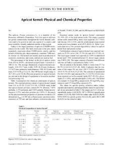
integrated). Consequently, the initial rate is affected more by non-defined factors, i.e. of mechanically destroyed cell surface or more sensitive membrane of apples grown by organic method (MERÉSZ et al., 2003). Table 4. The permeability of apricot (cv. Magyar kajszi) parenchyma at different ripening stage and harvesting date during storage (2–4 °C, 85–90% RH) Storage time (days) 0 3 6 10 “b” Coefficient “a” Value Correlation (R2)
Mature green (1) 1.9 2.8 1.6 2.4 52.8 81.6 0.9725
Ripeness stages Straw yellow (1) Bright orange (2) 2.0 2.6 2.8 3.7 1.8 3.3 3.0 3.6 54.7 61.6 32.7 21.9 0.9489 0.9601
Deep orange (2) 2.9 4.2 3.0 – – – –
The permeability is presented by equation y=b*ln(x)+a Harvest time was on 12 th July (1) and on 19 th July (2)
2.5. Ultrastructure The microstructural (SEM) changes of the apricot tissue structure are presented in Fig. 4. The mature green, straw yellow and bright orange samples showed a regular tissue structure, therefore in Fig. 4 only mature green (A and C) and deep orange (B and D) samples are presented. In the deep orange (overripe) apricots, middle lamellae are dissolved, cells are damaged. The tissue mostly consisted of collapsed and damaged cells as a consequence of cell wall break down (Fig. 4B). The surface of the mature green apricot is covered by waxy film (4C). The covering waxy film is gradually decreased and at the end disappeared from the fruits and the hair (4D). The epidermal and hypodermal layers were appropriate at every ripening stage. The cell wall and the middle lamella of the mature green apricots (TEM) were intact. There are circle-like endosomes in the vacuoles (Fig. 5A). There is a thin cytoplasm stripe at the cell wall (Fig. 5B) containing mainly mitochondria. Generally, there are intact cytoplasm membranes, but some injured cytoplasm parts were observed (Fig. 5C). In the similar cells, there are often mielinised structures surrounded by membranes in the vacuoles and vacuoles with smaller vesicules (Fig. 5D). Even in these cells, generally, the mitochondria have stable morphology.
Acta Alimentaria 37, 2008
KOVÁCS et al.: RIPENING AND MICROSTRUCTURE OF APRICOT
33
Fig. 4. SEM presented the tissue structure (4A–B) and the surface of the skin (4C–D) of apricot (mature green, deep orange) two days after harvesting. A and C: mature green; B and D: deep orange
In the straw yellow apricots, the cell wall started to loosen, the middle lamella lost pectic polysaccharides. A damaged plastid can be seen with a lot of lipid droplets (Fig. 6A). The structure of the cytoplasm is not recognised, the tonoplast and the cytoplasm membrane are injured. There are damaged citoplasm by the side of the cell wall with cell parts stringing of beads inside (Fig. 6B). In the cell wall, a bright stripe shows the absence of the middle lamella (Fig. 6C). Vesicules appeared in the cytoplasm, and endocytosis was observed. The changes in ultrastructure during senescence have been described, but mostly for senescing leaves. Some of the earliest structural changes indicative of senescence occur in the chloroplast, i.e. changes in the grana and also formation of lipid droplets (plastoglobuli). Polysomes and ribosomes in general decrease fairly early reflecting a decrease in protein synthesis. On the other hand, the nuclei and mitochondria generally show little structural change until later. Visible disintegration of the plasma and vacuolar membranes seem to be terminal (late) events. NOODÉN and co-workers (1997) divided senescence into different stages: (1) initiation, (2) degeneration (progression) and (3) terminal. The senescence initiators are environmental, disease and developmental, and all of these may be mediated by hormones.
Acta Alimentaria 37, 2008
34
KOVÁCS et al.: RIPENING AND MICROSTRUCTURE OF APRICOT
Fig. 5. The microstructure of mature green apricot tissue (TEM). A: intact cell wall part, cytoplasm residue at the cell wall, there are a huge mitochondrion in the foreground; B: cell wall with cytoplasm residue at the two sides of the wall; C: degradation of the cytoplasm, hurt plastids, mielinisation of membrane, intact mitochondria; D: part of cytoplasm, vacuoles, inclusions in the vacuole, the cell structure is not noticeable, the membranes are dissolved
Other changes occur, but they seem to vary somewhat among tissues and situations and are likely to be peripheral. At this point, it is difficult to say what these ultrastructural changes mean in the senescence process, except that nuclei and mitochondria may perform important functions that require their integrity until fairly late. The loss of integrity of the plasma (and probably also the vacuolar) membranes would mark death, the end of homeostasis. The relationship between senescence and apoptosis is an important issue. Apoptosis may be particularly important in the terminal phase of senescence (NOODÉN et al., 1997; DELORME et al., 2000; NOODÉN, 2004). While senescence and ripening are distinct development events, they clearly share overlapping physiological and biochemical pathways. A primary feature in both leaves and fruit is the controlled dismantling of the photosynthetic machinery, and degradation of starch and chlorophyll. A common characteristic of senescence and fruit ripening in climacteric fruits is the increase in respiration and ethylene production observed during these processes (JONES, 2004).
Acta Alimentaria 37, 2008
KOVÁCS et al.: RIPENING AND MICROSTRUCTURE OF APRICOT
35
Fig. 6. The microstructure of the cell wall and cytoplasm of the straw yellow apricot (TEM). A: The sign of the middle lamella damage is seen, the cytoplasm is degraded, hurt plastids and intact mitochondria are noticed; B: intact part of the cell wall with cytoplasm string; C: intensive cell wall break-down
The cell wall of the bright orange apricot is similar to that of the straw yellow ones (Fig. 7A–C). In this ripening stage, some characteristic metamorphoses were observed in the cytoplasm. There are empty vacuoles surrounding pseudo-double-membrane structures (Fig. 7A). There are a few cytoplasm residues among the large vacuoles. The cytoplasm is mixed with the vacuoles, therefore, the cell structure is not observed well (Fig. 7B). The sign of the necrotic cell damage is the loss of the cytoplasm membrane and the tonoplast. Among the subcellular components, the plastids are damaged early. The mitochondria are very stable. The residue of the peroxisomes (gerontosomes) means the scenescence of the cell. The cell content consists mainly of vacuoles, and the other subcellular components are not visible because of the endocytosis (Fig. 7C). Chloroplastids changed into gerontoplastids. Thylakoids and stroma components are progressively lost during chloroplastids senescence, but the envelope remains intact. The intactness of the envelope is important, because it has been identified as the site of chlorophyllase (MATILE et al., 1997).
Acta Alimentaria 37, 2008
36
KOVÁCS et al.: RIPENING AND MICROSTRUCTURE OF APRICOT
Fig. 7. The microstructure of the cell of the bright orange apricot (TEM). A: Damaged cytoplasm, vacuoles, pseudo double membrane; B: damaged cytoplasm can be seen with several vacuoles and mitochondria; C: huge vacuoles with a few cytoplasm residue
The solvation of symptoms of the cell wall is seen in the case of the deep orangeyellow, ripe, mellow, juicy apricot. The solubilisation and the change in fibrillar structure of the cell wall can be seen in Fig. 8A-C. In the beginning, the cell wall shows parallel arrangement, later fibrilles break and form V shape parts (Fig. 8D). The parallel network of fibrillar skeleton characterised the cell wall structure in apple cv. Empire and Golden Delicious. During the storage, this parallel network sustains a loss depending on the variety, the fibrilles show ramifications that come together with the fruit softening (KOVÁCS et al., 1997). The cell wall structure of tomato after cellulase treatment (CROOKES & GRIERSON, 1983) showed a very similar shape to the stored, deep orange apricot. Ultrastructural studies of fruit cell walls also suggested differences in the degree of cellulose microfibril disruption between species. The microfibrillar component in the walls of avocado (PLAT et al., 1991), apple and pear (BEN-ARIE & KISLEV, 1979) undergoes substantial ripening-related disruption, while immuno-localisation studies of kiwifruit suggested that cellulose remained stable during ripening (SUTHERLAND et al., 1999).
Acta Alimentaria 37, 2008
KOVÁCS et al.: RIPENING AND MICROSTRUCTURE OF APRICOT
37
Fig. 8. The microstructure of the cell wall of the deep orange apricot (TEM). A: Dissolving of cell; B: the middle lamella is not noticeable and the fibrillar structure of the cell wall is ragged; C, D: the microfibrilles of the cell wall
Given the current limited knowledge of microfibril architecture, it is difficult at this point to come to any firm conclusions regarding the extent and nature of cellulose microfibril degradation during ripening. In particular, the dynamics of the interaction between the glucan chains on the microfibril surface and associating hemicelluloses is likely to be an important factor in determining the structural properties of the hemicellulose-cellulose framework, but it is hard to elucidate. This is likely to remain the case until additional analytical tools and experimental approaches become available. The identification of enzymes and cellulose-interacting proteins that affect inter- and intramicrofibrillar associations, or that altering cellulose-hemicellulose interactions, may be one means of better understanding cellulose metabolism in vivo (ROSE et al., 2003). 3. Conclusions All investigated ripening related processes (the textural changes, pectin solubilisation, β-galactosidase, polygalacturonase, membrane permeability and ultrastructural changes) increased firstly in straw yellow apricot (cv. Magyar kajszi), and there were no
Acta Alimentaria 37, 2008
38
KOVÁCS et al.: RIPENING AND MICROSTRUCTURE OF APRICOT
differences between straw yellow and bright orange stages. In the straw yellow apricot, the cell wall started to loosen, the middle lamella lost pectic polysaccharides. The activity of β-galactosidase increased with the increasing ripeness stage, firstly in straw yellow stage, but the presence of the β-galactosidase does not mean that it is in function; as was seen in mature green apricot in immunological tests. Since apricots do not mature uniformly, selective harvesting involving two or more harvests is desirable. When the harvested fruits are inhomogeneous according to their colour, their ripening processes will be different during storage and these make results worse according to the fruits storability and marketability. * The project was supported by OTKA No. TO22838, MTA TKI Postharvest Research Group No. 13002. Thanks to Ms. J. FÉNYES and to Ms. R. FÁBIÁN technicians, Ms. E. RÓTH Ph.D student for assistance in the experiments. Thanks to Mr. J. LISZTES from Felsőörs for supplying the apricot samples.
References BARTLEY, I.M. (1974): β-Galactosidase activity in ripening apples. Phytochemistry, 13, 2107–2111. BEN-ARIE, R. & KISLEV, N. (1979): Ultrastructural changes in the cell walls of ripening apple and pear fruit. Plant Physiol., 64, 197–202. BLUMENKRANTZ, N. & ASBOE-HANSEN, G. (1973): New method for quantitative determination of uronic acids. Anal. Biochem., 54, 484–489. BOURANIS, D.L. & NIAVIS, C.A. (1992): Cell wall metabolism in growing and ripening stone fruits. Plant Cell. Physiol., 33, 999–1008. CHITARRA, A.B., LABAVITCH, J.M. & KADER, A.A. (1989): Canning-induced fruit softening and cell wall pectin solubilization in the ‘Patterson’ apricot. J. Fd Sci., 54, 990–992. CROOKES, P.R. & GRIERSON, D. (1983): Ultrastructure of tomato fruit ripening and the role of polygalacturonase isoenzymes in cell wall degradation. Plant Physiol., 72, 1088–1093. DELORME, V.G.R., MCCABE, P.F., KIM, D.J. & LEAVER, C.J. (2000): A matrix metalloproteinase gene is expressed at the boundary of senescence and programmed cell death in cucumber. Plant Physiol., 123, 917–927. DREYWOOD, R. (1946): Qualitative test for carbohydrate material. Ind. Eng. Chem. Analyt., 18, 499. FEMENIA, A., SÁNCHEZ, E.S., SIMAL, S. & ROSELLÓ, C. (1998a): Modification of cell wall composition of apricots (Prunus armeniaca) during drying and storage under modified atmospheres. J. agric. Fd Chem., 46, 5248–5253. FEMENIA, A., SÁNCHEZ, S.A.E., SIMAL, S. & ROSSELLÓ, C. (1998b): Developmental and ripening-related effects on the cell wall of apricot (Prunus armeniaca) fruit. J. Sci. Fd Agric., 77, 487–493. GOUBLE, B., BUREAU, S., GROTTE, M., REICH, M., RELING, P. & AUDERGON, J.M. (2004): Apricot postharvest ability in relation to ethylene production. Influence of the picking time and the cultivar. Acta Hortic., 682, 127–133. GROSS, K. & SAMS, C.E. (1984): Changes in cell wall neutral sugar composition during fruit ripening: a species survey. Phytochemistry, 23, 2457–2461. JONES, M.L. (2004): Changes in gene expression during senescence. -in: NOODÉN, L.D. (Ed). Plant cell death processes. Elsevier Academic Press, San Diego, California, USA. pp. 51–67. KOVÁCS, E. & NÉMETH-SZERDAHELYI, E. (2002): Activity of β-galactosidase and cell wall break down in apricot (Prunus armeniaca L.). J. Fd Sci., 67, 2004–2008.
Acta Alimentaria 37, 2008
KOVÁCS et al.: RIPENING AND MICROSTRUCTURE OF APRICOT
39
KOVÁCS, E., KERESZTES, Á. & KOVÁCS, J. (1988): Effect of gamma irradiation and calcium treatment on the ultrastructure of apples and pears. Fd Microstructure, 7, 1–14. KOVÁCS, E., VAN BUREN, J.P., PITIFER, L.A., HOCH, H.C. & TERHUNE, B.T. (1997): Effect of irradiation and storage on cell wall structure of Golden Delicious and Empire apples. Acta Alimentaria, 26, 171–190. KRAUSE, I., BELITZ, H.D. & KAISER, K.P. (1982): Nachweis von Kuhmilch in Schaf- und Ziegenmilch bzw. -käse durch isoelektrische Focussierung in harnstoffhaltigen polyacrylamidgelen. Z. Lebensm. Unters. Forsch., 174, 195–199. LOVÁSZ, T., MERÉSZ, P. & SASS, P. (1998): Postharvest permeability changes of the cell surface of apple tissue. Acta Alimentaria, 27, 207–219. MATILE, P., SCHELLENBERG, M. & VICENTINI, F. (1997): Localization of chlorophyllase in the chloroplast envelope. Planta, 201, 96–99. MBÉGUIÉ-A-MBÉGUIÉ, A., CHAHINE, H., GOMEZ, R.M., GOUBLE, B., REICH, M., AUDERGON, J.M., SOUTY, M., ALBAGNAC, G. & FILS-LYCAON, B. (1999): Molecular cloning and expression of a cDNA encoding 1aminocyclopropane-1-carboxylate (ACC) oxidase from apricot fruit (Prunus armeniaca). Physiol. Plantarium, 105, 294–303. MBÉGUIÉ-A-MBÉGUIÉ, D., GOUBLE, B., GOMEZ, R.M., AUDERGON, J.M., ALBAGNAC, G. & FILS-LYCAON, B. (2002): Two expansin cDNAs from Prunus armeniaca expressed during fruit ripening are differently regulated by ethylene. Plant Physiol. Biochem., 40, 445–452. MERÉSZ, P., KOVÁCS, E. & KŐVÁRI, E. (2003): Modelling of the fruit membrane permeability as a function of the postharvest physiological changes. Acta Hortic., 599, 429–433. MUSKOVICS, G., FELFÖLDI, J., KOVÁCS, E., PERLAKI, R. & KÁLLAY, T. (2006): Changes of physical properties during ripening in Hungarian sweet cherry (Prunus avium L.) cultivars. Postharvest Biol. Technol., 40(1) 56–63. NOODÉN, L.D. (2004): Plant cell death processes. Elsevier Academic Press, San Diego, California, USA, pp. 10–12. NOODÉN, L.D., GUIAMET, J.J. & JOHN, I. (1997): Senescence mechanism. Physiol. Plant., 101, 746–753. PLAT, D., BEN-SHALOM, N. & LEVI, A. (1991): Changes in pectic substances in carrots during dehydration with and without blanching. Fd Chem., 39, 1–12. REDGWELL, R.J. & PERCY, A.E. (1992): Cell wall changes during on-vine softening of kiwifruit. New Zealand J. Crop Hort. Sci., 20, 453–456. REDGWELL, R.J. & FISCHER, M. (2002): Fruit texture, cell wall metabolism and consumer perceptions. -in: KNEE, M. (Ed.) Fruit quality and its biological basis. CRC Press LLC, Boca Raton, FL. pp. 46–75. ROSE, J.K.C., CATALÁ, C., GONZALEZ-CARRANZA, Z.H. & ROBERTS, J.A. (2003): Cell wall disassembly. -in: ROSE, J.K.C. (Ed.) The plant cell wall. CRC Press Blackwell Publishing, New York, USA, pp. 264–324. SALTVEIT, M.E. (1989): A kinetic examination of ion leakage from chilled tomato pericarp disks. Acta Hortic, 27, 207–219. SUTHERLAND, P., HALLETT, I., REDGWELL, R., BENHAMOU, N. & MACRAE, E. (1999): Localization of cell wall polysaccharides during kiwifruit (Actinidia deliciosa) ripening. Int. J. Plant Sci., 160, 1099–1109. YOSHIOKA, H., KASHIMURA, Y. & KANEKO, K. (1995): Beta-D-galactosidase and alpha-L-arabinofuranosidase activities during the softening of apples. J. Jpn. Soc. Hortic. Sci., 63, 871–878.
Acta Alimentaria 37, 2008