Oberdan Leo, Jacques Urbain, Charlie R. Maliszewski,â and Muriel Moser ..... Shortman, K., McKenna, H. J. (1996) Dramatic increase in the numbers of.
Role of CD8a1 and CD8a2 dendritic cells in the induction of primary immune responses in vivo Roberto Maldonado-Lo´pez, Thibaut De Smedt, Bernard Pajak, Carlo Heirman,* Kris Thielemans,* Oberdan Leo, Jacques Urbain, Charlie R. Maliszewski,† and Muriel Moser De´partement de Biologie Mole´culaire, Universite´ Libre de Bruxelles, Rhode-Saint-Gene`se; *Laboratorium of Hematologie-Immunologie, Vrije Universiteit Brussel, Belgium; and †Immunex Corporation, Seattle, Washington
Abstract: Data from adoptive transfer of mature dendritic cells (DC) indicate that they are responsible for the induction of primary immunity. Two subclasses of DC have been recently identified in spleen that differ in their phenotype and in certain regulatory features. In vitro, both subsets have the capacity to activate naive T cells, although CD8a1 DC have been shown to induce T cell apoptosis and to stimulate lower levels of cytokines compared with CD8a2 DC. The objective of this study was to analyze the function of these distinct DC types in vivo. Our results show that both subsets, pulsed extracorporeally with antigen and injected in the footpads of syngeneic mice, sensitize an antigenspecific T cell primary response. However, CD8a1 cells trigger the development of Th1-type cells, whereas CD8a2 DC induce a Th2-type response. These observations suggest that the Th1/Th2 balance in vivo is regulated by the antigen-presentingcells of the primary immune responses. J. Leukoc. Biol. 66: 242–246; 1999. Key Words: immunity · tolerance · cell therapy · Flt3-ligand
INTRODUCTION Since the discovery of distinct T helper populations, it has become clear that the Th1/Th2 balance determines the efficiency of the immune response. A classical example has been provided by the infection of mice with Leishmania major. Early work has indeed shown that a Th1-type response leads to elimination of Leishmania and survival of the host, whereas a Th2-type response is ineffective and results in death. The essential role of Th differentiation on the outcome of parasitic infections is further illustrated by the observation that injection of cytokines or neutralizing antibodies, known to redirect the immune responses toward Th1 or Th2, often results in disease resolution or death [1–4]. The orientation of the immune response is of major interest for the design of new vaccines and for the immunotherapy of cancer. Several factors have been shown to influence the development of selected T helper populations, and include the MHC/peptide density [5], the nature of the antigen-presenting cell, and the cytokines released in the environment where T cells are primed [for review, see ref. 6]. In particular, we have 242
Journal of Leukocyte Biology
Volume 66, August 1999
recently shown that the type of antigen-presenting cell can influence the Th1/Th2 balance in vivo. Dendritic cells (DC), pulsed extracorporeally with antigen, induced the development of cells secreting interleukin-1 (IL-2), interferon-g (IFN-g), and IL-4 on rechallenge in vitro, whereas peritoneal macrophages sensitized cells that produced IL-4 but not IFN-g [7, 8]. Among the population of cells able to present antigens to class II-restricted CD41 T lymphocytes, DC display some unique migratory and functional properties that confer to them the capacity to optimally sensitize naive T lymphocytes in vivo [9]. In the mouse, two populations of DC have been identified that appear to belong to distinct lineages. The lymphoid DC express CD11c and CD8aa homodimers and have been shown to induce T cell apoptosis by a Fas-dependent mechanism and to trigger a limited T cell proliferation in vitro. The myeloidrelated DC are CD11c1 CD11b1 CD8a2 and optimally sensitize naive T cells to proliferate and produce IL-2 in a mixed lymphocyte reaction (MLR) assay [10, 11]. The objective of this study was to evaluate the immunostimulatory properties of lymphoid versus myeloid DC subset in vivo. Our data show that both subsets, pulsed with antigen in vitro, sensitize antigen-specific T cells in vivo and differentially regulate the development of T helper subpopulations. In addition, we compared the capacity of mature DC, isolated from untreated mice or generated in vivo by the administration of Flt3-ligand (Flt3L) [12, 13], to direct the development of T helper cells on transfer in vivo.
MATERIALS AND METHODS Mice BALB/c mice were purchased from Iffa-Credo. IL-12 p402/2 BALB/c mice were kindly provided by Dr. Jeanne Magram (Hoffmann-La Roche, Nutley, NJ) [14]. The mice were housed in our pathogen-free animal facility.
Abbreviations: DC, dendritic cell; Flt3L, Flt3 ligand; IL-2, interleukin-2; IFN-g, interferon-g; MLR, mixed lymphocyte reaction; rmGM-CSF, recombinant murine granulocyte-macrophage colony-stimulating factor; FITC, fluorescein isothiocyanate; PE, phycoerythrin; ELISA, enzyme-linked immunosorbent assay; KLH, keyhole limpet hemocyanin. Correspondence: Muriel Moser, De´partement de Biologie Mole´culaire; rue des Chevaux, 67; B-1640 Rhode-Saint-Gene`se, Belgium. E-mail: mmoser@dbm. ulb.ac.be Received February 10, 1999; revised March 16, 1999; accepted March 19, 1999.
http://www.jleukbio.org
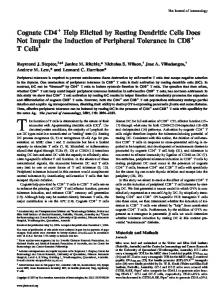
Fig. 1. Phenotype analysis of purified CD8a1 and CD8a2 DC. DC were purified from untreated or Flt3Linjected BALB/c mice. Top panel: cells were doublestained for CD8a expression in red and for CD11c expression in green. Bottom panel: cells were either unstained (dashed line) or stained for MHC class II (CLII), DEC-205, or CD86 expression (solid line). Cells were analyzed without gating.
Purification of DC subsets Spleen cells were digested with collagenase, further dissociated in Ca21-free media in the presence of EDTA, separated into low-and high-density fractions on a Nycodenz gradient, and cultured overnight with 15 ng/mL recombinant mouse granulocyte-macrophage colony-stimulating factor (rmGM-CSF). DC were pulsed with keyhole limpet hemocyanin (KLH; 30 µg/mL) during overnight culture. Nonadherent cells were incubated with anti-CD8a-coupled microbeads and separated according to CD8a expression by two passages over a MACS column (Miltenyi Biotec). The CD8a2 DC were further enriched by incubation with anti-CD11c-coupled microbeads and positive selection over a MACS column. The proportion of CD8a1 to CD8a2 DC at the end of the purification steps was 10–15% (untreated mice) and 30–40% (FLT3 L-treated mice) in all experiments performed.
Injections
anti-DEC-205). Some cells were double-stained for CD11c expression using FITC-conjugated N418 and for CD8a expression using biotin-conjugated anti-CD8a mAb (PharMingen) followed by phycoerythrin (PE)-streptavidin.
In vitro assays Lymph node cells were cultured in Click’s medium supplemented with 0.5% mouse serum and additives. Proliferation was measured by thymidine incorporation during the last 16 h of a 4-day culture. IFN-g was quantitated by a two-site enzyme-linked immunosorbent assay (ELISA) using mAb F1 and Db-1, kindly provided by Dr. Billiau (KUL, Leuven, Belgium), and P. H. Van Der Meide (TNO Health Research, Rijswijk, The Netherlands), respectively. IL-4 production was measured by ELISA (Genzyme Diagnostics, Cambridge, MA).
RESULTS
For Flt3L treatment, mice were injected daily with 10 µg of recombinant human Flt3L for 9 consecutive days. The immunization protocol was as follows: antigen-pulsed DC were washed in RPMI-1640 and administered at a dose of 3 3 105 cells into the hind footpads of syngeneic mice, in a volume of 50–100 µL, according to a protocol described by Inaba et al. [15]. Some groups were injected intraperitoneally with 0.2 µg recombinant mouse IL-12 on days 0, 1, 2, and 3.
Flow cytometry Cells were analyzed by flow cytometry with a FACSort (Becton-Dickinson, Mountain View, CA). Cells were incubated with 2.4G2 (a rat anti-mouse FcR mAb) for 10 min before staining to prevent antibody binding to FcR, and incubated with fluorescein isothiocyanate (FITC)-coupled GL1 (rat anti-IgG2a anti-CD86), MKD6 (murine IgG2a anti-I-Ad), or NLDC-145 (rat IgG2a
Phenotype analysis of mature CD8a1 and CD8a2 DC from untreated or Flt3L-injected mice DC were purified from spleens using a procedure that incorporates a step to dissociate DC-lymphocyte complexes [10, 11], pulsed with antigen (KLH) during overnight culture in the presence of GM-CSF, and further separated into CD8a1 and CD11c1 CD8a2 DC subsets by magnetic bead selection. In parallel, we purified both DC subclasses from mice treated with Flt3L because the availability of high numbers of DC of either subset would greatly facilitate the characterization of their function in vivo. Figure 1 shows the phenotype profile of
Maldonado-Lo´pez CD8a1 and CD8a2 DC and induction of primary immune responses
243
Fig. 2. CD8a1 and CD8a2 DC subsets induce the development of distinct T helper cells. BALB/c mice were either untreated or injected with CD8a1, CD8a2 (with or without IL-12) or the combination. DC were purified from spleens of uninjected or Flt3L-injected mice. Five days later, the lymph node cells were harvested and cultured with or without KLH. Proliferation, IFN-g, and IL-4 production were measured as indicated in Materials and Methods. Similar data were obtained in seven (untreated mice) and three (Flt3L) independent experiments.
representative populations isolated from mice either untreated or injected for nine consecutive days with 10 µg Flt3L and confirms purity at .95%. CD8a2 and CD8a1 DC expressed similar levels of CD11c, class II MHC, B7-1 (not shown), B7-2, and DEC-205 molecules. Of note, the treatment of mice with Flt3L did not seem to significantly affect the phenotype of splenic DC harvested after overnight culture.
CD8a1 and CD8a2 DC direct the development of distinct T helper cells DC (3 3 105) of either subset, pulsed with KLH during overnight culture, were injected into the hind footpads of syngeneic mice; the popliteal lymph nodes were harvested 5 days later. The data in Figure 2 indicate that administration of CD8a1 and CD8a2 DC resulted in similar priming of lymph node cells, as assessed by proliferation in culture on antigenic restimulation. We next measured the Th1 and Th2 cytokines released in the supernatants of these cultures. CD8a2 DC induced the activation of cells that secrete IL-4 and little IFN-g, whereas CD8a1 DC sensitized lymphocytes that produce IFN-g but no IL-4 on restimulation with the same antigen in vitro. Injection of a combination of both subsets resulted in the activation of cells secreting both lymphokines. The results in Figure 2 further show that similar lymphokine profiles were 244
Journal of Leukocyte Biology
Volume 66, August 1999
obtained, whether DC were purified from untreated animals or from Flt3L-treated mice.
The induction of Th1-type responses by CD8a1 DC is IL-12-dependent There is strong evidence that IL-12 is a major factor in the development of Th1 cells in vitro and in vivo [16, 17]. To evaluate the role of this cytokine in Th1 priming by CD8a1 DC, we compared the immune response induced by DC isolated from IL-12-deficient and wild-type mice. As shown in Figure 2, CD8a1 DC from IL-12-deficient mice [14] have a diminished capacity to sensitize cells that secrete IFN-g, compared with cells from wild-type mice. Of note, lymph node cells of mice primed with CD8a1 DC from IL-12 knockout mice produced more IL-4 than lymph node cells of mice injected with CD8a1 DC from control animals. Injection of CD8a2 DC from p40 knockout mice and recombinant IL-12 resulted in development of cells secreting IFN-g and no IL-4 (Fig. 2, right panel).
DISCUSSION The main finding of this work is that the putative lymphoid and myeloid subsets of DC, pulsed in vitro with antigen, efficiently http://www.jleukbio.org
prime specific T cells in vivo, and direct the differentiation of cells secreting distinct lymphokine profiles. The lymphoid subclass of DC, which expresses CD8a, sensitizes Th1-type cells, whereas the CD8a2 myeloid subset primes Th2-type cells. These observations provide a novel mechanism for the regulation of Th1/Th2 development in vivo. Similar results were recently reported by Pulendran et al. [18] who showed that the DC subsets primed antigen-specific T cells efficiently and induced distinct cytokines in T cells in vivo. However, the lymphokine profiles secreted by sensitized T lymphocytes are slightly different from the data reported here. These authors show that the lymphoid-related DC induced the Th1 cytokines IFN-g and IL-2, whereas the myeloid-related DC subset induced high levels of the Th2 cytokines in addition to the Th1 cytokines. The difference in cytokines produced by T cells primed by myeloid DC (Th1 and Th2 vs. Th2 in our experimental model) could be due to distinct functional properties of DC: indeed, Pulendran et al. used DC generated in vivo after Flt3L injection. The data presented here argue against this hypothesis: DC isolated from untreated or Flt3L-treated mice induce the development of the same T helper populations on transfer in vivo. However, it should be noted that our DC have spontaneously undergone a process of maturation in vitro, as previously shown. It would be of interest to characterize the phenotypic and functional properties of DC freshly isolated from Flt3L-injected mice, to determine whether this growth factor affects their function in vivo. The reason(s) for the difference between our observations and those of Pulendran et al. may reside in the differences in experimental setting, DC purification or maturation, and/or antigen. The cells that secrete IFN-g and IL-4 after in vivo injection of unseparated splenic DC could be either Th0-type lymphocytes that have been shown to be precursor cells [19] or, alternatively, could be a mixture of polarized Th1 and Th2, each producing a distinct cytokine profile. The observation that purified subsets of DC induce the development of different Th populations would support the latter hypothesis. However, reciprocal inhibition of T helper cells may lead to secretion of suboptimal levels of cytokines that would not drive full differentiation of T helper cells. Therefore, Th0 lymphocytes may be sensitized by default. Experiments are underway to visualize IFN-g and IL-4 production at the single cell level. Several factors have been shown to influence the Th1/Th2 balance in vivo. In particular, IL-12 appears to have a dominant role in triggering the development of Th1 cells [6, 16, 17]. Our data using IL-12-deficient mice indicate that the capacity of CD8a1 DC to direct Th1 differentiation is strictly dependent on IL-12. These observations are in agreement with two previous reports showing that, among DC, CD8a1 cells are the source of IL-12 [13, 20]. Similarly, we recently reported that CD8a1 DC produced high levels of IL-12 p70 heterodimers on stimulation, whereas CD8a2 DC secreted no detectable IL-12 in the same conditions [21]. We observed that CD8a1, but not CD8a2, DC produced IL-12 p40 without any intentional stimulation (data not shown). The major role of IL-12 is further illustrated by the observation that injection of IL-12 and CD8a2 DC resulted in Th1 priming. In addition, there is evidence that the density of the MHC/peptide complexes at the surface of the APC may
influence the Th1/Th2 balance [5]. Although the present report indicates that both CD8a1 and CD8a2 DC have the capacity to process an exogenous antigen during the purification steps and to subsequently present it to specific T lymphocytes, it is possible that the process of maturation that stabilizes the antigen/MHC complexes is differentially regulated in both subsets. A recent study by Pulendran et al. suggests that there is indeed substantial heterogeneity in the phagocytic capacities of the various DC subsets from Flt3L-treated mice [13]. We are presently testing whether both subsets of DC process and present antigen with similar efficiency to a costimulatory independent T cell hybridoma. It has been shown that the strength and the nature of the costimulation signal may also differentially regulate the development of Th1/Th2 populations [8, 22, 23]. However, the level of expression of B7-2 (this study), B7-1, and ICAM-1 (not shown) is similar at the surface of both subclasses of DC, suggesting that the differences in the capacity of CD8a1 and CD8a2 DC to polarize Th1 and Th2 cells is not based on differences in the maturation level of DC (as assessed by expression of MHC class II costimulatory and adhesion molecules). There is evidence that CD8a1 and CD8a2 DC belong to distinct lineages. They derive from different progenitors, require different cytokines for their development, and maintain their CD8a status in culture [24–26, and our unpublished observations]. In addition, Shortman and colleagues have recently reported that myeloid- and lymphoid-related DC subsets were differentially regulated by transcription factors [27]. The migration pathways of both subsets are still poorly understood. In the periphery, DC do not seem to express CD8a. It is generally admitted that Langerhans cells in the epidermis belong to the myeloid lineage and, when they undergo maturation, migrate to lymphoid organs where they become fully competent CD11c1 DC [9]. By contrast, lymphoid DC are described as resident cells [28]. It was recently proposed that myeloid DC, which migrate in response to adjuvants [29], are required for immunogenic antigen presentation to naive T cells in the T cell zones of secondary lymphoid organs; whereas the presentation by resident lymphoid DC would induce deletional tolerance [28]. Based on this model, Fazekas de St. Groth and colleagues further postulated that Th1 priming by injection of lymphoid DC in vivo would result from indirect modification of the response to immunogenic CD8a2 DC by CD8a1 DC [30]. However, we have recently obtained evidence [unpublished observations] that both subsets have migratory properties: (1) both subsets are located at the margin between the red and white pulp and in the T cell zone of the white pulp, although the majority of DC in contact with T cells expresses CD8a; (2) injection of lipopolysaccharide results in migration of CD8a1 and CD8a2 DC from the marginal zone to the T cell area; (3) transferred DC of both subclasses home to the draining lymph nodes. We therefore favor the simplest mechanism, i.e. that sensitization of Th1 and Th2 cells results from direct priming by transferred lymphoid and myeloid DC, respectively, in the draining lymph nodes. Using adoptive transfer of mature lymphoid and myeloid DC, we have shown that these subsets differentially regulate the
Maldonado-Lo´pez CD8a1 and CD8a2 DC and induction of primary immune responses
245
development of T helper cells in vivo. The physiological relevance of these findings in the regulation of the immune response remains unclear. In particular, additional studies are required to determine whether different antigens (self vs. foreign, intra- vs. extracellular) are captured and presented by CD8a1 and CD8a2 DC, and to evaluate the importance of the microanatomical segregation of both subclasses of DC.
ACKNOWLEDGMENTS The Laboratory of Animal Physiology was supported by grants of the Fonds National de la Recherche Scientifique (FNRS), by the Fonds de la Recherche Fondamentale Collective, by the European Commission (CEC TMR Network Contract FMRXCT96-0053), and by the Belgian Programme on Interuniversity Poles of Attraction initiated by the Belgian State, Prime Minister’s office, Science Policy Programming. R. M-L., T. D. S., and M. M. are supported by the FNRS. The authors are grateful to Dr. J. Magram for giving p40 knockout breeding pairs; Dr. K. Shortman, B. Fazekas de St. Groth, and B. Pulendran for interesting discussions and for sharing unpublished results, P. Veirman for animal care, and G. Dewasme, M. Swaenepoel, and F. Tielemans for technical assistance.
REFERENCES 1. Mosmann, T. R., Cherwinski, H., Bond, M. W., Giedlin, M. A.,Coffman, R. L. (1986) Two types of murine helper T cell clone. I. Definition according to profiles of lymphokine activities and secreted proteins. J. Immunol. 136, 2348–2357. 2. Holaday, B. J., Sadick, M. D., Wang, Z.-E., Reiner, S. L., Heinzel, F. P., Parslow, T. G., Locksley R. M. (1991) Reconstitution of Leishmania immunity in severe combined immunodeficient mice using Th1- and Th2-like cell lines. J. Immunol. 147, 1653–1658. 3. Mosmann, T. R., Coffman R. L. (1989) Th1 and Th2 cells: different patterns of lymphokine secretion lead to different functional properties. Annu. Rev. Immunol. 7, 145–173. 4. Sher, A., Coffman, R. L. (1992) Regulation of immunity to parasites by T cells and T cell-derived cytokines. Annu. Rev. Immunol. 10, 385–409. 5. Constant, S. L., Pfeiffer, C., Woodard, A., Pasqualini, T., Bottomly, K. (1995) Extent of T cell receptor ligation can determine the functional differentiation of naive CD41 T cells. J. Exp. Med. 182, 1591–1596. 6. O’Garra, A. (1988) Cytokines induce the development of functionally heterogeneous T helper cell subsets. Immunity 8, 275–283. 7. De Becker, G., Sornasse, T., Nabavi, N., Bazin, H., Tielemans, F., Urbain, J., Moser, M. (1994) Immunoglobulin isotype regulation by antigenpresenting-cells in vivo. Eur. J. Immunol. 24, 1523–1528. 8. De Becker, G., Moulin, V., Tielemans, F., De Mattia, F., Urbain, J., Leo, O., Moser, M. (1998) Regulation of T helper cell differentiation in vivo by soluble and membrane proteins provided by antigen-presenting-cells. Eur. J. Immunol. 28, 3161–3171. 9. Banchereau, J., Steinman, R. M. (1998) Dendritic cells and the control of immunity. Nature 392, 245–252. 10. Vremec, D., Zorbas, M., Scollay, R., Saunders, D. J., Ardavin, C. F., Wu, L., Shortman, K. (1992) The surface phenotype of dendritic cells purified from mouse thymus and spleen: investigation of the CD8 expression by a subpopulation of dendritic cells. J. Exp. Med. 176, 47–58.
246
Journal of Leukocyte Biology
Volume 66, August 1999
11. Su¨ss, G., Shortman, K. (1996). A subclass of dendritic cells kills CD4 T cells via Fas/Fas-ligand-induced apoptosis. J. Exp. Med. 183, 1789–1796. 12. Maraskovsky, E., Brasel, K., Teepe, M., Roux, E. R., Lyman, S. D., Shortman, K., McKenna, H. J. (1996) Dramatic increase in the numbers of functionally mature dendritic cells in Flt3 ligand-treated mice: multiple dendritic cell subpopulations identified. J. Exp. Med. 184, 1953–1962. 13. Pulendran, B., Lingappa, J., Kennedy, M. K., Smith, J., Teepe, M., Rudensky, A., Maliszewski, C. R., Maraskovsky, E. (1997) Developmental pathways of dendritic cells in vivo. Distinct function, phenotype, and localization of dendritic cell subsets in FLT3 ligand-treated mice. J. Immunol. 159, 2222–2231. 14. Magram, J., Connaughton, S. E., Warrier, R. R., Carvajal, D. M., Wu, C. Y., Ferrante, J., Stewart, C., Sarmiento, U., Faherty, D. A., Gately, M. K. (1996) IL-12 deficient mice are defective in IFN-g production and type-1 cytokine responses. Immunity 4, 471–481. 15. Inaba, K., Metlay, J. P., Crowley, M. T., Steinman, R. M. (1990) Dendritic cells pulsed with antigens in vitro can prime antigen-specific, MHCrestricted T cells in situ. J. Exp. Med. 172, 631–640. 16. Hsieh, C.-S., Macatonia, S. E., Tripp, C. S., Wolf, S. F., O’Garra, A., Murphy, K. M. (1993) Development of Th1 CD41 T cells through IL-12 produced by Listeria-induced macrophages. Science 260, 547–549. 17. Trinchieri, G. (1995) Interleukin-12: a proinflammatory cytokine with immunoregulatory functions that bridge innate resistance and antigenspecific adaptive immunity. Annu. Rev. Immunol. 13, 251–276. 18. Pulendran, B., Smith, J. L., Caspary, G., Brasel, K., Pettii, D., Maraskovsky, E., Maliszewski, C. R. (1999) Distinct dendritic cells subsets differentially regulate the class of immune response in vivo. Proc. Natl. Acad. Sci. USA 96, 1036–1041. 19. Seder, R. A., Paul, W. E. (1994) Acquisition of lymphokine-producing phenotype by CD41 T cells. Annu. Rev. Immunol. 12, 635–673. 20. Reis e Sousa, C., Hieny, S., Scharton-Kersten, T., Jankovic, D., Charest, H., Germain, R. N., Sher, A. (1997) In vivo microbial stimulation induces rapid CD40 ligand-independent production of interleukin 12 by dendritic cells and their redistribution to T cell areas. J. Exp. Med. 186, 1819–1829. 21. Maldonado-Lo´pez, R., De Smedt, T., Michel, P., Godfroid, J., Pajak, B., Heirman, C., Thielemans, K., Leo, O., Urbain, J., Moser, M. (1999) CD8a1 and CD8a2 subclasses of dendritic cells direct the development of distinct T helper cells in vivo. J. Exp. Med. 189, 587–592. 22. Sperling, A. I., Bluestone, J. A. (1996) The complexities of T-cell-costimulation: CD28 and beyond. Immunol. Rev. 153, 155–182. 23. Salomon, B., Bluestone, J. A. (1998) Cutting edge: LFA-1 interaction with ICAM-1 and ICAM-2 regulates Th2 cytokine production. J. Immunol. 161, 5138–5142. 24. Shortman, K., Wu, L. (1996) Early T lymphocyte progenitors. Annu. Rev. Immunol. 14, 29–47. 25. Wu, L., Li, C.-H., Shortman, K. (1996) Thymic dendritic cell precursor: relationship to the T lymphocyte lineage and phenotype of the dendritic cell progeny. J. Exp. Med. 184, 903–911. 26. Vremec, D., Shortman, K. (1997) Dendritic cell subtypes in mouse lymphoid organs: cross-correlation of surface markers, changes with incubation and differences among thymus, spleen and lymph nodes. J. Immunol. 159, 565–573. 27. Wu, L., D’Amico, A., Winkel, K. D., Suter, M., Lo, D., Shortman, K. (1998) RelB is essential for the development of myeloid-related CD8a2 dendritic cells but not of lymphoid-related CD8a1 dendritic cells. Immunity 9, 839–847. 28. Fazekas de St. Groth, B. (1998) The evolution of self-tolerance: a new cell arises to meet the challenge of self-reactivity. Immunol. Today 19, 448–454. 29. De Smedt, T., Pajak, B., Muraille, E., Lespagnard, L., Heinen, E., De Baetselier, P., Urbain, J., Leo, O., Moser, M. (1996) Regulation of dendritic cell numbers and maturation by lipopolysaccharide in vivo. J. Exp. Med 184, 1413–1424. 30. Smith, A. L., Fazekas de St. Groth, B. (1999) Antigen-pulsed CD8a1 dendritic cells generate an immune response after subcutaneous injection without homing to the draining lymph node. J. Exp. Med. 189, 593–598.
http://www.jleukbio.org