Jun 19, 1978 - and conversion of proinsulin to the release of newly synthesized insulinwas investigated in rat isolated is- lets exposed to colchicine (0.1 mM).
Role of Microtubules in the Synthesis, Conversion, and Release of (Pro)Insulin A BIOCHEMICAL AND RADIOAUTOGRAPHIC STUDY IN RAT ISLETS F. MALAISSE-LAGAE, M. AMHERDT, M. RAVAZZOLA, A. SENER, J. C. HUTTON, L. ORCI, and W. J. MALAISSE, Institute of Histology anid Embryology, Geneva University Medical School, Geneva, Switzerland, and Laboratory of Experimental Medicine, Brussels University, Brussels, Belgium
A B S T R A C T In the pancreatic B cell, microtubules are thought to be involved in the process of insulin release. Their possible participation in the sequence of events leading from the biosynthesis and conversion of proinsulin to the release of newly synthesized insulin was investigated in rat isolated islets exposed to colchicine (0.1 mM). When the islets were preincubated for 30 min with colchicine and [3H]leucine and, thereafter, incubated for two successive periods of 90 min each, still in the presence of colchicine, the release of preformed insulin was progressively inhibited and that of newly synthesized hormone delayed. When the islets were preincubated for 120 min with colchicine, subsequently pulse-labeled with [3H]leucine, and eventually examined by ultrastructural autoradiography, the export of newly synthesized proinsulin out of the rough endoplasmic reticulum, its transit through the Golgi complex, and its eventual packaging in secretory granules were all retarded. This situation was associated with a delayed conversion of proinsulin to insulin. Under the same experimental conditions, colchicine failed to affect the oxidation of glucose and adenylate charge in the islets. The effect of colchicine upon the release of preformed and newly synthesized insulin was not reproduced by lumicolchicine. It is concluded that colchicine interferes with the system controlling the intracellular transfer of secretory material from site of synthesis to site of release. This interference is likely to be linked to the effect of colchicine on microtubules. Parts of this work were reported in a preliminary form (1, 2). Receivedfor publication 19June 1978 and in revised form
22 Januarti 1979.
1284
INTRODUCTION
An array of ultrastructural, biochemical, functional, cinematographic, and pathological observations indicate that, in the pancreatic B cell, a microtubular-microfilamentous system is involved in the process of insulin release, as recently reviewed (3). However, only scanty data (4-7) are so far available concerning the participation of such a microtubularmicrofilamentous system in the regulation of proinsulin synthesis and conversion. This work examines the effect of colchicine in the sequence of events leading from the biosynthesis and conversion of proinsulin to the release of newly synthesized hormonal peptides (8-13).
METHODS All experiments were performed with isolatecl islets removed from fully fed albino rats (14). Colchicine was purchased from Sigma Chemical Co. (St. Louis, Mo.). For the preparation of lumicolchicine (15), a 2.5-mM solution of colchicine in 95% ethanol was irradiated for 50 h with a long-wave ultraviolet lamp (Universal UV Lampe; Camag, Muttenz, Switzerland). After partial evaporation of the solvent under a stream of N2, the residual solution was mixed with an equal volume of H20 and lyophilized. The absorbancy at 350 nm was reduced to a minimum of 5% of the original optical density. Effect of colchicine and lumicolchicine upon the release of preformned and newly synthesized insulin. Groups of 70 or 35 islets each were preincubated for 30 min (min 0-30) in 1.0 ml of bicarbonate-buffered medium (16) which contained bovine albumin (5.0 mg/ml), L-[4,5-3H(N)]leucine (100 ,uCi/ml, 1.7-2.4 ,uM, New England Nuclear, Boston, Mass.), glucose (16.7 mM), and, when required, either colchicine or lumicolchicine (0.1 mM). The islets were then submitted to four successive washes performed at room temperature (17). The washing medium (1.0 ml) was the same as that used for pre-
J. Clin. Invest. (© The American Society for Clinical Investigation, Inc.
0021-9738/79/06/1284/13 $1.00 Volumne 63 June 1979 1284 -1296
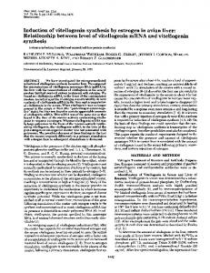
incubation except for the absence ofleucine. The length ofthe washing procedure did not exceed 15 min. The islets were then incubated for two successive periods of 90 min each (min 30-120 and min 120-210) in 1.0 ml of medium which again contained glucose (16.7 mM) and, as required, colchicine or lumicolchicine (0.1 mM). At the end of the second incubation, the islets were sonicated (17) in 1.0 ml of the bicarbonate-buffered solution. The technique used for the measurement of insulin release and insulin content in the islets is described in detail elsewhere (16, 18). Immunoreactive insulin (IRI)l -like tritiated peptides ([3H]IRI) were measured in both incubation media and islet homogenates with a method identical to that reported elsewhere (4). The principle of this method is to allow insulin to react with an excess of anti-insulin serum. Effect of colchicine on proinsulin synthesis and conversion. Groups of 25 islets each were preincubated for 120 min in 1.0 ml of a medium which contained glucose (16.7 mM) and, when required, colchicine (0.1 mM). The islets were then labeled with [3H]leucine (210 ,uCi/ml; 3.6 A±M) during a 10-min incubation performed in media (0.3 ml) of otherwise identical composition as those used for preincubation. The islets were washed twice at room temperature with 1.0 ml of a nonradioactive medium and, thereafter, incubated for 10, 25, 55, or 85 min in 1.0 ml of medium which contained glucose (16.7 mM), unlabeled leucine (1.0 mM), and, when required, colchicine (1.0 mM). The release of insulin was measured only during this last incubation period. After incubation, the islets were sonicated (17) at 0-4°C in 0.3 ml of acetic acid (2 M). The technique used for the separation of 3H-peptides in islet homogenates by polyacrylamide gel chromatography was previously described (17). Fig. 1 illustrates the pattern ofresults obtained by such a method. Effect of colchicine upon the radioautographic distribution of 3H-material. The experimental protocol was comparable tp that used to investigate the effect of colchicine upon proinsulin synthesis and conversion. Thus, the islets were preincubated for 120 min (40 islets/1.0 ml) in media that contained glucose (16.7 mM) with or without colchicine (0.1 mM); labeled over 5-min incubation (200 islets/1.0 ml) in the presence of [3H]leucine (125 ,uCi/ml; 2.5 ,uM), glucose, and, when required, colchicine; washed four times at room temperature with media containing unlabeled leucine (5.0 mM); and eventually incubated for 0, 10, 25, 55, or 85 min in media (40 islets/1.0 ml) again containing glucose (16.7 mM), unlabeled leucine (0.2 mM), and, when required, colchicine (0.1 mM). After the latter incubation, the islets were fixed with 2% glutaraldehyde in phosphate buffer (0.1 M, pH 7.4) and further processed for electron microscopy. Electron microscopic radioautography was performed on thin sections of isolated islets (19). Thin sections were coated with Ilford L-4 emulsion (Ilford Ltd., Ilford, Essex, England), exposed for 5-7 wk, and subsequently developed at 18°C for 4 min with Microdol (Eastman Kodak Co., Rochester, N. Y.) developer, or for 1-2 min with Phenidone (Geigy Chemical Corp., Ardsley, N. Y.) developer (20). The distribution of silver grains was determined on electron micrographs of the first 10-14 fields of B-cell cytoplasm containing radioautographic grains (332+26 per islet), according to the following five classes: rough endoplasmic reticulum (RER), including perinuclear cisternae, transition elements, and the surrounding cytoplasmic matrix; elements of the Golgi complex; secretory granules associated with the Golgi complex; secretory granules not associated
'Abbreviations used in this paper: IRI, immunoreactive insulin; PCG, percentage of grains; RER, rough endoplasmic reticulum.
15r
I 10 0
IaR I-
0 P
5
I
4
0 4c
0 5
10 15 20 FRRACTION NUMBER
25
FIGURE 1 Elution pattern after polyacrylamide gel chromatography of labeled material in homogenates of islets incubated for 90 min in the presence of glucose (16.7 mM) and [3H]leucine. Mean values (+SEM) for nonhormonal peptides (fraction 0-10), proinsulin (shaded area), and insulin (last peak) refer to six individual experiments are expressed in percentage of the total radioactivity eluted from the column.
with the Golgi complex; and other sites (mitochondria, nuclei, plasma membrane, lysosomes). The latter class only contained 8.5±0.6% (n = 60) of the total amount of silver grains. The method used to measure glucose oxidation (21) and the concentration of ATP, ADP, and AMP (22) in the islets are described elsewhere. All results are expressed as the mean (±SEM).
RESULTS
Effect of colchicine upon the release of preformed and newly synthesized insulin We have first investigated the extent to which a colchicine-induced alteration of the B-cell microtubular apparatus may affect the release of newly synthesized insulin. For this purpose, the islets were first exposed for 30 min to [3H]leucine, then washed to remove extracellular [3H]leucine, and eventually incubated for two successive periods of 90 min each in a leucine-free medium. Glucose (16.7 mM) and, when required, colchicine (0.1 mM) were present in the media throughout this procedure. The release of both insulin and [3HlIRI was measured during the two successive periods of incubation (min 30-120 and 120-210). After the second incubation, the islets were homogenized for measurement of their content of insulin, [3H]IRI, and TCAprecipitable tritiated material. In designing this experiment, we took into account the time- and dose-related effect of mitotic spindleinhibitors upon the B-cell microtubular apparatus (23), so that the labeling of the islets occurred at a time
Colchicine and Proinsulin Biosynthesis
1285
Effect of colchicine upon proinsulin synthesis. Colchicine reduced the amount of [3H]leucine incorporated in the integrated amount of [3H]IRI, i.e., that both released in the media and recovered in the islets (-20.9 ±7.8% by paired comparison; P < 0.05). The specific activity (counts per minute per microunit) of the integrated amount of insulin was consequently also reduced after exposure to colchicine (P < 0.01). These data, however, do not distinguish whether this reduction is related to the "antitubulin" property of colchicine or to a decreased uptake of [3H]leucine (24), an abnormality in the intracellular compartmentation of this amino acid, or a true reduction in the B-cell biosynthetic activity. Whatever the explanation, it should be stressed that colchicine failed to affect the preferential stimulant effect of glucose upon the biosynthesis of proinsulin as distinct from that of other islet proteins (5, 17). Thus, the ratio of [3H]IRI to total TCA-precipitable 3H-material in the final insular homogenate was not lower after exposure to colchicine (0.25±0.02) than under control conditions (0.22±0.02). Effect of colchicine upon [3H]IRI release. To correct the above-mentioned effect of colchicine upon [3H]leucine incorporation in insular proteins, the presentation of results dealing with the effect of the mitotic spindle-inhibitor upon release of newly synthesized peptides will be restricted to those data which illustrate
(min 0-30) when only a modest decrease in the number of microtubules had presumably been achieved. Effect of colchicine upon insulin release. The release of insulin evoked by glucose during the first incubation (30th-120th min) averaged 334+±20 uU/islet (Table I). It fell slightly during the second incubation (120th-210th min) to 79.4+7.4% of its initial paired value. This behavior confirms previous observations (4, 5). Colchicine inhibited glucose-induced insulin secretion. Relative to its paired control value found in the absence of colchicine during the same periods of incubation, the release of insulin evoked by glucose in the presence of colchicine averaged 83.3±3.8% (P < 0.01) and 58.9±6.7% (P < 0.001), over the first and second period of incubation, respectively. Such a timerelated increase in the inhibitory effect of colchicine is also obvious when considering the fractional rather than absolute values for insulin release (Table I). As expected from the findings so far outlined, the residual insulin content of the islets exposed to colchicine was somewhat higher than that of the islets incubated in the absence of the mitotic spindle-inhibitor, the latter difference being significant (P < 0.001) judged from the increase in the fractional content (+8.6 ± 1.8). The total amount of insulin released in the media and recovered in the islets was almost identical in the presence or absence of colchicine.
TABLE I
Effect of Colchicine upon the Release of Preformed and Newly Synthesized Insulin Parameter
Coichicine
Control
% of total
AUIislet
Insulin Released from min 30 to 120 Released from min 120 to 210 Final content Integrated amount
334±20 258±23
276±13 150±17
1,159+42 1,751+68
1,310+110 1,735±120
19.1±1.1 14.5±0.8 66.3±1.1 100.0
Integrated amount
226±20 155±35 971±78
58±16 87±18 920±99
1,352±88
16.4±1.1 8.7±0.9 74.9±1.4 100.0
% of total
cpmlislet
[3H]IRI Released from min 30 to 120 Released from min 120 to 210 Final content
Coichicine
Control
1,064±111
17.5±2.1 11.1±2.3 71.4±3.0 100.0
5.2±1.4 8.0±1.6 86.8±2.3 100.0
cpm/cpm
[3H]IRI/3H-protein Final content
0.22±0.02
0.25±0.02
cpm/,uU
[3H]IRI/insulin Released from min 30 to 120 Released from min 120 to 210 Final content Integrated data
0.67±0.03 0.59±0.12 0.83±0.05 0.77±0.03
0.20±0.06 0.53±0.11 0.71±0.04 0.61±0.04
ratio
0.87±0.08
0.77±0.16 1.08±0.04 1.00
Mean values (±SEM) refer to 10 individual observations.
1286
offractional values
Malaisse-Lagae, Amherdt, Ravazzola, Sener, Hutton, Orci, and Malaisse
0.30±0.08 0.87±0.18 1.14±0.03 1.00
TABLE II Effect of Colchicine upon Proinsulin Conversion Control
Colchicine
1.85+0.25 (12)
1.04±0.15 (12)
.:
:: .? . :.> . f:
::;: : : : :.: X
:^\
- sl _52 ff
:.
_. __ A_ .A
U
w_
w w1
*
B'''' ;'':' .. t
. '%:
i. . . ,S
S Af'ye
::
K:
* ;>:
*.
v
a,
.e :.
S
:t
_ _
.:
q-
..
,ip 5; .F.'
*. ;:,,;
*::1:
^
wr
f .::
0
0
~~~~~~~~~~~~~~~~~~~~~~~~~~~~~~..
..
0
4-
4-
_
*:
.wS
.,: * *
_jR
.:,,
x Za
-r
_
_.
-.
I.
"
?f4: .W
F.
0
i:
^
*'
-
_
_...
.J
.
a
..
_
-
^ RER #
AW),. ...ws,.
..st"b.
~ *.1_
MF
s;§2.it-'
a::-
,.
::::
*:
._-
,
.F'
'W
jlillidb .,_, ._.
::
ii:.
|w
|F:
::
...
.:
Vt,
i
CW
:k'
4 >e, i'.
...i
::
s.
ss
...
1288
i:
Malaisse-Lagae, Amherdt, Ravazzola, Sener, Hutton, Orci, and Malaisse
'.
the presence of unlabeled leucine (1.0 mM) for either 10, 25, 55, or 85 min (chase-incubation period). Glucose (16.7 mM) and, when required, colchicine (0.1 mM) were present in the media throughout the preincubation, pulse-labeling, and chase-incubation periods. The release of insulin was measured during the final incubation, whereas the islets were eventually homogenized in 0.3 ml of acetic acid (2.0 M) for separation of 3Hpeptides by polyacrylamide gel chromatography. Effect of colchicine upon insulin release. The mean rate of glucose-induced insulin release in the islets incubated for 25, 55, and 85 min after labeling was significantly lower (P < 0.01) in the islets exposed to colchicine than the cointrol islets (Table II). Relative to the mean control value, the rate of secretion in the islets first exposed for 120 min to colchicine averaged 56.1±7.9%, a value close to that found after the same length of pretreatment with colchicine in the first series of experiments, in which glucose-induced insulin release averaged, between the 120th and 210th min, 58.9+±6.7% of its paired control value. Effect of colchicine upon proinsulin synthesis. The rate of [3H]leucine incorporation in the islet proteins averaged, in the control islets, 4.45±0.36 fimol/islet per min, a value close to that found, with a different technique, in the first series of experiments (4.07+0.23 fmol/islet per min). The incorporation of radioactive leucine in the islet proteins was slightly but significantly reduced by colchicine, the colchicine-induced reduction averaging 22.6±6.2% of the mean control value (Table II). The relative magnitude of such a reduction was almost identical to that observed for the [3H]IRI in the first series of experiments (20.9±7.8%). Once again, however, colchicine failed to affect the preferential incorporation of [3H]leucine in hormonal pepti(les; the ratio of hormonal to total 3H-peptides averaging 0.66±0.03 and 0.62±0.02 in the absence and presence of colchicine, respectively. Such a ratio remained fairly stable throughout the final incubation. Effect of colchicine upon proinsulin conversion. In the control islets, the fractional amount of proinsulin converted to insulin and C-peptide progressively increased from 4.2±1.0 to 91.3+1.3% between the 10th and 85th min of the final incubation period (Table II). In the islets exposed to colchicine, the conversion of proinsulin was considerably delayed, 25.6+±3.7% of the
hormonal precursor being still presenit in the islets at the 85th min of incubation (Table II). When plotted in semilogarithmic coordinates, the line relating the residual relative amount of proinsulin to time suggested that, between the 25th and 85th min, the meani appaient half-life for the process of proinsulin conversioin to insulin was increased by colchicine friom 19 to 31 min
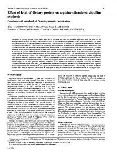
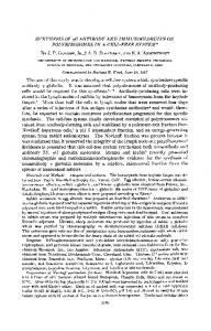
(Fig. 2). Effect of colchicine upon the morphology of B cells and upon the radioautographic distribution of 3H-material In this study, as well as in previous reports dealing with microtubule inhibitors (2, 25, 26), the most consistent alterations observed in the B cells exposed to colchicine concerned the RER and the Golgi region (Figs. 3 and 4). RER cisternae were often dilated and contained a variable amount of pale flocculent material. The Golgi region was characterized by the presence of large accumulations of microvesicles between and around Golgi cisternae. Numerous transition elements were seen contributing microvesicles. These changes were associated with a virtual absence of microtubules. The procedure used to follow, at the ultrastructuiral level, the fate of 3H-peptides in the islets was almost identical with that used for the study of proinsulin conversion (Table III), except that the exposure time to [3H]leucine was reduced from 10 to 5 min and the concentration of unlabeled leucine in the final incubation medium increased from 1.0 to 5.0 mM. The distributioni of silver grains, as determined on the electron micrographs (Figs. 3 and 4), is expressed as the percentage of grains (PCG) found over each type of organelle(s). RER. Throughout the 90-min period of observation, more radioactivity was found in the RER of colchicinetreated than control B cells (Fig. 5). The integrated mean value (Sth-9Oth min) averaged 35.9+2.1 PCG, after exposure to colchicine and 27.1± 1.8 PCG in control islets, respectively (n = 30; P < 0.005). Between the 5th and 30th min, the amount of radioactivity located in the RER progressively fell from 37.8+3.8 to 19.7 ±2.8 PCG in the control islets, and from 47.3±4.9 to 32.2±6.2 PCG in colchicine-treated islets (n = 6). When corrected for an apparent phenomenion of recirculation of 3H-material in the RER, the half-life for
FIGURE 3 Radioautographies (x21,000) of B cells. Emulsion developed with Phenidone. (a) Control B cell, preincubated for 2 h, pulse-labeled with [3H]leucine for 5 min, and further incubated for 25 min at 16.7 mM glucose. The majority of radioautographic grains overlie the Golgi area, where they are seen mostly accumulated over maturing secretory granules. The arrows indicate microtubules. (b) B cell: same protocol as in a, except for the continuous presence of colchicine (0.1 mM). Radioautographic grains are found in the Golgi region as well as over dilated elements of the RER. The asterisk indicates accumiiulation of microvesicles. Te, transition elements of the RER. The bars indicate 1 am.
Colchicine and Proinsulin Biosynthesis
1289
AL >: a ris s>'''
.i,~ ~ ~ ~ E F
0
_
*
,..'tS
T, . .S, .e, Mt'>
F.s'
.
S
_
A
i; i; > rr sB
*
A
;
*
w
_j,
^
l
2_
;
4
*;9~~~~~~~~~~~~~A
;
bs......................................................... .'';9A S.t1
;
=
i
w
'>
t
A | j ; 1~~~~~~~~~~~~~~~~~~4
|
1
!Nz
---
ff~~~~~i
ref 4iCeM M v ;xEsC?d Y98.s;t iiMi~~~~~~~~~~~~~~~~~-A % d Wi
, ls ,.>w
All*ts j