Roles of Apoptosis in Airway Epithelia Yohannes Tesfaigzi Lovelace Respiratory Research Institute, Albuquerque, New Mexico
The airway epithelium functions primarily as a barrier to foreign particles and as a modulator of inflammation. Apoptosis is induced in airway epithelial cells (AECs) by viral and bacterial infections, destruction of the cytoskeleton, or by exposure to toxins such as high oxygen and polycyclic hydrocarbons. Various growth factors and cytokines including TGF-, IFN-␥, or the activators of the death receptors, TNF-␣ and FasL, also induce apoptosis in AECs. However, cell death is observed in maximally 15% of AECs after 24 h of treatment. Preincubation with IFN-␥ or a zinc deficiency increases the percentage of apoptotic AECs in response to TNF-␣ or FasL, suggesting that AECs have mechanisms to protect them from cell death. Apoptosis of AECs is a major mechanism in reducing cell numbers after hyperplastic changes in airway epithelia that may arise due to major injuries in response to LPS or allergen exposures. Resolution of hyperplastic changes or changes during prolonged exposure to an allergen is primarily regulated by the Bcl-2 family of proteins. Fas and FasL are both expressed in AECs, and their main function may be to control inflammation by inducing Fasinduced death in inflammatory cells without inducing apoptosis in neighboring cells. Furthermore, AECs engulf dying eosinophils to clear them by phagocytosis. Therefore, in the airway epithelium apoptosis serves three main roles: (1 ) to eliminate damaged cells; (2 ) to restore homeostasis following hyperplastic changes; and (3 ) to control inflammation, and thereby support the barrier and antiinflammatory functions. Keywords: cytokines; infection; inflammation; phagocytosis; mucus cell hyperplasia
The airway epithelium (AE) is developmentally programmed to protect itself from environmental toxins and injury by forming a barrier, trapping foreign particles that enter the airway, and allowing their removal by ciliary action. This primary role explains why secretory and ciliated cells are the major cell types within the airway epithelium. In recent years, it has become increasingly clear that the AE also is key modulator of inflammatory responses and plays a crucial role for normal immune function. For this purpose, the AE not only produces a broad range of cytokines and chemokines but also generates lipids and peptide mediators (enzymes and enzyme inhibitors) and reactive oxygen species (ROS) that enhance the infiltration of the lung airspaces with leukocytes (1). Mediators secreted by airway epithelial cells (AECs) and infiltrating leukocytes in turn affect the differentiation of the AE and the cell death processes of epithelial cells. This review focuses on what is known about the apoptotic signaling mechanisms in AECs in the context of the various roles airway epithelial cell death contributes to the normal func-
(Received in original form January 10, 2006 and in final form January 29, 2006) This research was supported by grants from the National Heart, Lung, and Blood Institute (HL068111) and from the National Institutes of Environmental Health Sciences (ES09237). Correspondence and requests for reprints should be addressed to Yohannes Tesfaigzi, Lovelace Respiratory Research Institute, 2425 Ridgecrest Drive, SE, Albuquerque, NM 87108. E-mail:
[email protected] Am J Respir Cell Mol Biol Vol 34. pp 537–547, 2006 Originally Published in Press as DOI: 10.1165/rcmb.2006-0014OC on January 26, 2006 Internet address: www.atsjournals.org
tioning of the epithelium. Because of the different natures of the type I and type II alveolar cells, apoptosis related to alveolar epithelial cells will not be discussed in the present review.
THE BASIC PATHWAYS OF APOPTOSIS To make this review on apoptotic pathways in airway epithelial cell death easily understandable for readers not familiar with this subject, a brief summary of the major pathways known to elicit programmed cell death in many cell types is provided. The studies summarized were primarily conducted in cells of the hematopoietic compartment and have laid the foundation for studies in neuronal and epithelial cells from various organs (2). Two pathways broadly categorize the sequence of events that culminate in the activation of aspartate-specific cysteine proteases, caspases (cysteinyl, aspartate-specific proteases): the extrinsic and the intrinsic pathways. The extrinsic pathway can be initiated by one of several cell surface death receptors when bound by the appropriate ligand (3). TNF receptor 1 (TNFR-1) and Fas receptors contain death domains (DDs) and recruit the DD-containing adaptor molecules, TNFR-1–associated death domain (TRADD), and Fasassociated death domain (FADD), respectively. Homotypic interaction between the DDs of Fas and FADD induces the recruitment and self-activation of pro–caspase-8 (4). In TNF signaling, TRADD recruits FADD after formation and release of a TNFR-1 complex to initiate pro–caspase-8 activation (5, 6). The receptors for TNF-related apoptosis-inducing ligand (TRAIL), TRAIL-R1 (also known as death receptor-5, DR-5), also recruit and activate pro–caspase-8 (7) in a FADD-dependent manner (8). The intrinsic pathway is characterized by the permeabilization of the outer mitochondrial membrane and the release of several pro-apoptotic factors into the cytosol. These factors include cytochrome c (9, 10), Smac/Diablo (11, 12), apoptosis-inducing factor (AIF) (13), endonuclease G (14), and HtrA2/Omi (15). The release of these mediators is regulated by the Bcl-2 family (16). Once released, cytochrome c binds to an adaptor protein, Apaf-1, which self-oligomerizes and recruits pro–caspase-9 to form the apoptosome complex (17). This promotes the autoprocessing of caspase-9, which in turn recruits and cleaves pro–caspase-3, which degrades proteolytically targeted substrates and activates DNases. Proteins in the Bcl-2 family are major regulators of the intrinsic pathway. Family members include both pro- and antiapoptotic proteins, and they share homology in four conserved regions, termed Bcl-2 homology (BH) domains 1–4 (18). The family can be divided into three main subclasses: 1. The anti-apoptotic proteins that include Bcl-2 and Bcl-xL are all multi-domain, sharing homology throughout all four BH domains. Bcl-2 and Bcl-xL enhance cell survival by inhibiting apoptosis induced under a wide variety of circumstances. Bcl-2 and Bcl-xL extend cell survival by preventing cell death in different cell types and in response to different stimuli, suggesting that Bcl-2 and Bcl-xL act at a central control point in the pathway to apoptotic cell death (18, 19, 2). 2. The multidomain pro-apoptotic proteins such as Bax and Bak possess sequence homology in at least three BH
538
AMERICAN JOURNAL OF RESPIRATORY CELL AND MOLECULAR BIOLOGY VOL 34
domains. Bax and Bak have overlapping roles in the regulation of apoptosis during mammalian development and tissue homeostasis (20). Activation of these “multidomain” pro-apoptotic members is an essential gateway required for cell death in response to diverse stimuli (21). 3. The third subset of this family includes Bid, Bad, Noxa, Bik, Bmf, Blk, Hrk, Bnip-3, Puma, and Bim, all of which lack BH-1, -2, and -4 (22). BH-3–only proteins are proapoptotic and share sequence homology within the amphipathic ␣-helical BH-3 region (23–25). Bid is cleaved by caspase-8 to generate active truncated Bid (tBid), which leads to the Bax-dependent release of pro-apoptotic factors from mitochondria. Thus, this event integrates the extrinsic and intrinsic pathways (26). The BH3 region is required for the pro-apoptotic members to cause binding to multidomain Bcl-2 family members and induce death (27). BH-3 molecules display some selectivity for multiple domain Bcl-2 members (24, 28). Inactivation of the antiapoptotic proteins Bcl-2 and Bcl-xL by BH-3 domain-only members results in the activation of the “multidomain” pro-apoptotic members Bax and Bak (29). The pro-apoptotic activity of BH-3–only molecules is kept in check by either p53-dependent transcriptional control (25), posttranslational modification, or by binding to the dynein light chain in myosin V filamentous actin, thereby being sequestered from binding to Bcl-2 (30, 31). Association of BH-3 proteins with actin filaments enables them to sense death signals and initiate anoikis (30, 32). When cells receive a stimulus to undergo apoptosis, the BH-3–only members translocate to inactivate Bcl-2 or to enable Bax and Bak and initiate a cascade of events leading to activation of caspases and DNases. Members of all three classes of the Bcl-2 family can localize to the endoplasmic reticulum (ER) membrane and influence ER homeostasis (33). Calcium release from the ER can either directly activate effectors of caspases or sensitize mitochondria to a variety of extrinsic and intrinsic death stimuli.
APOPTOSIS AS A MECHANISM OF REMOVING DAMAGED AECs The epithelial barrier represents a critical line of defense against the environment, so AECs are likely designed to be refractory to a number of potentially apoptotic stimuli, including cell death receptor activators such as TNF-␣ and FasL. The relative resistance of AECs to apoptosis is likely helpful in maintaining the integrity of the epithelial barrier during an inflammatory response when immune cells that express or secrete these death receptor ligands are trafficking through the lung. These characteristics may be the basis for the inherent properties of the airway epithelium, which is a stable mucosal surface with relatively low rates of cell proliferation under normal conditions (34). Cell Death by Viral Infection
Apoptosis in response to viral infection likely occurs as part of the host response in an attempt to limit virus replication and to prevent persistent infection and spread of the virus. Thus, in the context of a virus infection, damage to the epithelial barrier may be the lesser evil. As a consequence, many viruses have evolved various mechanisms to inhibit or evade apoptosis. In addition, death by apoptosis enhances the efficiency of capture of viral antigens by antigen-presenting cells and presentation to T cells. Infection of AECs with the adenoviral vector Ad-CMV-lacZ (35), or with influenza A virus (36), induces apoptotic cell death. Respiratory syncytial virus (RSV)-induced apoptosis of airway
2006
epithelial cells has been reported; however, the Fas pathway appears to not be important (37). Other studies have implicated RSV in inducing anti-apoptotic pathways. Treatment of RSVinfected cells with an inhibitor of phosphatidyl-inositol 3-kinase (PI3-k) resulted in more rapid apoptosis, implying that under normal conditions signaling through the PI3-k pathway mediates an inhibition of RSV-induced apoptosis (38). This is consistent with another report showing that apoptosis is inhibited or delayed in RSV-infected cells because a minimal cytopathic effect was observed in response to RSV infection in primary human AECs that had differentiated into a pseudostratified mucocilliary epithelium in vitro (39). Analysis of apoptotic genes expressed in RSV-infected cells showed that cells may be susceptible to killing through the TRAIL pathway and Mcl-1 might account for the delayed induction of apoptosis in RSV-infected cells (40). Cell Death by Bacterial Infection
Under normal conditions, primary AECs in culture form tight junctions that render them resistant to apoptosis by an inoculum of Pseudomonas aeruginosa PAO1 (only 5–10% of cells showed mitochondrial permeability changes) at doses that are sufficient to activate proinflammatory cytokine expression. Resistance is lost, and up to 50% of the population undergo apoptosis after 6 h of exposure once the tight junctions are disrupted with EGTA or in cell lines such as 9HTEo- cells that do not form tight junctions (41). Specific adhesions, complete LPS, and functional type II secretion system by the P. aeruginosa are necessary to evoke apoptosis even in the susceptible cells that were unable to form tight junctions. Cystic fibrosis transmembrane conductance regulator (CFTR) mutation is attributed to the accumulation of CFTR in the ER and activation of the NF-B, which is anti-apoptotic (42). In addition, caspase activity may be inhibited by impaired pH regulation in CFTR cells (43). However, AECs with CFTR dysfunction (⌬F508 – W1282X or ⌬F508) show no differences in apoptosis compared with normal cells after treatment with P. aeruginosa (41). Similarly, CFTR ⫹/⫹ and ⫺/⫺ mice show few apoptotic cells in the airway epithelium after P. aeruginosa infection. It is possible that P. aeruginosa toxins induce apoptosis by disturbing actin polymerization and cell–cell contacts, and may not be related to CFTR (44). Vernooy and coworkers (45) report that instillation of LPS induces apoptosis in bronchial epithelial cells within 2 h and peaking at 24 h and that TNF-␣ is not involved in this cell death process. However, this report does not exclude the possibility that TUNEL positivity in the AE could stem from dying inflammatory cells. P. aeruginosa stimulates transcription of IL-8, the major attractant for polymorphonuclear leukocytes (PMN) by stimulating translocation of NF-B. NF-B also inhibits cell death by inducing TRAF-1 and TRAF-2 and cIAP1 and cIAP2 to suppress caspase-8 activation (46, 47), and also regulates expression of genes that inhibit cell death in part by inducing the anti-apoptotic Bcl-2 (48). Therefore, other pro-apoptotic pathways may be activated to override the anti-apoptotic function of NF-B to induce cell death in primary AECs (41). Bacterial exoproducts (especially exotoxin, ETA) from P. aeruginosa induce death in 16HBE14o⫺ cells 24 and 48 h after treatment with 1,000 ng/ml as measured by the dimethylthiazole 2,5 diphenyl tetrazolium bromide (MTT) assay. This cell death is not affected by zVAD-fmk (a broad spectrum inhibitor for caspases), condensed nuclei are not observed, and mitochondria of dying cells were highly condensed (49). The absence of mitochondrial swelling suggests that mitochondrial permeability transition played a minor role in this cell death because large transmembrane pores are likely to open in response to mitochondrial dysfunction. The authors conclude that ETA induces cell
A Tribute to Carol Basbaum
death in AECs by a mechanism different than that classically described as apoptosis. Because ETA did not induce apoptosis in primary cultures of human nasal polyp epithelial cells, and the role of tight junctions was not tested, the significance of ETA to cause cell death under physiologic conditions is not clear. Cell Death by Destruction of Cytoskeletal Support
Disruption of the elongation or the aggregation of actin filament with cytochalasin D (0.5 g/ml) or with jasplakinolide (3 M), respectively, induces apoptosis as measured by TUNEL positivity in 30–50% 1HAEo⫺ and in 10–15% primary normal human bronchial epithelial (NHBE) cells within 5 h (50). This death was abrogated by z-VAD-fmk but not by blocking the Fas receptor. Pro–caspase-8 was cleaved by cytochalasin D but not by jasplankinolide, suggesting that disruption of actin filament integrity by the two agents causes cell death by different mechanisms. Further support for the significance of actin filaments in affecting cell death stems from inhibition studies of Rho kinase, which regulates the formation of stress fibers and focal adhesions (51). Inactivating Rho kinase with Y-27632 or HA1077 or by overexpressing a mutant Rho kinase causes membrane ruffling and apoptosis (as measured by TUNEL staining in 15–25% of primary AECs). While the signaling of cell death induced by destruction of actin filaments is not known, there is increasing evidence that in other cell types disruption of microtubule turnover by toxol or vincristine (which phosphorylate Raf-1 and Bcl-2) leads to apoptosis and detachment (52, 53). Activation of caspase-3 cleaves the actin filament, severing the protein gelsolin and leading to morphologic changes of the dying cells (54). However, it is not known whether the cleavage of actin cytoskeletal network itself causes apoptosis or if this is just one of the manifestations of the cell’s shape. Furthermore, BH-3–only proteins are associated with actin filaments, and disruption of their interaction may enable them to sense death signals and initiate detachment of AECs (30, 32). Cell Death by Oxygen Exposure
AECs are constantly exposed to high oxygen pressure and have developed an antioxidant capacity that is sustained with antioxidant enzymes to detoxify reactive oxygen species (ROS). However, when this protective system is overwhelmed, apoptosis is induced with a minimal amount of necrosis (measured by the release of lactic dehydrogenase, LDH), as shown for primary small airway epithelial cells (SAEs) exposed to 95% O2 for 2 d (55). However, A549 cells primarily undergo necrosis, which is why it is necessary to test the effect of each stimulus in primary cells to validate their importance under physiologic conditions. When adult male C57Bl/6J mice are exposed to 95% O2 for 72 h, AECs (most prominently the AECs of the distal bronchiolar epithelium) and alveolar cells show increased p53 protein levels and loss of DNA integrity as detected by TUNEL (56). DNA damage induced by ionizing radiation, bleomycin, or drugs that inhibit DNA toposisomerase activity prolong p53 protein half-life, and this accumulation of p53 induces a G1 arrest (57, 58). However, when DNA damage is extensive, p53 can induce cell death by inducing Bax (59) or by directly damaging mitochondrial integrity (60). Death of AECs by IFN-␥, TNF-␣, and Fas
Exposure of NHBEs to IFN-␥ causes Bax translocation to the ER, calcium release (data not shown), and caspase activation (61). Although IFN-␥ has induced Fas in keratinocytes (62), we did not observe induction of Fas in NHBEs exposed to IFN-␥. Co-treatment with Fas-antibody does not enhance IFN-␥– induced cell death in NHBEs. IFN-␥ has been shown to induce
539
apoptosis in a variety of cell types, including colon adenocarcinoma cells (63, 64), primary human keratinocytes (62), HeLa cells (65), breast tumor cells (66), and fibroblasts (67). IFN-␥– enhanced signaling proteins that are responsible for cell death remain largely unknown (68), and various pathways appear to be involved in IFN-␥–induced apoptosis. TNFR-1 and -2 are expressed in AECs, and their levels are increased in response to TNF-␣ (69); however, preincubation of these cells with TNF-␣, IL-1, or IFN-␥ did not effect expression of Fas or the mRNA for the adaptor proteins FADD, TRADD, or caspase-8 (70). AECs also express Fas receptor and FasL (69, 71, 72). Therefore, several studies have tested whether Fas or TNF-␣ induce cell death in AECs. Only 10–15% of primary AECs or A549 cells undergo cell death in response to anti-Fas ligation of the receptor (73, 74, 72) or to TNF-␣ (69). While all SAEs expressed Fas receptor to a similar extent as did NHBEs, 40% of distal SAEs showed cell death, and only 10% of proximal NHBEs showed TUNEL positivity when exposed to soluble FasL (500 ng/ml) (70). While NHBEs are refractory to Fas or TNF-␣, they can be sensitized by other cytokines to readily undergo cell death to these mediators. Activated T cells and eosinophils from healthy and atypic individuals induce apoptosis in AECs, and cell death was blocked when activated T cells and eosinophils were preincubated with antibodies to IFN-␥ and TNF-␣ (69). However, eosinophils induced apoptosis in AECs only when cells were pretreated with IFN-␥. Interestingly, apoptosis induced in AECs by T cell supernatants was prevented by caspase inhibitors but was not affected by Fas or FasL, suggesting that the FasL–Fas pathway plays a less important role in the death of AECs. A549 (72), NT-1, and the cystic fibrosis cell lines CFT-1 and CFT-2 (75) are also not susceptible to anti-Fas mAb, although they express the Fas receptor. However, up to 74% of these cell lines, primary NHBEs, and SAEs show cell death when pretreated with 10 ng/ml IFN-␥ for 24 h (69). A minimum of 6–8 h of pretreatment was sufficient to increase cell death in A549 cells by anti-Fas (72). Collectively, these studies suggest that IFN-␥ sensitizes AECs to TNF-␣– or FasL-induced cell death. However, the mechanisms of this sensitization process are not known. TGF- and Death of AECs
In NHBEs, TGF- induces apoptosis by increasing the p38 MAP kinase phosphorylation, but also by increasing the phosphorylation of ERK 1/2 and JNK. This cell death was inhibited by budesonide or with inhibitors of the MAP kinases P38, ERK 1/2, and JNK (76). Another report showed that TGF-1 protected NHBEs from Fas-induced cell death that was in the range of 10–15%, and this protection was blocked by inhibition of the Smad pathway (73). These contradictory findings may suggest that the definition of inducing apoptosis in a cell population is somewhat vague, or that future studies need to clarify whether TGF- has a protective role when it is present together with other inducers of apoptosis. Corticosteroids and Apoptosis of AECs
Corticosteroids elicit apoptosis in eosinophils and in T lymphocytes in a rapid and massive manner (77–79). Several studies from the same group report that corticosteroids elicit apoptosis in AECs where the magnitude of apoptosis in response to dexamethasone treatment for 24 h reached a maximum ⵑ 5–10% as measured by TUNEL positivity (80–82). The authors acknowledge that this proportion is small compared with apoptosis elicited by the same agents in hematopoietic cells, but we propose that epithelial damage over prolonged treatment with corticosteroids may lead to a significant degree of airway mucosal damage. These studies used concentrations of corticosteroids at the high
540
AMERICAN JOURNAL OF RESPIRATORY CELL AND MOLECULAR BIOLOGY VOL 34
end of what might be achieved in a clinical setting (80). While apoptosis of AECs in the AE of an individual with asthma can be detected after collection by endobronchial biopsy, other studies have not observed epithelial cell apoptosis by individuals with asthma (83). Therefore, the physiologic significance still remains to be elucidated. The -adrenergic agonist, albuterol, and both PKA activators, forskolin and dibutyryl cAMP, significantly inhibited the 5% apoptosis induced by corticosteroids. And an inhibitor of PKA, H-89, also blocked the protective effect of albuterol, suggesting that the inhibition was through the PKA pathway. Because treatment with these -agonists for longer than 4 h after treatment with corticosteroids was not protective, the authors recognize that the timing of the protective effect of albuterol (an agent that is relatively short in half-life for biological effect when inhaled) may be a concern. While the mitochondrial pathway and cytochrome c release is suggested, Bcl-xL does not affect this cell death, and absence of blocking experiments make a conclusive understanding of the mechanism of action impossible at present. Fas receptor–activated cell death of AECs was not affected by -adrenergic agonists, indicating that the pathways for the corticosteroid receptor and Fas receptor–induced cell death are different. However, the Fasinduced cell death (5–15% of cells showed TUNEL positivity at 24 h) is also relatively minor. Corticosteroid treatment of BALB/c mice over prolonged periods of up to 4 wk caused increased presence of AECs in the lavage fluid. Furthermore, the concentration of dexamethasone at 1 mg/kg, sufficient to reduce the allergen-induced inflammation, did not reverse airway epithelial shedding. Analysis of the epithelial cells by histology showed increased TUNEL positivity and also increased detection of cleaved p85 by immunohistochemistry. Dorscheid and colleagues (84) concluded that corticosteroid treatment over prolonged periods may induce apoptotic cell death in airway epithelium in vivo, and while sufficient to reduce the allergen inflammatory response, did not alter the number of AECs that are shed during an asthmatic phenotype. In contrast to these studies, several reports show that glucocorticoids inhibit cell death induced by cytokines, such as IFN-␥ or TGF-. Dexamethasone at a concentration of 1 mM induced the hIAP mRNA significantly and inhibited cell death in A549 cells by IFN-␥ from 38% to 7% and by IFN-␥ plus anti-Fas from 81% to 10% (72). IFN-␥ further increased dexamethasoneinduced hIAP. Cell death by IFN-␥ and Fas was successfully inhibited by zVAD-fmk, suggesting that caspases are involved in this cell death process. IFN-␥ induced IL-1–converting enzyme (ICE) or caspase-1; however, the cleavage product of caspase-1 was not detected. Dexamethasone did not affect IFN-␥–induced caspase-1 expression. Because zVAD-fmk inhibited IFN-␥ and anti-Fas–induced apoptosis the authors conclude that caspase 3 and possibly caspase 7 were involved in this cell death process. Whether dexamethasone-induced hIAP suppresses apoptosis by IFN-␥ and Fas is not clear. Also, the TGF-–induced MAP kinase activation and apoptosis in AECs are effectively inhibited by budesonide, suggesting that inhaled glucocorticoids have a protective role against epithelial injury (76). Zinc as Anti-Apoptotic Factor
When primary AECs are deprived of zinc, a nominal increase in caspase activation and apoptosis is observed in 10% of cells; however, the barrier function of differentiated cultures is not disrupted (85). Zinc depletion combined with treatment with TNF-␣, IFN-␥, and FasL abruptly decreases trans epithelial resistance and increases apoptosis to 60–70% as measured by caspase-3 activity. This apoptosis was suppressed by adding zinc or magnesium sulfates. Interestingly, zinc increased phosphory-
2006
lation of Akt within 2 h of treatment, and suppression of Akt phosphorylation with LY249002 suppressed the protective function of zinc. This study suggests that subacute zinc deficiency may place the AE at a disadvantage during periods of inflammation by reducing the refractory state of the AE. While zinc activates PI3 kinase signaling it is not clear whether other signaling pathways may be activated by zinc to protect AECs from cell death. Identification of the pathways that make a normal AE resistant to many inducers of apoptosis is a crucial field of study to fully understand deficiencies that may provide sustained damage and inflammation in chronic diseases.
APOPTOSIS TO RESTORE HOMEOSTASIS AFTER HYPERPLASTIC CHANGES IN THE AIRWAY EPITHELIUM Exposure of the lungs to harmful substances such as bacteria or cigarette smoke induces acute lung inflammation characterized by infiltration with neutrophils and macrophages (86–88), followed by proliferation of nonciliated columnar epithelial cells (89–93). Inflammatory cells secrete a number of proteins (including cytokines, chemokines, and proteases) that promote the differentiation of proliferating and pre-existing epithelial cells into mucous cells by inducing mucin biosynthesis (94–96). Therefore, after injury, the airway epithelium presents goblet cell metaplasia and hyperplasia (97). Goblet cell metaplasia (GCM) can occur as a result of preexisting Clara or serous cells that begin to express MUC5AC (98) in the absence epithelial cell proliferation. Another study (92) reported that 10% of mucous cells proliferate in conjunction with a compensatory decrease in Clara and ciliated cell numbers. Our studies show an increase in total AECs together with increased GCM (93, 99, 100). The difference between the three studies appears to be the extent of injury and inflammatory response due to length of allergen exposure or dose of LPS administered. The resolution of GCM under conditions where epithelial cells have not proliferated is associated with downregulation of MUC5AC expression, which leads to the reversion of mucous cells to Clara or serous cells. When injury is extensive and epithelial cell proliferation has occurred, in the absence of further insult to the epithelium, inflammation is cleared, and mucous cell numbers are reduced by programmed cell death (74, 101, 102). Resolution of Hyperplastic Changes after a Single Injury to the Airway Epithelium
After a single intratracheal instillation of LPS, the number of AECs per millimeter of basal lamina exceeds the number found in normal epithelium by 30–40% (93, 99). Resolution of these hyperplastic changes involves reduction of AEC numbers by 30% together with downregulation of MUC5AC expression and differentiation of mucus-producing cells into Clara, serous, or other types of cells. Classic apoptotic epithelial cells are not obvious during the recovery of the airway epithelium; whether some of the metaplastic cells slough off during this process is still under investigation. It is possible that selected cells are removed by extrusion and may be sloughed off before the classic apoptotic morphology appears. Cell death involving detachment from the basement membrane is also called anoikis (103). In an attempt to understand the mechanisms involved in reducing the number of metaplastic mucous cells, we analyzed the expression of regulators of apoptosis and found that metaplastic mucous cells transiently express Bcl-2, an inhibitor of apoptosis (104, 102). Bcl-2 is expressed in 30% of LPS-induced metaplastic goblet cells in airway epithelia (102) or in ⵑ 50% of ozone-induced metaplastic goblet
A Tribute to Carol Basbaum
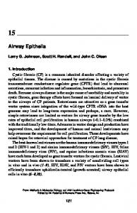
cells in epithelia lining the maxillo-turbinates (104). Metaplastic goblet cells are heterogeneous in their ability to express Bcl-2, which suggests that mechanisms may be in place to selectively eliminate AECs. However, these selection mechanisms are currently unknown. Bcl-2 is expressed in proliferating and nonproliferating mucous cells, as was shown by identifying proliferating AECs using BrdU (93). The presence of Bcl-2 in BrdU-negative cells shows that Bcl-2 is expressed in pre-existing, nonproliferating mucous cells and that it does not have cell cycle–regulatory functions as was shown in other cell systems (105–107). The following observations show that Bcl-2 expression sustains GCM. (1 ) After a single intratracheal instillation of LPS to F344/N or brown Norway rats, the percentage of Bcl-2–positive mucous cells decreases to background levels at least 2 d before the decrease of GCM (99, 102). These data suggest that antiapoptotic Bcl-2 must be downregulated before mucous cell numbers can be reduced. (2 ) Downregulation of Bcl-2 mRNA levels, as shown by in situ hybridization, reduces epithelial mucosubstances in organ cultures and rats in vivo. The reduction of mucous cell numbers when Bcl-2 levels are decreased directly links Bcl-2 to the sustenance of metaplastic mucous cells. Conversely, sustained expression of Bcl-2 in transgenic mice caused increased LPS-induced GCM over a prolonged period compared with wildtype mice (99). Treatment of rats with antisense oligodeoxynucleotide (ASODN) inhibits Bcl-2 expression and reduces GCM (99), indicating that pro-apoptotic pressures are present and initiate cell death once Bcl-2 levels are reduced. We have identified BH-3 domain-only proteins that are important in airway epithelial cell death, but their importance in the resolution process remains to be elucidated. This family of proteins is also known to sense death signals associated with detachment-induced cell death (30, 32) (Figure 1). Disruption of this recovery process may persistently elevate mucous cell numbers and contribute to mucous hypersecretion and airway obstruction found in chronic lung diseases such as CF, chronic bronchitis, or asthma (108, 109). Various studies show the importance of goblet cell hyperplasias as the major source of mucin in the tracheobronchial tree (110, 111). Therefore, the possible significance of Bcl-2 expression in chronic diseases associated with chronic mucin hypersecretion was studied. Horses with reactive airway obstruction (RAO) have increased mucous secretions (112), and persistent mucus overproduction is an important component of airway obstruction (113). These RAO horses have a significantly higher percentage of Bcl2–positive mucous cells compared with control horses (personal observation). Furthermore, a significantly higher percentage of mucous cells immunostain with Bcl-2 antibodies in humans with CF compared
Figure 1. Regulation of the anti-apoptotic activity of Bcl-2 by expression levels and by BH-3 domain-only proteins that sense the death signals from the actin-myosin filaments.
541
with control subjects without disease and to subjects with other lung diseases (99), suggesting that GCM may be sustained and not transient if the expression of Bcl-2 is dysregulated. Another group of researchers used microarray approaches to identify Bcl-2 as one of the genes expressed after a smoke-induced injury to human AECs (114). Collectively, these studies demonstrate that Bcl-2 expression in mucous cells is observed in a variety of species, including human, horse, rat, and mouse, and that such regulation is conserved among different species. Studies of how the epithelium removes excess epithelial cells after inflammatory responses may help identify molecular deficiencies that lead to excess mucous cell numbers under pathologic conditions. While O’Reilly and coworkers (56) describe the cell death that occurs directly in response to hyperoxia, it is also known that relief of oxidant stress is often associated with significant epithelial cell proliferation and differentiation to replace sick and dying cells that were injured during the exposure period (115). Because hyperoxia alters cell proliferation and survival, the proliferating cells may be enhanced to result in increased cell numbers per millimeter of basal lamina compared with normal epithelia. Therefore, reduction of these cell numbers is important during the recovery process. These observations support the notion that in chronic inflammation proliferation and apoptosis are balanced in airway epithelia. Activation of cell proliferation may also activate the cellular apoptotic program that may counter-regulate survival signals. These signals may be provided externally by nearby cells providing cytokines such as TNF-␣ and IFN-␥ that cause cell death or by growth factors such as EGF that inhibit apoptosis by inducing anti-apoptotic proteins. Resolution of Allergen-Induced Hyperplastic Changes
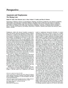
In rodents, chronic exposure to an allergen initially induces inflammation, which decreases over time (116, 117), whereas in humans with allergic asthma, inflammation persists and is chronic during their lifetime (118). This transition from antigen sensitization to immunologic tolerance is accompanied by a shift in the lymphocyte content in the lung tissue and bronchial lavage fluid (BALF) (119, 116). Antigen-specific regulatory T cells are believed to produce IL-10 transiently and to thereby inhibit the asthma phenotype during the development of tolerance (120, 121). Prolonged exposure of mice to an allergen causes the cytokine IL-13 that is secreted by T helper cell type 2 (Th2) to decrease and IFN-␥, which is secreted by Th1 cells, to increase in the bronchoalveolar lavage fluid (61). IFN-␥ mediates the reduction of IL-9– and IL-13–induced GCM by inducing programmed cell death (74) (Figure 2). While Bcl-2 is not detected, the percentage of Bax-immunopositive mucous cells increases from ⵑ 3–25%, while the number of metaplastic mucous cells decreases (61). The fact that Bcl-2 expression is associated with the appearance of LPS-induced GCM and that Bcl-2 is not detected in allergen-induced GCM in mice suggests different resolution mechanisms in these two experimental systems. Overall, ⵑ 25–35% of mucous cells express Bax after repeated exposure to allergen for 15 d. Trifilieff and colleagues (122) found that by 3 d after allergen exposure, ⵑ 30% of epithelial cell nuclei are BrdU-positive, a marker for cells that undergo DNA synthesis during the cell cycle. Taken together, the observed Bax positivity in ⵑ 25–35% of mucus cells during the resolution of allergen-induced GCM suggests that the Bax-positive mucous cells may represent cells that must be eliminated to reconstitute the original cell number of the repaired epithelium. One could speculate that humans with allergic asthma are deficient in developing such immune deviation to specific compounds or in IFN-␥ signaling to reduce GCM. In some instances, serum levels of IFN-␥ can be increased in severe asthma
542
AMERICAN JOURNAL OF RESPIRATORY CELL AND MOLECULAR BIOLOGY VOL 34
2006
Figure 2. Allergic inflammation induces epithelial cell hyperplasias by IL-9 and IL-13, and the resolution occurs when IFN-␥ is present at increased concentrations compared with the Th2 cytokines.
cases (123), and IFN-␥ levels in BALF are sometimes found even in mild asthma (124, 125). Although these data seem paradoxical, a reasonable hypothesis would explain a failure of IFN-␥ in these patients with asthma to drive the resolution of airway hyperreactivity and GCM. For instance, IFN-␥ levels may not be high enough to reverse the Bax inhibitory effects of IL-13. Supporting this hypothesis, the instillation of 100 ng IFN-␥ causes reduction of GCM, while 50 ng does not (74). It is also possible that in a subpopulation of individuals with asthma, a deficiency in the IFN-␥–signaling pathway may render IFN-␥ incapable of inducing cell death in epithelial cells. This hypothesis is supported by recent reports that polymorphisms in genes encoding for IFN-␥ and IFN regulatory factor-1 (IRF-1) confer genetic susceptibility to allergic asthma in Japanese children (126) and that Stat 1 is constitutively activated in epithelial cells of individuals with asthma (127). These alterations may contribute to deficient signaling of IFN-␥ and sustained increased levels of GCM even in the presence of IFN-␥. Supporting this hypothesis, mice deficient in Stat 1 (an obligatory protein for IFN-␥ signaling) do not resolve GCM after repeated and prolonged exposure to an allergen (74). These animals may represent appropriate models to study chronic inflammatory airway disease that resembles human asthma and to test immunomodulatory therapies to reverse established airway remodeling.
AECS CONTROL INFLAMMATION BY APOPTOTIC MECHANISMS Fas and FasL on AECs Reduce Inflammation
Several studies have confirmed by flow cytometry and immunohistochemistry that Fas and FasL are expressed in AECs (69, 71). Fas and FasL expression are similar in both the SAEs and NHBEs (70). Coexpression of Fas and FasL in the same cells is uncommon for mammalian cells and has only been described for corneal epithelium (128) and crypt cells in ulcerative colitis (129). The functional role of this coexpression of epithelial cells
is still not clear. A possible role of the FasL in causing adjacent cell death should be manifested by rapid cell turnover. However, AE has estimated turnover rates of ⬍ 1% in normal tissue (130). AECs must be refractory to FasL-induced death signals. Fas signaling may have functions unrelated to the cell death process (131), but whether these functions are important in AECs remains to be determined. Another role for FasL expression in AECs may be to control the numbers of inflammatory cells by inducing cell death. Support for the anti-inflammatory role of FasL is found from observation in mice and from reports showing that the AE is capable of controlling the number of inflammatory cells and to actively reduce the inflammatory response once the insult is removed (132). In mice, expression of FasL, as shown by in situ hybridization, was found to be primarily restricted to bronchial Clara cells and nonciliated columnar AECs (71). Some squamous cells of the upper airways also showed FasL expression. In gld mutant mice, where FasL is nonfunctional, a diffuse accumulation of mononuclear inflammatory cells is observed in the lung that is different from inflammation typified by bronchus-associated lymphoid tissues (BALT). The finding is in agreement with other reports showing that in lpr or gld mice that have nonfunctional Fas and FasL, respectively, lymphocytes accumulate in epithelial tissues such as the small intestine, female genital tract, the lung, and T cells express markers of chronic activation including CD69 (133). These findings support the hypothesis that FasL has an immune regulatory role in all epithelial tissues. FasL mRNA expression was reduced in mice exposed to allergen where airway inflammation is increased, suggesting that FasL on AECs control the infiltration by T cells of the lung (71). Fas and FasL in Diseased Airway Epithelia
FasL expression is markedly more intense in ciliated and submucosal gland of CF airways compared with that of normal subjects and in the CF cell line (CF-T2) compared with the non-CF cell line (NT) (134). However, expression of Fas is similar in both
A Tribute to Carol Basbaum
airway tissues and cell lines from subjects with CF and control subjects. The findings are consistent with others reporting Fas and FasL expression in bronchial epithelial cells (135–137). Increased FasL expression in patients with CF could be due to various reasons: (1 ) chronic inflammation (although IFN-␥ and TNF-␣ did not increase Fas and FasL expression in their studies, it is possible that other cytokines may be responsible); (2 ) extracellular matrix consisting of subepithelial fibrosis and degradation of collagenic and elastic fibers in CF may induce expression (138). Whether induction of FasL is directly signaled by CFTR deficiency or by upregulating cytokine expression is not known. In serum of patients with CF, soluble FasL was significantly lower than in serum obtained from control subjects (138), although the significance of this finding is unclear. Expression of Fas and FasL in the same epithelium is also found in autoimmune diseases, where their interaction leads to epithelial destruction. FasL may also induce apoptosis in inflammatory immune cells (71, 135, 136). FasL may confer immune suppression in malignancy by inducing apoptosis of Fas bearing immune cells such as lymphocytes and dendritic cells (139). FasL can also be proinflammatory by activating PMNs (140). Because PMNs are predominant phagocytes in CF (141), death of PMNs may increase DNA that alters viscosity of CF airway secretions (142). Hamann and coworkers (135) report that Fas expression appears to be greater in epithelial regions where some epithelial damage was present. Overexpression of Fas may be proinflammatory in chronic inflammation such as asthma and chronic bronchitis and, as a result, may play a role in the overall epithelial integrity. This observation may explain why increased epithelial cell turnover has been documented in chronic bronchitis (130). On the other hand, the expression of FasL may function to prevent infiltration of Fas-bearing inflammatory cells such as eosinophils in the epithelial layer. A role for Fas and FasL in immune privilege has been suggested for other tissues including testes (143), brain (144), and placenta (145). Decreased FasL expression or the inheritance of a weak form of FasL may predispose to increased inflammation and a weakening of this immune barrier, leading to conditions of chronic inflammation such as asthma. AECs as Phagocytes
Walsh and coworkers (146) and Sexton and colleagues (147) showed by electron microscopy and confocal microscopy that resting and cytokine-stimulated human SAEs and NHBEs recognize and ingest apoptotic eosinophils. Furthermore, the same group showed that A549 also phagocytose apoptotic eosinophils by recognizing integrin, lectin, and phosphatidylserine membrane receptors, and that this process can be upregulated by dexamethasone (148). Treatment with cytochalasin D at 10 M concentration completely abolishes the uptake of apoptotic eosinophils by SAEs, NHBEs, or macrophages. This apoptosis was confirmed by the lack of adhesive interactions between viable eosinophils and neutrophils with airway epithelial cells. Interestingly, the uptake of eosinophils was inhibited by preincubation of apoptotic eosinophils with the amino sugars. Ligation of CD44 receptors on SAEs or NHBEs or treatment of these cells with IL-1 (this study did not differentiate whether this was IL-1␣ or IL-1) increased the phagocytosis ability. However, apoptotic neutrophils were not subjected to phagocytosis by AECs even in the presence of cytokines such as IL-1. Interestingly, cell lines of mammary epithelium (ZR-75–1) and colon epithelium (HT-60) also revealed no ingestion of apoptotic neutrophils but of apoptotic eosinophils. These studies suggest that AECs are active participants in the removal of apoptotic eosinophils and may be important in the resolution of eosinophilic inflammation in the asthmatic lung.
543
SUMMARY AND FUTURE DIRECTIONS Many studies have shown that T lymphocytes, neutrophils, and eosinophils undergo cell death in massive numbers within 3–6 h after cytokine deprivation or stimulation with death agonists. However, AECs undergo cell death in significant numbers only 24 h after exposure to cell death agonists, and in most instances the percentage of dying cells is in the range of 5–15%. We find that 60% of AECs die 4 d after exposure to IFN-␥ (74). These findings suggest that cell death in AECs is either not mediated by a direct pathway and requires protein synthesis or checkpoints to stop direct signaling to activate effector caspases are in place. Elimination of these checkpoints such as by pretreatment with IFN-␥ may increase susceptibility and the appearance of massive cell death to Fas or TNF-␣. It is also feasible that the airway epithelium has developed various mechanisms as protection from undergoing cell death in response to various injurious substances: formation of tight junctions (which may give stability by coordinating the structural actin filaments) or the readiness to activate IB, which causes the proinflammatory cytokine expression and also expression of pro-survival proteins including the caspase inhibitors c-IAP1 (149) and c-IAP-2 (150). Many environmental toxins, including PAHs and their derivatives such as PAH epoxides, cause release of calcium from the ER. This ER calcium can mimic signaling for cell proliferation. However, it is possible that once AECs are sensitized to undergo cell death by certain conditions, or when Bax is in the right cytosolic compartment, the same signal may induce cell death. It is conceivable that the refractory nature of AECs to apoptosis may stem from their ability to successfully sequester proapoptotic mediators so they need to have other signals to activate them for activating downstream caspases. In physiologic settings, the slow induction of cell death in AECs is critical because rapid cell death would not allow for closure of the basement membrane by neighboring cells. One could postulate that neighboring cells have mechanisms to sense the death of AECs to cover the open space created by the dying cell(s). Furthermore, it is still not clear whether dying AECs are removed by phagocytosis by neighboring AECs or macrophages or by sloughing off into the airway lumen and undergoing detachment-induced cell death, also called anoikis. It is crucial to understand the mechanisms that make AECs refractory to cell death. Fas and FasL are expressed, but AECs do not undergo cell death by this pathway. In fact, our studies suggest that FasL induces proliferation of AECs in mice that have been exposed to allergen (personal observation). SAEs appear to be more sensitive to Fas ligation than NHBEs (70), suggesting that one needs to be very careful when reporting what cells are being used in the cell death process and indicate that proximal and distal airway epithelial cells may activate different pathways to their cell death process. Furthermore, while Hagimoto and coworkers (151) report that activation of Fas with recombinant human FasL induces IL-8 secretion in NHBEs, Nakamura and colleagues (70) did not find increased IL-8 by activating Fas with Fas antibody. These studies suggest that various means of activation of Fas could result in different outcomes. It is possible that GCM could be reduced by targeting cell death processes in cases where intrinsic changes in airway epithelia sustain GCM independent of inflammation. However, in chronic diseases, GCM may be a result of continuous inflammation and repeated injury and repair to the airway epithelium. Therefore, it is important to determine how altering the halflife of goblet cells without removal of the continuous inflammation may affect the dynamics of the airway epithelia. It may be crucial to use Bcl-2 and/or the pro-apoptotic players to reduce GCM as part of a general therapeutic effort to reduce
544
AMERICAN JOURNAL OF RESPIRATORY CELL AND MOLECULAR BIOLOGY VOL 34
inflammation. Chronic inflammation in animal models needs to be established to adequately address these complex issues of GCM in chronic inflammation. In the context of apoptosis to restore homeostasis in hyperplastic epithelium, it remains unclear how the epithelium determines the number of cells that must be eliminated to restore the original condition. Is it possible that epithelial cells sense intercellular pressure and signal cell death to eliminate cells and create enough space? Furthermore, it is unknown whether the discarded cells represent abnormal or damaged epithelial cells and whether there are signals that determine which cells should be discarded from the airway epithelium. Finally, how the epithelium restores the normal proportions of epithelial cell types is unclear. Why do a certain number of the remaining cells differentiate into serous, Clara, ciliated, and mucous cells to reconstitute the proportions found in normal epithelia? Future studies will also focus on the ability of the AE to control inflammation. Deficiency in this ability may be crucial for sustained inflammation in airways and could provide novel targets for developing new therapies. Conflict of Interest Statement : Y.T. does not have a financial relationship with a commercial entity that has an interest in the subject of this manuscript.
References 1. Jyonouchi H. Airway epithelium and apoptosis. Apoptosis 1999;4:407– 417. 2. Thornberry NA, Lazebnik Y. Caspases: enemies within. Science 1998; 281:1312–1316. 3. Locksley RM, Killeen N, Lenardo MJ. The TNF and TNF receptor superfamilies: integrating mammalian biology. Cell 2001;104:487–501. 4. Chinnaiyan AM, O’Rourke K, Tewari M, Dixit VM. FADD, a novel death domain-containing protein, interacts with the death domain of Fas and initiates apoptosis. Cell 1995;81:505–512. 5. Hsu H, Shu HB, Pan MG, Goeddel DV. TRADD-TRAF2 and TRADD-FADD interactions define two distinct TNF receptor 1 signal transduction pathways. Cell 1996;84:299–308. 6. Hsu H, Huang J, Shu HB, Baichwal V, Goeddel DV. TNF-dependent recruitment of the protein kinase RIP to the TNF receptor-1 signaling complex. Immunity 1996;4:387–396. 7. Pan G, O’Rourke K, Chinnaiyan AM, Gentz R, Ebner R, Ni J, Dixit VM. The receptor for the cytotoxic ligand TRAIL. Science 1997;276: 111–113. 8. Schneider T, van Velzen D, Moqbel R, Issekutz AC. Kinetics and quantitation of eosinophil and neutrophil recruitment to allergic lung inflammation in a brown Norway rat model. Am J Respir Cell Mol Biol 1997;17:702–712. 9. Kluck RM, Bossy-Wetzel E, Green DR, Newmeyer DD. The release of cytochrome c from mitochondria: a primary site for Bcl-2 regulation of apoptosis. Science 1997;275:1132–1136. 10. Yang J, Liu X, Bhalla K, Kim CN, Ibrado AM, Cai J, Peng TI, Jones DP, Wang X. Prevention of apoptosis by Bcl-2: release of cytochrome c from mitochondria blocked. Science 1997;275:1129– 1132. 11. Du C, Fang M, Li Y, Li L, Wang X. Smac, a mitochondrial protein that promotes cytochrome c-dependent caspase activation by eliminating IAP inhibition. Cell 2000;102:33–42. 12. Verhagen AM, Ekert PG, Pakusch M, Silke J, Connolly LM, Reid GE, Moritz RL, Simpson RJ, Vaux DL. Identification of DIABLO, a mammalian protein that promotes apoptosis by binding to and antagonizing IAP proteins. Cell 2000;102:43–53. 13. Susin SA, Lorenzo HK, Zamzami N, Marzo I, Snow BE, Brothers GM, Mangion J, Jacotot E, Costantini P, Loeffler M, et al. Molecular characterization of mitochondrial apoptosis-inducing factor. Nature 1999;397:441–446. 14. Li LY, Luo X, Wang X. Endonuclease G is an apoptotic DNase when released from mitochondria. Nature 2001;412:95–99. 15. Suzuki Y, Imai Y, Nakayama H, Takahashi K, Takio K, Takahashi R. A serine protease, HtrA2, is released from the mitochondria and interacts with XIAP, inducing cell death. Mol Cell 2001;8:613–621. 16. Willis S, Day CL, Hinds MG, Huang DC. The Bcl-2-regulated apoptotic pathway. J Cell Sci 2003;116:4053–4056.
2006
17. Zou H, Li Y, Liu X, Wang X. An APAF-1.cytochrome c multimeric complex is a functional apoptosome that activates procaspase-9. J Biol Chem 1999;274:11549–11556. 18. Adams JM, Cory S. The Bcl-2 protein family: arbiters of cell survival. Science 1998;281:1322–1326. 19. Cryns V, Yuan J. Proteases to die for. Genes Dev 1998;12:1551–1570. 20. Lindsten T, Ross AJ, King A, Zong WX, Rathmell JC, Shiels HA, Ulrich E, Waymire KG, Mahar P, Frauwirth K, et al. The combined functions of proapoptotic Bcl-2 family members bak and bax are essential for normal development of multiple tissues. Mol Cell 2000;6:1389–1399. 21. Wei MC, Zong WX, Cheng EH, Lindsten T, Panoutsakopoulou V, Ross AJ, Roth KA, MacGregor GR, Thompson CB, Korsmeyer SJ. Proapoptotic BAX and BAK: a requisite gateway to mitochondrial dysfunction and death. Science 2001;292:727–730. 22. Opferman JT, Korsmeyer SJ. Apoptosis in the development and maintenance of the immune system. Nat Immunol 2003;4:410–415. 23. Tong Y, Yang Q, Vater C, Venkatesh LK, Custeau D, Chittenden T, Chinnadurai G, Gourdeau H. The pro-apoptotic protein, Bik, exhibits potent antitumor activity that is dependent on its BH3 domain. Mol Cancer Ther 2001;1:95–102. 24. Oda E, Ohki R, Murasawa H, Nemoto J, Shibue T, Yamashita T, Tokino T, Taniguchi T, Tanaka N. Noxa, a BH3-only member of the Bcl-2 family and candidate mediator of p53-induced apoptosis. Science 2000;288:1053–1058. 25. Villunger A, Michalak EM, Coultas L, Mullauer F, Bock G, Ausserlechner MJ, Adams JM, Strasser A. p53- and drug-induced apoptotic responses mediated by BH3-only proteins puma and noxa. Science 2003;302:1036–1038. 26. Luo X, Budihardjo I, Zou H, Slaughter C, Wang X. Bid, a Bcl2 interacting protein, mediates cytochrome c release from mitochondria in response to activation of cell surface death receptors. Cell 1998;94:481–490. 27. Coultas L, Huang DC, Adams JM, Strasser A. Pro-apoptotic BH3-only Bcl-2 family members in vertebrate model organisms suitable for genetic experimentation. Cell Death Differ 2002;9:1163–1166. 28. Letai A, Bassik MC, Walensky LD, Sorcinelli MD, Weiler S, Korsmeyer SJ. Distinct BH3 domains either sensitize or activate mitochondrial apoptosis, serving as prototype cancer therapeutics. Cancer Cell 2002; 2:183–192. 29. Gillissen B, Essmann F, Graupner V, Starck L, Radetzki S, Dorken B, Schulze-Osthoff K, Daniel PT. Induction of cell death by the BH3only Bcl-2 homolog Nbk/Bik is mediated by an entirely Bax-dependent mitochondrial pathway. EMBO J 2003;22:3580–3590. 30. Puthalakath H, Villunger A, O’Reilly LA, Beaumont JG, Coultas L, Cheney RE, Huang DC, Strasser A. Bmf: a proapoptotic BH3-only protein regulated by interaction with the myosin V actin motor complex, activated by anoikis. Science 2001;293:1829–1832. 31. Day CL, Puthalakath H, Skea G, Strasser A, Barsukov I, Lian LY, Huang DC, Hinds MG. Localization of dynein light chains 1 and 2 and their pro-apoptotic ligands. Biochem J 2004;377:597–605. 32. Marani M, Hancock D, Lopes R, Tenev T, Downward J, Lemoine NR. Role of Bim in the survival pathway induced by Raf in epithelial cells. Oncogene 2004;23:2431–2441. 33. Breckenridge DG, Germain M, Mathai JP, Nguyen M, Shore GC. Regulation of apoptosis by endoplasmic reticulum pathways. Oncogene 2003;22:8608–8618. 34. Bowden DH. Cell turnover in the lung. Am Rev Respir Dis 1983;128: S46–S48. 35. Teramoto S, Johnson LG, Huang W, Leigh MW, Boucher RC. Effect of adenoviral vector infection on cell proliferation in cultured primary human airway epithelial cells. Hum Gene Ther 1995;6:1045–1053. 36. Brydon EW, Smith H, Sweet C. Influenza A virus-induced apoptosis in bronchiolar epithelial (NCI-H292) cells limits pro-inflammatory cytokine release. J Gen Virol 2003;84:2389–2400. 37. Bitko V, Barik S. An endoplasmic reticulum-specific stress-activated caspase (caspase-12) is implicated in the apoptosis of A549 epithelial cells by respiratory syncytial virus. J Cell Biochem 2001;80:441–454. 38. Thomas KW, Monick MM, Staber JM, Yarovinsky T, Carter AB, Hunninghake GW. Respiratory syncytial virus inhibits apoptosis and induces NF-kappa B activity through a phosphatidylinositol 3-kinasedependent pathway. J Biol Chem 2002;277:492–501. 39. Zhang L, Peeples ME, Boucher RC, Collins PL, Pickles RJ. Respiratory syncytial virus infection of human airway epithelial cells is polarized, specific to ciliated cells, and without obvious cytopathology. J Virol 2002;76:5654–5666. 40. Kotelkin A, Prikhod’ko EA, Cohen JI, Collins PL, Bukreyev A. Respiratory syncytial virus infection sensitizes cells to apoptosis mediated by
A Tribute to Carol Basbaum
41.
42.
43.
44. 45.
46.
47.
48.
49.
50.
51.
52.
53. 54.
55.
56.
57. 58.
59.
60.
61.
tumor necrosis factor-related apoptosis-inducing ligand. J Virol 2003; 77:9156–9172. Rajan S, Cacalano G, Bryan R, Ratner AJ, Sontich CU, van Heerckeren A, Davis P, Prince A. Pseudomonas aeruginosa induction of apoptosis in respiratory epithelial cells: analysis of the effects of cystic fibrosis transmembrane conductance regulator dysfunction and bacterial virulence factors. Am J Respir Cell Mol Biol 2000;23:304–312. DiMango E, Ratner AJ, Bryan R, Tabibi S, Prince A. Activation of NF-kappaB by adherent Pseudomonas aeruginosa in normal and cystic fibrosis respiratory epithelial cells. J Clin Invest 1998;101:2598– 2605. Gottlieb RA, Dosanjh A. Mutant cystic fibrosis transmembrane conductance regulator inhibits acidification and apoptosis in C127 cells: possible relevance to cystic fibrosis. Proc Natl Acad Sci USA 1996;93: 3587–3591. Aktories K. Bacterial toxins that target Rho proteins. J Clin Invest 1997; 99:827–829. Vernooy JH, Dentener MA, van Suylen RJ, Buurman WA, Wouters EF. Intratracheal instillation of lipopolysaccharide in mice induces apoptosis in bronchial epithelial cells: no role for tumor necrosis factor-alpha and infiltrating neutrophils. Am J Respir Cell Mol Biol 2001;24:569–576. Wang CY, Mayo MW, Korneluk RG, Goeddel DV, Baldwin AS Jr. NF-kappaB antiapoptosis: induction of TRAF1 and TRAF2 and c-IAP1 and c-IAP2 to suppress caspase-8 activation. Science 1998;281: 1680–1683. Van Antwerp DJ, Martin SJ, Kafri T, Green DR, Verma IM. Suppression of TNF-alpha-induced apoptosis by NF-kappaB. Science 1996;274:787–789. Catz SD, Johnson JL. Transcriptional regulation of bcl-2 by nuclear factor kappa B and its significance in prostate cancer. Oncogene 2001;20:7342–7351. Plotkowski MC, Povoa HC, Zahm JM, Lizard G, Pereira GM, Tournier JM, Puchelle E. Early mitochondrial dysfunction, superoxide anion production, and DNA degradation are associated with non-apoptotic death of human airway epithelial cells induced by Pseudomonas aeruginosa exotoxin A. Am J Respir Cell Mol Biol 2002;26:617–626. White SR, Williams P, Wojcik KR, Sun S, Hiemstra PS, Rabe KF, Dorscheid DR. Initiation of apoptosis by actin cytoskeletal derangement in human airway epithelial cells. Am J Respir Cell Mol Biol 2001;24:282–294. Moore M, Marroquin BA, Gugliotta W, Tse R, White SR. Rho kinase inhibition initiates apoptosis in human airway epithelial cells. Am J Respir Cell Mol Biol 2004;30:379–387. Blagosklonny MV, Giannakakou P, el-Deiry WS, Kingston DG, Higgs PI, Neckers L, Fojo T. Raf-1/bcl-2 phosphorylation: a step from microtubule damage to cell death. Cancer Res 1997;57:130–135. Haldar S, Basu A, Croce CM. Bcl2 is the guardian of microtubule integrity. Cancer Res 1997;57:229–233. Kothakota S, Azuma T, Reinhard C, Klippel A, Tang J, Chu K, McGarry TJ, Kirschner MW, Koths K, Kwiatkowski DJ, et al. Caspase-3generated fragment of gelsolin: effector of morphological change in apoptosis. Science 1997;278:294–298. Jyonouchi H, Sun S, Abiru T, Chareancholvanich S, Ingbar DH. The effects of hyperoxic injury and antioxidant vitamins on death and proliferation of human small airway epithelial cells. Am J Respir Cell Mol Biol 1998;19:426–436. O’Reilly MA, Staversky RJ, Stripp BR, Finkelstein JN. Exposure to hyperoxia induces p53 expression in mouse lung epithelium. Am J Respir Cell Mol Biol 1998;18:43–50. Elledge SJ. Cell cycle checkpoints: preventing an identity crisis. Science 1996;274:1664–1672. Yonish-Rouach E, Grunwald D, Wilder S, Kimchi A, May E, Lawrence JJ, May P, Oren M. p53-mediated cell death: relationship to cell cycle control. Mol Cell Biol 1993;13:1415–1423. Venot C, Maratrat M, Dureuil C, Conseiller E, Bracco L, Debussche L. The requirement for the p53 proline-rich functional domain for mediation of apoptosis is correlated with specific PIG3 gene transactivation and with transcriptional repression. EMBO J 1998;17:4668– 4679. Chipuk JE, Kuwana T, Bouchier-Hayes L, Droin NM, Newmeyer DD, Schuler M, Green DR. Direct activation of Bax by p53 mediates mitochondrial membrane permeabilization and apoptosis. Science 2004; 303:1010–1014. Tesfaigzi Y, Fischer MJ, Green FHY, De Sanctis GT, Wilder JA. Bax is crucial for IFNg-induced resolution of allergen-induced mucous cell metaplasia. J Immunol 2002;169:5919–5925.
545 62. Trautmann A, Akdis M, Kleemann D, Altznauer F, Simon HU, Graeve T, Noll M, Brocker EB, Blaser K, Akdis CA. T cell-mediated Fasinduced keratinocyte apoptosis plays a key pathogenetic role in eczematous dermatitis. J Clin Invest 2000;106:25–35. 63. Ossina NK, Cannas A, Powers VC, Fitzpatrick PA, Knight JD, Gilbert JR, Shekhtman EM, Tomei LD, Umansky SR, Kiefer MC. Interferongamma modulates a p53-independent apoptotic pathway and apoptosisrelated gene expression. J Biol Chem 1997;272:16351–16357. 64. Coulter KR, Doseff A, Sweeney P, Wang Y, Marsh CB, Wewers MD, Knoell DL. Opposing effect by cytokines on Fas-mediated apoptosis in A549 lung epithelial cells. Am J Respir Cell Mol Biol 2002;26:58–66. 65. Deiss LP, Feinstein E, Berissi H, Cohen O, Kimchi A. Identification of a novel serine/threonine kinase and a novel 15-kD protein as potential mediators of the gamma interferon-induced cell death. Genes Dev 1995;9:15–30. 66. Ruiz-Ruiz C, Munoz-Pinedo C, Lopez-Rivas A. Interferon-gamma treatment elevates caspase-8 expression and sensitizes human breast tumor cells to a death receptor-induced mitochondria- operated apoptotic program. Cancer Res 2000;60:5673–5680. 67. Shen Y, Devgan G, Darnell JE Jr, Bromberg JF. Constitutively activated Stat3 protects fibroblasts from serum withdrawal and UV-induced apoptosis and antagonizes the proapoptotic effects of activated Stat1. Proc Natl Acad Sci USA 2001;98:1543–1548. 68. Barber GN. The interferons and cell death: guardians of the cell or accomplices of apoptosis? Semin Cancer Biol 2000;10:103–111. 69. Trautmann A, Schmid-Grendelmeier P, Kruger K, Crameri R, Akdis M, Akkaya A, Brocker EB, Blaser K, Akdis CA. T cells and eosinophils cooperate in the induction of bronchial epithelial cell apoptosis in asthma. J Allergy Clin Immunol 2002;109:329–337. 70. Nakamura M, Matute-Bello G, Liles WC, Hayashi S, Kajikawa O, Lin SM, Frevert CW, Martin TR. Differential response of human lung epithelial cells to fas-induced apoptosis. Am J Pathol 2004;164:1949– 1958. 71. Gochuico BR, Miranda KM, Hessel EM, De Bie JJ, Van Oosterhout AJ, Cruikshank WW, Fine A. Airway epithelial Fas ligand expression: potential role in modulating bronchial inflammation. Am J Physiol 1998;274:L444–L449. 72. Wen LP, Madani K, Fahrni JA, Duncan SR, Rosen GD. Dexamethasone inhibits lung epithelial cell apoptosis induced by IFN-gamma and Fas. Am J Physiol 1997;273:L921–L929. 73. Undevia NS, Dorscheid DR, Marroquin BA, Gugliotta WL, Tse R, White SR. Smad and p38-MAPK signaling mediates apoptotic effects of transforming growth factor-beta1 in human airway epithelial cells. Am J Physiol Lung Cell Mol Physiol 2004;287:L515–L524. 74. Shi ZQ, Fischer MJ, De Sanctis GT, Schuyler M, Tesfaigzi Y. IFNg but not Fas mediates reduction of allergen-induced mucous cell metaplasia by inducing apoptosis. J Immunol 2002;168:4764–4771. 75. Amsellem C, Durieu, Chambe MT, Peyrol S, Pacheco Y. In vitro expression of fas and CD40 and induction of apoptosis in human cystic fibrosis airway epithelial cells. Respir Med 2002;96:244–249. 76. Pelaia G, Cuda G, Vatrella A, Fratto D, Grembiale RD, Tagliaferri P, Maselli R, Costanzo FS, Marsico SA. Effects of transforming growth factor- and budesonide on mitogen-activated protein kinase activation and apoptosis in airway epithelial cells. Am J Respir Cell Mol Biol 2003;29:12–18. 77. Meagher LC, Cousin JM, Seckl JR, Haslett C. Opposing effects of glucocorticoids on the rate of apoptosis in neutrophilic and eosinophilic granulocytes. J Immunol 1996;156:4422–4428. 78. Ohta K, Yamashita N. Apoptosis of eosinophils and lymphocytes in allergic inflammation. J Allergy Clin Immunol 1999;104:14–21. 79. Compton MM, Cidlowski JA. Rapid in vivo effects of glucocorticoids on the integrity of rat lymphocyte genomic deoxyribonucleic acid. Endocrinology 1986;118:38–45. 80. Dorscheid DR, Wojcik KR, Sun S, Marroquin B, White SR. Apoptosis of airway epithelial cells induced by corticosteroids. Am J Respir Crit Care Med 2001;164:1939–1947. 81. Tse R, Marroquin BA, Dorscheid DR, White SR. Beta-adrenergic agonists inhibit corticosteroid-induced apoptosis of airway epithelial cells. Am J Physiol Lung Cell Mol Physiol 2003;285:L393–L404. 82. White SR, Dorscheid DR. Corticosteroid-induced apoptosis of airway epithelium: a potential mechanism for chronic airway epithelial damage in asthma. Chest 2002;122:278S–284S.
546
AMERICAN JOURNAL OF RESPIRATORY CELL AND MOLECULAR BIOLOGY VOL 34
83. Vignola AM, Chanez P, Campbell AM, Souques F, Lebel B, Enander I, Bousquet J. Airway inflammation in mild intermittent and in persistent asthma. Am J Respir Crit Care Med 1998;157:403–409. 84. Dorscheid DR, Low E, Conforti A, Shifrin S, Sperling AI, White SR. Corticosteroid-induced apoptosis in mouse airway epithelium: effect in normal airways and after allergen-induced airway inflammation. J Allergy Clin Immunol 2003;111:360–366. 85. Bao S, Knoell DL. Zinc modulates airway epithelium susceptibility to death receptor-mediated apoptosis. Am J Physiol Lung Cell Mol Physiol 2005;290:L433–L441. 86. Raetz CR, Ulevitch RJ, Wright SD, Sibley CH, Ding A, Nathan CF. Gram-negative endotoxin: an extraordinary lipid with profound effects on eukaryotic signal transduction. FASEB J 1991;5:2652–2660. 87. Sandstrom T, Bjermer L, Rylander R. Lipopolysaccharide (LPS) inhalation in healthy subjects increases neutrophils, lymphocytes and fibronectin levels in bronchoalveolar lavage fluid. Eur Respir J 1992;5:992– 996. 88. Tesfaigzi J, Johnson NF, Lechner JF. Induction of EGF receptor and erbB-2 during endotoxin-induced alveolar type II cell proliferation in the rat lung. Int J Exp Pathol 1996;77:143–154. 89. Wells AB. The kinetics of cell proliferation in the tracheobronchial epithelia of rats with and without chronic respiratory disease. Cell Tissue Kinet 1970;3:185–206. 90. Rutten AA, Beems RB, Wilmer JW. Effects of all-trans retinol and cigarette smoke condensate on hamster tracheal epithelium in organ culture: II. A histomorphological study. Virchows Arch B Cell Pathol 1988;55:177–186. 91. Herard AL, Zahm JM, Pierrot D, Hinnrasky J, Fuchey C, Puchelle E. Epithelial barrier integrity during in vitro wound repair of the airway epithelium. Am J Respir Cell Mol Biol 1996;15:624–632. 92. Reader JR, Tepper JS, Schelegle ES, Aldrich MC, Putney LF, Pfeiffer JW, Hyde DM. Pathogenesis of mucous cell metaplasia in a murine asthma model. Am J Pathol 2003;162:2069–2078. 93. Tesfaigzi Y, Harris JF, Hotchkiss JA, Harkema JR. DNA synthesis and Bcl-2 expression during the development of mucous cell metaplasia in airway epithelium of rats exposed to LPS. Am J Physiol Lung Cell Mol Physiol 2004;286:L268–L274. 94. Basbaum C, Jany B. Plasticity in the airway epithelium. Am J Physiol 1990;259:L38–L46. 95. Stassen M, Muller C, Arnold M, Hultner L, Klein-Hessling S, Neudorfl C, Reineke T, Serfling E, Schmitt E. IL-9 and IL-13 production by activated mast cells is strongly enhanced in the presence of lipopolysaccharide: NF-kappa B is decisively involved in the expression of IL-9. J Immunol 2001;166:4391–4398. 96. Kim JH, Lee SY, Bak SM, Suh IB, Shin C, Shim JJ, In KH, Kang KH, Yoo SH. Effects of matrix metalloproteinase inhibitor on LPSinduced goblet cell metaplasia. Am J Physiol Lung Cell Mol Physiol 2004;287:L127–L133. 97. Tesfaigzi Y. Processes involved in the repair of injured airway epithelia. Arch Immunol Ther Exp (Warsz) 2003;51:283–288. 98. Evans CM, Williams OW, Tuvim MJ, Nigam R, Mixides GP, Blackburn MR, DeMayo FJ, Burns AR, Smith C, Reynolds SD, et al. Mucin is produced by clara cells in the proximal airways of antigen-challenged mice. Am J Respir Cell Mol Biol 2004;31:382–394. 99. Harris JF, Fischer MJ, Hotchkiss JA, Monia BP, Randell SH, Harkema JR, Tesfaigzi Y. Bcl-2 sustains increased mucous and epithelial cell numbers in metaplastic airway epithelium. Am J Respir Crit Care Med 2005;171:764–772. 100. Pierce J, Wilder JA, Strasser A, Tesfaigzi Y. Loss of pro-apoptotic Bim promotes accumulation of pulmonary T lymphocytes but not of epithelial cells during prolonged exposure of mice to allergen. Am J Physiol (In press) 101. Blyth DI, Pedrick MS, Savage TJ, Bright H, Beesley JE, Sanjar S. Induction, duration, and resolution of airway goblet cell hyperplasia in a murine model of atopic asthma: effect of concurrent infection with respiratory syncytial virus and response to dexamethasone. Am J Respir Cell Mol Biol 1998;19:38–54. 102. Tesfaigzi Y, Fischer MJ, Martin AJ, Seagrave J. Bcl-2 in LPS- and allergen-induced hyperplastic mucous cells in airway epithelia of Brown Norway rats. Am J Physiol Lung Cell Mol Physiol 2000;279: L1210–L1217. 103. Grossmann J. Molecular mechanisms of “detachment-induced apoptosis–Anoikis”. Apoptosis 2002;7:247–260. 104. Tesfaigzi J, Hotchkiss JA, Harkema JR. Expression of Bcl-2 during mucous cell metaplasia and remodeling in F344/N rats. Am J Respir Cell Mol Biol 1998;18:794–799.
2006
105. Mazel S, Burtrum D, Petrie HT. Regulation of cell division cycle progression by bcl-2 expression: a potential mechanism for inhibition of programmed cell death. J Exp Med 1996;183:2219–2226. 106. Linette GP, Li Y, Roth K, Korsmeyer SJ. Cross talk between cell death and cell cycle progression: BCL-2 regulates NFAT-mediated activation. Proc Natl Acad Sci USA 1996;93:9545–9552. 107. O’Reilly LA, Harris AW, Tarlinton DM, Corcoran LM, Strasser A. Expression of a bcl-2 transgene reduces proliferation and slows turnover of developing B lymphocytes in vivo. J Immunol 1997;159:2301– 2311. 108. Lemjabbar H, Li D, Gallup M, Sidhu S, Drori E, Basbaum C. Tobacco smoke-induced lung cell proliferation mediated by tumor necrosis factor alpha-converting enzyme and amphiregulin. J Biol Chem 2003; 278:26202–26207. 109. Maestrelli P, Saetta M, Mapp CE, Fabbri LM. Remodeling in response to infection and injury: airway inflammation and hypersecretion of mucus in smoking subjects with chronic obstructive pulmonary disease. Am J Respir Crit Care Med 2001;164:S76–S80. 110. Heidsiek JG, Hyde DM, Plopper CG, St George JA. Quantitative histochemistry of mucosubstance in tracheal epithelium of the macaque monkey. J Histochem Cytochem 1987;35:435–442. 111. Ordonez CL, Khashayar R, Wong HH, Ferrando R, Wu R, Hyde DM, Hotchkiss JA, Zhang Y, Novikov A, Dolganov G, et al. Mild and moderate asthma is associated with airway goblet cell hyperplasia and abnormalities in mucin gene expression. Am J Respir Crit Care Med 2001;163:517–523. 112. Jefcoat AM, Hotchkiss JA, Gerber V, Harkema JR, Basbaum CB, Robinson NE. Persistent mucin glycoprotein alterations in equine recurrent airway obstruction. Am J Physiol Lung Cell Mol Physiol 2001;281:L704–L712. 113. Robinson NE, Derksen FJ, Olszewsky MA, Buechner-Maxwell VA. The pathogenesis of chronic pulmonary disease of horses. Br Vet J 1996;152:283–306. 114. Yoneda K, Peck K, Chang MM, Chmiel K, Sher YP, Chen J, Yang PC, Chen Y, Wu R. Development of high-density DNA microarray membrane for profiling smoke- and hydrogen peroxide-induced genes in a human bronchial epithelial cell line. Am J Respir Crit Care Med 2001;164:S85–S89. 115. Tryka AF, Witschi H, Gosslee DG, McArthur AH, Clapp NK. Patterns of cell proliferation during recovery from oxygen injury: species differences. Am Rev Respir Dis 1986;133:1055–1059. 116. Yiamouyiannis CA, Schramm CM, Puddington L, Stengel P, BaradaranHosseini E, Wolyniec WW, Whiteley HE, Thrall RS. Shifts in lung lymphocyte profiles correlate with the sequential development of acute allergic and chronic tolerant stages in a murine asthma model. Am J Pathol 1999;154:1911–1921. 117. Haczku A, Moqbel R, Elwood W, Sun J, Kay AB, Barnes PJ, Chung KF. Effects of prolonged repeated exposure to ovalbumin in sensitized Brown Norway rats. Am J Respir Crit Care Med 1994;150:23–27. 118. Howarth PH. The airway inflammatory response in allergic asthma and its relationship to clinical disease. Allergy 1995;50:13–21. 119. Cui ZH, Joetham A, Aydintug MK, Hahn YS, Born WK, Gelfand EW. Reversal of allergic airway hyperreactivity after long-term allergen challenge depends on gammadelta T cells. Am J Respir Crit Care Med 2003;168:1324–1332. 120. Stock P, Akbari O, Berry G, Freeman GJ, Dekruyff RH, Umetsu DT. Induction of T helper type 1-like regulatory cells that express Foxp3 and protect against airway hyper-reactivity. Nat Immunol 2004;5: 1149–1156. 121. Akbari O, Freeman GJ, Meyer EH, Greenfield EA, Chang TT, Sharpe AH, Berry G, DeKruyff RH, Umetsu DT. Antigen-specific regulatory T cells develop via the ICOS-ICOS-ligand pathway and inhibit allergen-induced airway hyperreactivity. Nat Med 2002;8:1024–1032. 122. Trifilieff A, El-Hashim A, Bertrand C. Time course of inflammatory and remodeling events in a murine model of asthma: effect of steroid treatment. Am J Physiol Lung Cell Mol Physiol 2000;279:L1120– L1128. 123. Corrigan CJ, Kay AB. CD4 T-lymphocyte activation in acute severe asthma: relationship to disease severity and atopic status. Am Rev Respir Dis 1990;141:970–977. 124. Cembrzynska-Nowak M, Szklarz E, Inglot AD, Teodorczyk-Injeyan JA. Elevated release of tumor necrosis factor-alpha and interferon-␥ by bronchoalveolar leukocytes from patients with bronchial asthma. Am Rev Respir Dis 1993;147:291–295. 125. Holtzman MJ, Sampath D, Castro M, Look DC, Jayaraman S. The onetwo of T helper cells: does interferon-␥ knock out the Th2 hypothesis for asthma? Am J Respir Cell Mol Biol 1996;14:316–318.
A Tribute to Carol Basbaum 126. Nakao F, Ihara K, Kusuhara K, Sasaki Y, Kinukawa N, Takabayashi A, Nishima S, Hara T. Association of IFN-gamma and IFN regulatory factor 1 polymorphisms with childhood atopic asthma. J Allergy Clin Immunol 2001;107:499–504. 127. Sampath D, Castro M, Look DC, Holtzman MJ. Constitutive activation of an epithelial signal transducer and activator of transcription (STAT) pathway in asthma. J Clin Invest 1999;103:1353–1361. 128. Wilson SE, Li Q, Weng J, Barry-Lane PA, Jester JV, Liang Q, Wordinger RJ. The Fas-Fas ligand system and other modulators of apoptosis in the cornea. Invest Ophthalmol Vis Sci 1996;37:1582–1592. 129. Iwamoto M, Koji T, Makiyama K, Kobayashi N, Nakane PK. Apoptosis of crypt epithelial cells in ulcerative colitis. J Pathol 1996;180:152–159. 130. Demoly P, Simony-Lafontaine J, Chanez P, Pujol J, Lequeux N, Michel F, Bousquet J. Cell proliferation in the bronchial mucosa of asthmatics and chronic bronchitics. Am J Respir Crit Care Med 1994;150:214–217. 131. Ma Y, Liu H, Tu-Rapp H, Thiesen HJ, Ibrahim SM, Cole SM, Pope RM. Fas ligation on macrophages enhances IL-1R1-Toll-like receptor 4 signaling and promotes chronic inflammation. Nat Immunol 2004;5: 380–387. 132. Gilroy DW, Lawrence T, Perretti M, Rossi AG. Inflammatory resolution: new opportunities for drug discovery. Nat Rev Drug Discov 2004;3:401–416. 133. Ibraghimov AR, Lynch RG. T cell specialization at environmental interfaces: T cells from the lung and the female genital tract of lpr and gld mice differ from their splenic and lymph node counterparts. Eur J Immunol 1994;24:1848–1852. 134. Durieu I, Amsellem C, Paulin C, Chambe MT, Bienvenu J, Bellon G, Pacheco Y. Fas and Fas ligand expression in cystic fibrosis airway epithelium. Thorax 1999;54:1093–1098. 135. Hamann KJ, Dorscheid DR, Ko FD, Conforti AE, Sperling AI, Rabe KF, White SR. Expression of Fas (CD95) and FasL (CD95L) in human airway epithelium. Am J Respir Cell Mol Biol 1998;19:537–542. 136. Druilhe A, Wallaert B, Tsicopoulos A, Lapa e Silva JR, Tillie-Leblond I, Tonnel AB, Pretolani M. Apoptosis, proliferation, and expression of Bcl-2, Fas, and Fas ligand in bronchial biopsies from asthmatics. Am J Respir Cell Mol Biol 1998;19:747–757. 137. Martin TR, Hagimoto N, Nakamura M, Matute-Bello G. Apoptosis and epithelial injury in the lungs. Proc Am Thorac Soc 2005;2:214–220. 138. Durieu I, Peyrol S, Gindre D, Bellon G, Durand DV, Pacheco Y. Subepithelial fibrosis and degradation of the bronchial extracellular matrix in cystic fibrosis. Am J Respir Crit Care Med 1998;158:580–588.
547 139. Niehans GA, Brunner T, Frizelle SP, Liston JC, Salerno CT, Knapp DJ, Green DR, Kratzke RA. Human lung carcinomas express Fas ligand. Cancer Res 1997;57:1007–1012. 140. Chen JJ, Sun Y, Nabel GJ. Regulation of the proinflammatory effects of Fas ligand (CD95L). Science 1998;282:1714–1717. 141. Khan TZ, Wagener JS, Bost T, Martinez J, Accurso FJ, Riches DW. Early pulmonary inflammation in infants with cystic fibrosis. Am J Respir Crit Care Med 1995;151:1075–1082. 142. Shah PL, Scott SF, Knight RA, Marriott C, Ranasinha C, Hodson ME. In vivo effects of recombinant human DNase I on sputum in patients with cystic fibrosis. Thorax 1996;51:119–125. 143. Bellgrau D, Gold D, Selawry H, Moore J, Franzusoff A, Duke RC. A role for CD95 ligand in preventing graft rejection. Nature 1995;377: 630–632. 144. Weller M, Fontana A. The failure of current immunotherapy for malignant glioma. Tumor-derived TGF-beta, T-cell apoptosis, and the immune privilege of the brain. Brain Res Brain Res Rev 1995;21:128–151. 145. Hunt JS, Vassmer D, Ferguson TA, Miller L. Fas ligand is positioned in mouse uterus and placenta to prevent trafficking of activated leukocytes between the mother and the conceptus. J Immunol 1997;158: 4122–4128. 146. Walsh GM, Sexton DW, Blaylock MG, Convery CM. Resting and cytokine-stimulated human small airway epithelial cells recognize and engulf apoptotic eosinophils. Blood 1999;94:2827–2835. 147. Sexton DW, Al-Rabia M, Blaylock MG, Walsh GM. Phagocytosis of apoptotic eosinophils but not neutrophils by bronchial epithelial cells. Clin Exp Allergy 2004;34:1514–1524. 148. Sexton DW, Blaylock MG, Walsh GM. Human alveolar epithelial cells engulf apoptotic eosinophils by means of integrin- and phosphatidylserine receptor-dependent mechanisms: a process upregulated by dexamethasone. J Allergy Clin Immunol 2001;108:962–969. 149. You M, Ku PT, Hrdlickova R, Bose HR Jr. ch-IAP1, a member of the inhibitor-of-apoptosis protein family, is a mediator of the antiapoptotic activity of the v-Rel oncoprotein. Mol Cell Biol 1997;17:7328–7341. 150. Chu ZL, McKinsey TA, Liu L, Gentry JJ, Malim MH, Ballard DW. Suppression of tumor necrosis factor-induced cell death by inhibitor of apoptosis c-IAP2 is under NF-kappaB control. Proc Natl Acad Sci USA 1997;94:10057–10062. 151. Hagimoto N, Kuwano K, Kawasaki M, Yoshimi M, Kaneko Y, Kunitake R, Maeyama T, Tanaka T, Hara N. Induction of interleukin-8 secretion and apoptosis in bronchiolar epithelial cells by Fas ligation. Am J Respir Cell Mol Biol 1999;21:436–445.