Jun 8, 2008 - We investigated whether Schistosoma mansoni infection or injection of parasite ...... Schistosoma japonicum egg antigens stimulates CD4CD25.
INFECTION AND IMMUNITY, Jan. 2009, p. 98–107 0019-9567/09/$08.00⫹0 doi:10.1128/IAI.00783-07 Copyright © 2009, American Society for Microbiology. All Rights Reserved.
Vol. 77, No. 1
Schistosoma mansoni Antigens Modulate Experimental Allergic Asthma in a Murine Model: a Major Role for CD4⫹ CD25⫹ Foxp3⫹ T Cells Independent of Interleukin-10䌤 Lucila G. G. Pacífico,1 Fa´bio A. V. Marinho,1 Cristina T. Fonseca,1,2 Michele M. Barsante,1 Vanessa Pinho,3 Policarpo A. Sales-Junior,4 Luciana S. Cardoso,5 Maria Ilma Arau ´jo,5 5 6 1 Edgar M. Carvalho, Geovanni D. Cassali, Mauro M. Teixeira, and Sergio C. Oliveira1* Departamento de Bioquímica e Imunologia e Instituto para Investigac¸a ˜o em Imunologia-Instituto Mileˆnio, Universidade Federal de Minas Gerais, Belo Horizonte, MG, Brazil1; Laborato ´rio de Esquistossomose, CPqRR, FIOCRUZ, MG, Brazil2; Departamento de Morfologia, ICB, Universidade Federal de Minas Gerais, Belo Horizonte, MG, Brazil3; Hospital Santa Casa de Miserico ´rdia de Belo Horizonte, Nu ´ cleo de Po ´s-Graduac¸a ˜o e Pesquisa, Belo Horizonte, MG, Brazil4; Servic¸o de Imunologia, Hospital Universita ´rio Professor Edgard Santos, Salvador, Bahia, Brazil5; and Departamento de Patologia Geral, ICB, Universidade Federal de Minas Gerais, Belo Horizonte, MG, Brazil6 Received 8 June 2008/Returned for modification 18 July 2008/Accepted 15 September 2008
In areas where schistosomiasis is endemic, a negative correlation is observed between atopy and helminth infection, associated with a low prevalence of asthma. We investigated whether Schistosoma mansoni infection or injection of parasite eggs can modulate airway allergic inflammation in mice, examining the mechanisms of such regulation. We infected BALB/c mice with 30 S. mansoni cercariae or intraperitoneally injected 2,500 schistosome eggs, and experimental asthma was induced by ovalbumin (OVA). The number of eosinophils in bronchoalveolar lavage fluid was higher in the asthmatic group than in asthmatic mice infected with S. mansoni or treated with parasite eggs. Reduced Th2 cytokine production, characterized by lower levels of interleukin-4 (IL-4), IL-5, and immunoglobulin E, was observed in both S. mansoni-treated groups compared to the asthmatic group. There was a reduction in the number of inflammatory cells in lungs of S. mansoni-infected and egg-treated mice, demonstrating that both S. mansoni infection and the egg treatment modulated the lung inflammatory response to OVA. Only allergic animals that were treated with parasite eggs had increased numbers of CD4ⴙ CD25ⴙ Foxp3ⴙ T cells and increased levels of IL-10 and decreased production of CCL2, CCL3, and CCL5 in the lungs compared to the asthmatic group. Neutralization of IL-10 receptor or depletion of CD25ⴙ T cells in vivo confirmed the critical role of CD4ⴙ CD25ⴙ Foxp3ⴙ regulatory T cells in experimental asthma modulation independent of IL-10. and helminth infections associated with poverty and lack of basic sanitary conditions (21). Both helminth infections and allergic diseases are associated with Th2 cytokines and high levels of IgE and eosinophilia. Though they appear to have similar immune responses, a negative correlation between helminth infection and allergic disease has been observed. Epidemiological studies have shown that some parasites are associated with reduced risk of atopy in children (20, 31). Medeiros et al. (24) found decreased prevalence and severity of asthma in individuals living in areas where schistosomiasis is endemic. Another study showed an inverse correlation between Schistosoma mansoni infection and positive responses to aeroallergens in skin prick tests (3). In Gabon, school children infected with Schistosoma haematobium had a lower prevalence of skin reactivity to house dust mites than noninfected children (32). The mechanism involved in asthma modulation is currently being clarified, using a murine model. Mice infected with the gastrointestinal nematode Heligmosomoides polygyrus are able to modulate allergic inflammation in the lung through a mechanism that involves IL-10 (17). In the case of schistosomiasis, the modulatory effect of infection seems to depend on the intensity and chronicity of infection (28). Additionally, recent studies have demonstrated that animals infected with schistosomes modulate experimentally induced allergy through IL-10 production (22, 28). We have demonstrated that mice infected with S. mansoni or animals that were treated with eggs of this parasite had a significantly decreased eosinophilic
The prevalence of allergic diseases such as asthma has increased markedly over the past few decades (5). The immune response to allergens is characterized by eosinophilic inflammation of the airways, airway hyperreactivity, and immunoglobulin E (IgE) production by B cells (39). The immune etiology of asthma is complex. Genetic and immunological analyses of atopic individuals have revealed that Th2 cytokines are usually associated with allergies (23, 25). Furthermore, Th2 cells which produce interleukin-4 (IL-4), IL-5, and IL-13 mediate the inflammatory reaction in the lung. Production of IL-5 increases differentiation, recruitment, and survival of eosinophils and therefore plays an important role in the development of pulmonary eosinophilia during allergic disorders (26). Moreover, IL-13 is important for IgE production, mucus hyperplasia, and eosinophilia (34). The levels of these cytokines are higher in allergic patients and play a direct role in the inflammatory response. It has been suggested that people in developing countries suffer less from allergic disease than those who live in industrialized countries because the former are frequently exposed to bacteria
* Corresponding author. Mailing address: Departamento de Bioquímica e Imunologia, ICB, Universidade Federal de Minas Gerais, Av. Anto ˆ nio Carlos, 6627, Pampulha, Belo Horizonte, MG, Brazil 31270-901. Phone and fax: 55-31-34992666. E-mail: scozeus@icb .ufmg.br. 䌤 Published ahead of print on 29 September 2008. 98
S. MANSONI ANTIGENS MODULATE EXPERIMENTAL ASTHMA
VOL. 77, 2009
response in the airways, lower IL-4 and IL-5 production, and lower levels of antiovalbumin (anti-OVA)-specific IgE antibodies, and they became more resistant to experimental asthma. Ours is the first study that demonstrates that asthma protection by S. mansoni antigens is associated with CD4⫹ CD25⫹ Foxp3⫹ T cells independent of IL-10 activity, which is consistent with the hypothesis that parasite-induced regulatory T cells (Tregs) can downmodulate Th2 allergic inflammation. MATERIALS AND METHODS Mice, parasites, and egg preparation. Female BALB/c mice, 6 to 8 weeks old, were obtained from the Federal University of Minas Gerais animal facility. The mice were treated with an antihelminthic drug 14 days before the experiments started. Cercariae of S. mansoni (LE strain) were maintained routinely on Biomphalaria glabrata snails at Rene Rachou Research Center (Fiocruz/MG-Brazil) and obtained by exposing infected snails to light for 1 hour to induce shedding. Cercarial numbers and viability were determined using a light microscope. S. mansoni eggs were isolated from frozen livers of infected mice and washed several times in saline buffer. Antibodies. The following antibodies were used for flow cytometry analysis: anti-mouse CD4–fluorescein isothiocyanate (FITC) (clone H129.19; BD Bioscience), anti-mouse CD25–biotin (clone 7D4; BD Bioscience), anti-mouse IL10–phycoerythrin (PE) (clone JES5-16E3; BD Bioscience), anti-mouse Foxp3–PE (clone FJK-16S; eBioscience), and anti-mouse Foxp3–biotin (clone FJK-16S; eBioscience). Anti-mouse CD16/CD32, Fc Block (BD Bioscience), and streptavidin-PE-Cy5 (BD Bioscience) reagents were also used for flow cytometry assays. Anti-IL-10 receptor (anti-IL-10R) clone 1B1.3a (BD Bioscience), control monoclonal antibody (MAb) (GL113; BD Bioscience), and anti-CD25 clone PC61 (27) were used in the neutralization and depletion protocols. Infection with cercariae, S. mansoni egg administration, and experimental asthma induction. Experimental allergy was induced in mice at 8 weeks after infection and 2 weeks after intraperitoneal egg injection. Briefly, mice were infected with 30 cercariae from S. mansoni strain LE by percutaneous exposure of abdominal skin for 1 hour. The animals were sensitized to OVA (SigmaAldrich; 10 g in 1 mg of alum) twice at days 35 and 49 postinfection and then challenged with aerosolized OVA or saline from days 56 to 60 postinfection. At day 61, the mice were sacrificed. In the S. mansoni egg administration protocol, each animal was injected intraperitoneally with 2,500 parasite eggs 7 days after subcutaneous OVA sensitization (10 g in 1 mg of alum). At day 14, 7 days after egg treatment, the mice were given a second subcutaneous injection of OVA, and from days 21 to 25 the animals were challenged with aerosolized OVA or saline. At day 26, the mice were sacrificed. Animals were grouped according to treatment as follows: phosphate-buffered saline (PBS) group, noninfected, not egg treated, and saline challenged; asthma group, noninfected, not egg treated, and OVA challenged; INF/asthma group, infected with cercariae and OVA challenged; egg/asthma group, egg treated and OVA challenged. Bronchoalveolar lavage (BAL). The tracheas of euthanized mice were cannulated and the airway lumens washed three times with 1-ml aliquots of PBS. The recovered fluid was centrifuged, and cell pellets were resuspended in 1 ml of PBS containing 3% bovine serum albumin. Total leukocytes were counted with a hemocytometer, and the percentages of different leukocytes were determined using standard morphological criteria, examining cytospin slides by May-Grunwald and Giemsa staining. Determination of EPO activity. The eosinophil peroxidase (EPO) assay was performed as previously described (29). Briefly, 100 mg of tissue from each lung was weighed, homogenized in 1.9 ml of PBS, and centrifuged at 12,000 ⫻ g for 10 min. The supernatant was discarded, and the erythrocytes were lysed. The samples were then centrifuged, the supernatant was discarded, and the pellet was suspended in 1.9 ml of 0.5% hexadecyltrimethyl ammonium bromide in PBS saline. The samples were frozen three times in liquid nitrogen and centrifuged at 4°C at 12,000 ⫻ g for 10 min. The supernatant was used in the enzymatic assay. Briefly, -phenylenediamine (OPD) (10 mg) was dissolved in 5.5 ml distilled water, and then 1.5 ml of OPD solution was added to 8.5 ml of Tris buffer (pH 8.0), followed by the addition of 7.5 l of H2O2. Using a 96-well plate, 100 l of substrate solution was added to 50 l of each sample. After 30 min, the reaction was stopped with 50 l of 1 M H2SO4 and the absorbance was read at 492 nm. Measurement of cytokine and chemokine levels in lungs. In order to evaluate the levels of cytokines and chemokines in lung tissues, 100 mg of each lung was homogenized in 1 ml of PBS (0.4 m NaCl and 10 mM NaPO4) containing protease inhibitors (0.1 mM phenylmethylsulfonyl fluoride, 0.1 mM benzetho-
99
nium chloride, 10 mM EDTA, and 2 l aprotinin A) and 0.05% Tween 20. The samples were then centrifuged for 10 min at 3,000 ⫻ g, and the supernatant was immediately used to detect cytokines and chemokines. The concentrations of cytokines (IL-5, IL-4, and IL-10) and chemokines (CCL2, CCL3, and CCL5) in lungs of mice were measured using commercially available kits (OptEIA; BD Bioscience for cytokines and R&D Diagnostics for chemokines) according to the manufacturer’s instructions. Lung pathology. The lungs were collected 24 h after asthma induction and fixed in 10% buffered formalin. The fragments were then dehydrated, cleared, and embedded in paraffin. Serial sagittal sections of the whole lung (3 to 4 m thick) were made, stained with Gomori trichrome, and examined for cell infiltration as previously described (10). Measurement of anti-OVA-specific IgE antibodies. Quantification of antiOVA-specific IgE antibodies was done by enzyme-linked immunosorbent assay (ELISA). Maxisorp 96-well microtiter plates (Nunc, Denmark) were coated with rat anti-mouse unlabeled IgE (1:250; Southern Biotechnology) in pH 9.6 carbonate-bicarbonate buffer for 12 to 16 h at 4°C and then blocked for 1 hour at room temperature with 200 l/well of 0.25% PBS-casein. Fifty microliters of each serum was added per well and incubated for 2 hours at room temperature. The samples were then incubated with 50 l/well of OVA-biotin (1 mg/ml; Sigma) at room temperature for 1 hour. Plate-bound antibody was detected by treatment with 50 l/well of streptavidin-horseradish peroxidase (1:10,000; Southern Biotechnology) for 1 hour at room temperature. The color reaction was developed by adding 100 l/well of 200 pmol of OPD (Sigma) in pH 5.0 citrate phosphate buffer plus 0.04% H2O2 for 10 min and stopped with 50 l of 5% sulfuric acid per well. The plates were read at 492 nm in an ELISA reader (Bio-Rad, Hercules, CA). In vivo MAb treatment. Mice were injected intraperitoneally with 1 mg of anti-CD25 (PC61) or control MAb (GL113) in PBS 1 day before egg treatment or S. mansoni cercariae infection and 1 day before the first OVA subcutaneous injection and OVA challenge. Fluorescence-activated cell sorter analysis of lung cells confirmed 85% depletion of CD25⫹ T cells (data not shown). For the IL-10 blocking experiment, mice were injected with 1 mg of anti-mouse IL-10R (BD Bioscience) or isotype control antibody (GL113) 1 day before OVA challenge and 2 days after OVA challenge. Cell staining and flow cytometry. The lungs of five mice per group were removed and treated with 100U/ml of collagenase from Clostridium histolyticum (Sigma) for 30 min at 37°C. Subsequently, the digested lung tissue was filtered through a 70-m cell strainer and the red blood cells were lysed with ACK buffer. The cell suspension was washed twice in RPMI 1640 and adjusted to 1 ⫻ 106 cells per well for ex vivo staining and to 2.5 ⫻ 105 cells for the intracellular cytokine experiment. For CD4⫹ CD25⫹ Foxp3⫹ ex vivo staining, the cells were generally blocked with anti-mouse CD16/CD32 MAbs (Fc-Block) and stained for surface markers using FITC-labeled anti-mouse CD4 and biotin-labeled anti-mouse CD25 (BD Bioscience) MAbs or their isotype controls, which were incubated for 20 min at 4°C with antibody dilution solution (PBS 0.15 M, 0.5% bovine serum albumin, 2 mM NaN3). The cells were then washed with 0.15 M PBS and incubated with strepatavidin-PE-Cy5 (1:200) for an additional 20 min at 4°C. Surface-stained cells were washed twice with 0.15 M PBS and incubated with fixation/permeabilization buffer (eBioscience) for 30 min at 4°C. Anti-Foxp3-PE-labeled antibodies in permeabilization buffer (eBioscience) were added to cells and then incubated for 30 min at 4°C. Cells were washed twice with 150 l of permeabilization buffer (eBioscience) and fixed with 2% paraformaldehyde. For IL-10 and Foxp3 intracellular staining, 2 ⫻ 105 cells per well were cultured for 14 h in medium and concanavalin A (1 g/ml) or soluble egg antigen (SEA; (25 g/ml) from S. mansoni. After this stimulation period, 1 mg/ml of brefeldin A was added to the cell culture, which was incubated for an additional 4 hours in a CO2 incubator at 37°C. Before CD4 staining, the cells were treated with anti-CD16/CD32 (Fc Block). Cell surface and intracellular staining were performed as described above for ex vivo experiments; however, the cells were stained for CD4, IL-10, and Foxp3 using anti-CD4 FITC-labeled, anti-IL-10 PE-labeled, and antiFoxp3 biotin-labeled, plus streptavidin-PE-Cy5, antibodies. Data acquisition was performed using FACSCan (Becton Dickinson, San Jose, CA). Data analysis was performed using a FlowJO interface. Statistical analysis. Statistical analysis was performed using Student’s t test and/or analysis of variance with the software package GraphPad Prism 5.0 (GraphPad Software, San Diego, CA).
RESULTS Schistosoma mansoni infection or egg intraperitoneal injection modulates OVA-induced inflammation in mouse lungs. In order to determine if S. mansoni infection or parasite eggs
100
PACI´FICO ET AL.
INFECT. IMMUN.
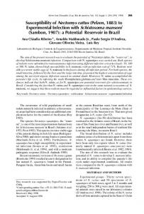
FIG. 1. Effect of Schistosoma mansoni or parasite egg administration in eosinophilic airway inflammation. The numbers of eosinophils in BAL fluid (A) and levels of EPO in lungs (B) decreased in mice infected with S. mansoni or treated with parasite eggs. Five mice per group were used in three independent experiments. *, P ⬍ 0.05 compared to values for mice treated with OVA only (asthma). Error bars indicate standard deviations. O.D., optical density.
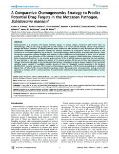
FIG. 2. Effect of Schistosoma mansoni or parasite eggs in the modulation of Th2 cytokine responses in the lung. Cytokine levels in the lungs of mice infected with S. mansoni or of mice injected with parasite eggs and in which asthma was induced were analyzed. Levels of IL-4 (A) and IL-5 (B) produced by S. mansoni-infected mice or animals treated with parasite eggs were determined for five individual mice per group. *, P ⬍ 0.05, compared to values for mice treated with OVA only (asthma). Error bars indicate standard deviations.
affect eosinophilic airway inflammation, mice were infected with S. mansoni cercariae or injected intraperitoneally with S. mansoni eggs and were then OVA challenged. The number of eosinophils recovered in BAL fluid was analyzed, and EPO activity was measured in lungs as an indirect measurement of eosinophils in this organ. There were more eosinophils in the asthmatic group than in the control group (PBS) (P ⬍ 0.0001). Mice infected with cercariae (P ⬍ 0.0001) or that received S. mansoni eggs (P ⬍ 0.0005) and were then challenged with the OVA aerosol had significantly fewer eosinophils than did mice in the asthmatic group (Fig. 1A). Furthermore, mice in the INF/asthma (P ⬍ 0.0001) and egg/asthma (P ⬍ 0.048) groups had more eosinophils than the PBS-treated control mice. We also detected reduced EPO levels in the lungs of mice in the INF/asthma (P ⬍ 0.005) and egg/asthma (P ⬍ 0.006) groups compared with asthmatic animals (Fig. 1B). These findings show that S. mansoni infection and parasite egg treatment modulate eosinophil-mediated lung inflammation. IL-4 and IL-5 Th2 cytokines are downregulated by S. mansoni infection and by parasite egg treatment. In allergic disorders, high levels of Th2 cytokines, such as IL-4 and IL-5, are observed at the inflammatory site. To determine if the reduction in eosinophil numbers observed in the BAL fluid and lungs of infected and egg-treated mice was associated with a decrease in Th2 cytokine production, we evaluated IL-4 and IL-5 production in the lungs of the mice. Lower levels of IL-4 and IL-5 were detected in S. mansoni-infected and in parasite egg-injected mice than in asthmatic mice (Fig. 2A and B).
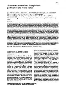
Decreased levels of anti-OVA IgE are detected in S. mansoniinfected and in parasite egg-injected animals. IgE is one of the immunological components responsible for mast cell degranulation and allergy; it is directly correlated with IL-4, which is involved in an IgE isotype switch. We measured OVA-specific IgE levels by ELISA. Significantly lower anti-OVA IgE levels were observed in mice infected with S. mansoni and in those that were injected with parasite eggs than in asthmatic animals (Fig. 3). Reduced inflammatory cell recruitment into lungs of infected and parasite egg-treated mice. Histological sections stained with Gomori trichrome were used to evaluate the inflammatory process in the lung. An increased inflammatory response, characterized by an intense inflammatory cell infiltrate (Fig. 4B), was observed in the lungs of asthmatic mice compared with animals infected with S. mansoni or injected with parasite eggs (Fig. 4C and D, respectively). Schistosoma mansoni egg-treated mice have increased IL-10 production in the lungs. IL-10 is an anti-inflammatory cytokine that seems to be involved in asthma modulation. We measured IL-10 levels in lung homogenates by ELISA. Mice that were injected with S. mansoni eggs and challenged with OVA produced higher levels of IL-10 than asthmatic mice; however, no difference in IL-10 synthesis was observed between S. mansoniinfected and asthmatic animals (Fig. 5). Schistosoma mansoni egg treatment increases the numbers of CD4ⴙ CD25ⴙ Foxp3ⴙ IL-10ⴚ cells. We measured the numbers of CD4⫹ CD25⫹ Foxp3⫹ T cells in the lungs of asthmatic animals by flow cytometry. Both mouse groups that were in-
VOL. 77, 2009
S. MANSONI ANTIGENS MODULATE EXPERIMENTAL ASTHMA
FIG. 3. OVA-specific IgE level in sera of mice infected with Schistosoma mansoni or of mice that received S. mansoni eggs and in which experimental asthma was induced. Values represent means for five mice per group. An asterisk denotes differences of INF/asthma (P ⬍ 0.0001) and egg/asthma (P ⬍ 0.0014) groups compared to values obtained for mice treated with OVA only (asthma). The level of OVAspecific IgE measured in the asthma group is also significant compared to the PBS control group (P ⬍ 0.0001). Error bars indicate standard deviations. O.D., optical density.
fected with S. mansoni or injected with parasite eggs had significantly more CD4⫹ CD25⫹ cells than did PBS-treated or asthmatic mice (Fig. 6A). However, only the mice that were injected with S. mansoni eggs had more CD4⫹ CD25⫹ Foxp3⫹ T cells (Fig. 6B). In order to determine if CD4⫹ CD25⫹ Foxp3⫹ T cells were the source of the IL-10 detected in the lungs of these animals, lung cell suspensions were stained with anti-CD4–FITC, anti-IL-10–PE, and anti-Foxp3–biotin plus streptavidin-PE-Cy5. Although significant IL-10 production was detected in the lungs of mice that were injected with
FIG. 4. Histological analysis of lung tissue from mice treated with PBS, OVA, Schistosoma mansoni infection, or parasite eggs. Animals were sacrificed and their lungs washed with PBS and stored in formaldehyde until histological procedures. The lungs were sliced and stained with Gomory trichrome. (A) lung from the PBS control group; (B) lung from the asthma group; (C) lung from the S. mansoni-infected group; (D) lung from mice that were injected with parasite eggs. Lung tissue analysis revealed reduced pulmonary inflammation in OVAsensitized S. mansoni-infected or parasite egg-injected mice compared to mice from the asthmatic group. Five animals per group were analyzed. Magnification, ⫻40; bars, 20 m.
101
FIG. 5. Effect of Schistosoma mansoni or parasite egg administration on IL-10 production. IL-10 in lungs of mice in the PBS, asthma, INF/asthma, and egg/asthma groups was measured by ELISA. Five individual mice per group were used; an asterisk denotes differences compared to values obtained for mice treated with OVA only (asthma) (P ⬍ 0.05). Error bars indicate standard deviations.
parasite eggs, this cytokine did not come from CD4⫹ Foxp3⫹ T cells (Fig. 7). Schistosoma mansoni eggs reduce the production of chemokines. CCL2, CCL3, and CCL5 are important chemokines for the recruitment of eosinophils (2). We examined how levels of these chemokines affected the inflammatory response in lungs. Mice that were treated with S. mansoni eggs produced lower levels of these chemokines than asthmatic mice (Fig. 8). However, no significant difference was observed between the INF/ asthma group and asthmatic animals (data not shown). These data suggest a possible role for these chemokines in S. mansoni egg modulation of experimental asthma. CD25ⴙ cell depletion, but not IL-10R neutralization, restores eosinophil numbers in the BAL fluid of egg-treated mice. Since IL-10 and CD4⫹ CD25⫹ Foxp3⫹ T cells are the main candidates for asthma modulation, we depleted CD25⫹ T cells and neutralized IL-10R in order to determine their role in this regulation. Mice that were depleted with anti-CD25 had more eosinophils than the control group (GL113), and the number of eosinophils was similar to that found in asthmatic mice (Fig. 9). In contrast, neutralization of IL-10R had no effect on eosinophil numbers in the BAL fluid of parasite egg-treated animals. Effect of anti-CD25 and anti-IL-10R on Th2 cytokine and chemokine production in lungs. To determine the role of CD4⫹ CD25⫹ T cells and IL-10 in Th2 cytokine and chemokine production, we measured IL-4, IL-5, CCL2, CCL3, and CCL5 levels in the lungs of egg/asthma anti-CD25-treated and egg/asthma anti-IL-10R-treated mice. IL-4 and IL-5 (Fig. 10A) and chemokines (Fig. 10B) were significantly increased in mice depleted of CD25⫹ cells and in animals with neutralized IL10R compared with the isotype control group (GL113). CD25ⴙ T cells control tissue pathology in S. mansoni eggtreated mice. Mouse lungs were stained with hematoxylin-eosin for inflammatory cell analysis. The asthmatic group (Fig. 11B) had numerous inflammatory cells and a dense cell layer in the bronchioles compared to the PBS (Fig. 11A) and egg/asthma GL113 (Fig. 11C) groups. Additionally, treatment with anti-CD25 antibodies resulted in more inflammatory cells (Fig. 11D) than in the egg/asthma GL113 group (Fig. 11C). However, when IL-10R was neutralized, no alteration in the inflammatory cell profile was observed in the lungs (Fig. 11E). These findings are in agreement with the results of the BAL fluid assay.
102
PACI´FICO ET AL.
INFECT. IMMUN.
FIG. 6. Schistosoma mansoni egg antigens induce an increase in the number of lung CD4⫹ CD25⫹ Foxp3⫹ T cells. Cell suspensions isolated from lungs of mice in the PBS, asthma, INF/asthma, and egg/asthma groups were stained for surface expression of CD4⫹ and CD25⫹ cells and intracellularly for Foxp3⫹ expression and then analyzed by flow cytometry. (A) Frequency of CD4⫹ CD25⫹ T cells in the lymphocyte region of the forward/side scatter plot. Differences in CD4⫹ CD25⫹ frequency compared to PBS are denoted by an asterisk and those compared to the asthmatic group by # (P ⬍ 0.05). (B) Frequency of CD4⫹ CD25⫹ Foxp3⫹ T cells within the CD4⫹ T-cell population. Differences in CD4⫹ CD25⫹ Foxp3⫹ compared to the PBS group, the asthmatic group, or the S. mansoni-infected group are denoted by an asterisk (P ⬍ 0.05).
DISCUSSION Parasite infection continues to be a problem worldwide, and helminths currently infect billions of individuals (7). The major burden of this disease falls on inhabitants of developing countries, given that over the last 4 to 5 decades, helminth infec-
tions have been disappearing in industrialized societies. In developing countries, helminths are a major source of immunomodulatory signals (9). However, few studies have tried to explain how helminth infection modulates allergic responses (3, 38). It is clear now that the protective effect of helminth
VOL. 77, 2009
S. MANSONI ANTIGENS MODULATE EXPERIMENTAL ASTHMA
103
FIG. 7. CD4⫹ Foxp3⫹ T cells are not the source of IL-10 produced in lungs of schistosome egg-treated mice. Cell suspensions isolated from the lungs of mice in the PBS (dark bars), asthma (white bars), and egg/asthma (gray bars) groups were cultured for 18 h in medium, concanavalin A (ConA) (1 g/ml), or SEA (25 g/ml), stained for surface expression of CD4⫹ and intracellularly for Foxp3⫹ and IL-10 expression, and then analyzed by flow cytometry. (A) The gate used for the analysis of CD4⫹ Foxp3⫹ IL-10⫹ frequency. The lymphocyte region was determined in the forward/side scatter plot, and the CD4⫹ subpopulation was represented in a histogram for CD4 expression; then a dot plot showing the frequency of CD4⫹ cells expressing Foxp3 and producing IL-10 was constructed. (B) Mean frequency (⫾ standard deviation) of CD4⫹ Foxp3⫹ IL-10⫹ cells. Differences in CD4⫹ Foxp3⫹ IL-10⫹ frequency between stimulated cells and nonstimulated cells were evaluated using Student’s t test. Asterisks represents significant differences (P ⬍ 0.05) compared to unstimulated cells.
infection against allergy development cannot be explained simply in terms of the absence of Th1 stimuli. This had led to a reworking of the hygiene hypothesis, in which the regulatory mechanisms induced by inflammatory responses have been reviewed (21, 33). A study by van den Biggelaar et al. (31) demonstrated that the inverse association between degree of infection with schistosomes and positive skin prick tests had to
do with the amount of IL-10 in the serum. This work led to the suggestion that IL-10 interferes with allergic effector mechanisms either by inhibiting mast cell degranulation or by inhibiting Th2 cell proliferation (38). Further, the possibility that Tregs inhibit allergic disease has received growing support from both animal and human studies (1, 30). Here we have provided direct experimental evidence to support the hypoth-
104
PACI´FICO ET AL.
FIG. 8. Effect of schistosome egg injection on chemokine production. The levels of CCL2, CCL3, and CCL5 in the lungs of mice that received Schistosoma mansoni eggs were decreased compared with those in animals from the asthmatic group. The data shown are the means ⫾ standard deviations for five mice per group (PBS, asthma, egg, and egg/asthma). *, P ⬍ 0.05 compared to values obtained for mice treated with OVA only (asthma).
esis that S. mansoni antigens downregulate allergic reactions through the action of Tregs. We have shown here that S. mansoni infection and schistosome egg injection modulate allergic inflammation in OVAsensitized mice by reducing eosinophil numbers in the lungs. During the allergic response, eosinophils appear at the site of inflammation, and they produce a variety of inflammatory mediators (8). In the asthmatic group, the number of eosinophils and the EPO level were much higher than those in mice infected with S. mansoni cercariae or injected with parasite eggs. Furthermore, the levels of Th2 effector cytokines IL-4 and IL-5 were reduced in infected or egg-injected animals. When we examined IgE responses, the INF/asthma and egg/asthma groups had significantly reduced levels of anti-OVA IgE along with decreased IL-4 production. Similar findings using other helminths have been reported. Itami et al. (14) and Lima et al. (18) showed that mice protected against experimental asthma by Ascaris suum components had suppressed cellular migration, and the EPO activity correlated significantly with the reduction in the levels of IL-4 and IL-5 in the lungs. Eosinophilic inflammation in the bronchial mucosa has been recognized as a prominent pathological feature of bronchial asthma. Th2 cells have been implicated in the local infiltration and activation of eosinophils (26). In our study, IL-5 and IL-4 levels were significantly lower in the lungs of the INF/asthma and egg/asthma animals versus asthmatic animals. Studies with IL-4 deficient (IL-4⫺/⫺) mice have shown substantially fewer eosinophils in BAL fluid and much less peribronchial inflammation than in wild-type mice, indicating that IL-4 is a central mediator of allergic airway inflammation (6). The finding that eosinophils also store IL-5 strongly supports the idea that eosinophil-derived eotaxin and IL-5 contribute to the local accumu-
INFECT. IMMUN.
FIG. 9. Depletion of CD25⫹ T cells but not IL-10R neutralization increases eosinophilic inflammation. This graph demonstrates the number of eosinophils in BAL fluid from mice that received anti-IL10R, anti-CD25 antibodies, or isotype control antibody (GL113). Five mice per group were used; the protocol for treatment with anti-CD25 or anti-IL-10R is described in Materials and Methods. This is representative of three independent experiments. *, P ⬍ 0.05 compared to values obtained for mice treated with OVA only (asthma); #, P ⬍ 0.05 compared to values obtained for the control group (egg/asthma/ GL113). Error bars indicate standard deviations.
lation of eosinophils at inflammatory loci and to their enhanced survival (12). The production of eosinophil-specific chemotactic factors during the allergic airway response may be a pivotal event resulting in eosinophil accumulation, activation, and airway damage. The migration of eosinophils as well as Th2 cells is controlled by chemokines, suggesting a crucial role of these molecules in the pathogenesis of bronchial asthma (26). Alam et al. (2) reported that chemokines that induce chemotaxis of inflammatory cells, such as CCL2, CCL3, and CCL5, had increased levels in BAL fluid of asthmatic patients compared with healthy subjects. Lukacs et al. (19) found that CCL3 has a role in eosinophil accumulation in the lung and airways during allergic airway inflammation. They also showed that CCL3 and CCL5 are major eosinophil chemotactic factors that are produced during airway responses. We have shown here that mice treated with parasite eggs and in which asthma was induced had significantly lower levels of CCL2, CCL3, and CCL5 than the asthmatic group. In contrast to what was observed in egg-treated mice, we did not find reduced chemokine levels in mice infected with S. mansoni. However, we cannot rule out the role of these molecules in Th2 cell recruitment in mice infected with S. mansoni, since the kinetics of the experiments with worm infection and egg treatment were different. Regulatory cytokines, such as IL-10, are thought to be one of the main mechanisms of regulation of Th2 cell-mediated allergic inflammation (16). Additionally, studies performed by Kaviratne et al. (15) demonstrated no major regulatory role for transforming growth factor  in murine schistosomiais. It has been shown that Nippostrongylus brasiliensis infection suppresses allergen-induced airway eosiniphilia via IL-10 (36). When we infected mice with S. mansoni, we did not observe an increase in IL-10 production. Recently, Smits et al. (28) reported that in cases of chronic S. mansoni infection, suppressive mechanisms that regulate immune reactions to allergens are IL-10 dependent. Suppression of OVA-induced airway eo-
VOL. 77, 2009
S. MANSONI ANTIGENS MODULATE EXPERIMENTAL ASTHMA
105
FIG. 10. Effect of anti-CD25 and anti-IL-10R antibody treatment on Th2 cytokine and chemokine production in lungs. Measurements of IL-4 and IL-5 (A) and of CCL2, CCL3, and CCL5 (B) were also used for characterization of the inflammatory process. Mice that received eggs and anti-IL-10R antibodies or that received eggs and anti-CD25 antibodies showed higher levels of IL-4, IL-5, CCL2, CCL3, and CCL5 than the control group (egg/asthma/GL113) (#, P ⬍ 0.05). The control group also had a decrease in cytokine and chemokine levels compared with asthmatic mice (*, P ⬍ 0.05). Five mice per group were analyzed. Data are representative of three independent experiments. All statistical analysis was done using analysis of variance (P ⬍ 0.05). Error bars indicate standard deviations.
sinophilia observed in the chronic phase was also correlated with infection intensity. Therefore, we speculate that the lack of enhanced IL-10 production that we observed might be because we induced acute instead of chronic S. mansoni infection in BALB/c mice. Since only the schistosome egg treatment enhanced IL-10 production, we decided to investigate the regulatory mechanisms involved in parasite egg suppression. We treated egg/asthma group mice with blocking IL-10R antibodies or anti-CD25 to study the roles of IL-10 and Tregs in asthma modulation. Treatment with anti-CD25 antibodies fully reversed suppression of eosinophilic airway inflammation,
whereas blocking IL-10R had no effect on lung eosiniphilia. Although IL-10R blockage led to increases in levels of IL-4, IL-5, and chemokines (CCL2, CCL3, and CCL5), similar to levels found in asthmatic animals, treatment with anti-IL-10R antibodies did not significantly change eosinophil numbers in BAL fluid and inflammatory cell numbers in lungs. Therefore, our main hypothesis is that Treg activity is responsible for experimental asthma modulation, based on our finding that anti-CD25 antibody treatment reversed suppression. Further, we found that Foxp3⫹ Tregs are an essential part of this reg-
106
PACI´FICO ET AL.
INFECT. IMMUN.
FIG. 11. CD25⫹ T cells control tissue pathology in lungs. Lung sections were stained with hematoxylin-eosin. (A) PBS group; (B) asthma group, (C) egg/asthma GL113-treated group, (D) egg/asthma anti-CD25-treated group; (E) egg/asthma anti-IL-10R-treated group. The lungs from the asthma group (B) showed numerous inflammatory cells with a dense cellular layer within the bronchioles compared with the PBS (A) and the egg/asthma GL113 (C) groups. Lungs from mice that were injected with parasite eggs and anti-CD25 antibodies had more inflammatory cells than those from the egg/asthma GL113 control group (D). In parallel with the results of the BAL fluid assay demonstrating fewer eosinophils, panel E shows similar numbers of inflammatory cells in lungs of mice treated with eggs and anti-IL-10R antibodies and in lungs of mice from the control group (egg/asthma GL113). Five animals per group were analyzed. Magnification, ⫻40; bars, 20 m.
ulatory network and that the effect of their suppression is independent of IL-10. Recently Yang et al. (37) showed that Schistosoma japonicum egg antigen treatment induces a significant increase in CD4⫹ CD25⫹ T cells in BALB/c mice when there is a combination of SEA injection and oral administration of dead S. japonicum eggs. They demonstrated that IL-10-producing CD4⫹ CD25⫹ T cells stimulated by S. japonicum egg antigen treatment modulate airway inflammation in a murine model of asthma. Therefore, in our study the mechanisms of modulation appear to be different from those found by Yang et al. (37). We observed a regulatory function for Tregs in schistosome egginduced asthma modulation, independent of IL-10. Unexpectedly, we found that most Foxp3⫹ CD4⫹ T cells did not produce IL-10. Further, we found that egg-induced Foxp3⫹ Tregs are not the main source of IL-10. Recently, CD4⫹ CD25⫹ T cells were identified as a major source of IL-10 in schistosomeinfected mice (13). Additionally, Yang et al. (37) demonstrated that IL-10 producing CD4⫹ CD25⫹ T cells modulate airway inflammation. This discrepancy in IL-10 production by Tregs might be due to different protocols for egg antigen administration and/or to egg antigens present in S. mansoni and S. japonicum that could differentially stimulate Tregs to produce IL-10. More recently, using a bead-induced pulmonary granuloma model, it was found that only CD4⫹ CD25⫹ Foxp3⫺ T cells produce large amounts of IL-10, while CD4⫹ CD25⫹ Foxp3⫹ T cells do not (11). Similar to what we found, Baumgart et al. (4) demonstrated that S. mansoni egg-induced Foxp3⫹ Tregs are not the main source for IL-10. Therefore, we speculate that S. mansoni egg-induced CD4⫹ CD25⫹ Tregs modulate experimental allergic asthma via cell-cell contactdependent mechanisms (4). Baumgart et al. (4) showed that
increased expression of ␣E7 integrin can improve the contact between CD103⫹ Tregs and E-cadherin-expressing target cells, thereby increasing modulation. Recently, Wilson et al. (35) demonstrated that mesenteric lymph node cells from H. polygyrus-infected IL-10 knockout mice transfer suppression to OVA-sensitized hosts, indicating that IL-10 is not the primary modulator of the allergic response. In summary, we demonstrated that mice infected with male or female worms and mice injected with S. mansoni eggs develop a significantly decreased eosinophilic response in the airways, reduced IL-4 and IL-5 production, and reduced levels of anti-OVA-specific IgE antibodies and that they become more resistant to experimental asthma. Asthma protection was associated mostly with CD4⫹ CD25⫹ Foxp3⫹ T cells, which is consistent with the hypothesis that parasite-induced Tregs can downmodulate Th2 allergic inflammation. These findings can help explain reactions found in humans, since individuals with active helminth infections appear to be less responsive to allergen provocation than individuals who are free of infection (3, 24). Finally, the identification of parasite molecules that induce protection against allergic diseases could lead to the development of new asthma therapies. ACKNOWLEDGMENTS This work was supported by CNPq, FINEP/SEBRAE, and FAPEMIG. We thank Rodrigo Correˆa-Oliveira of CPqRR-FIOCRUZ for providing S. mansoni cercariae for the challenge infection experiments. We also thank Tatiani Maioli, Danielle Foschetti, Francsineia Assis, and Ana Maria F. Caetano for helping us with the IgE ELISA. REFERENCES 1. Akbari, O., P. Stock, R. H. DeKruyff, and D. T. Umetsu. 2003. Role of regulatory T cells in allergy and asthma. Curr. Opin. Immunol. 15:627–633.
VOL. 77, 2009
S. MANSONI ANTIGENS MODULATE EXPERIMENTAL ASTHMA
2. Alam, R., J. York, M. Boyars, S. Stafford, J. A. Grant, J. Lee, P. Forsthe, T. Sim, and N. Ida. 1996. Increased MCP-1, RANTES, and MIP1-alpha in bronchioalveolar lavage fluid of allergic asthmatic patients. Am. J. Respir. Crit. Care Med. 153:1398–1404. 3. Arau ´jo, M. I., A. A. Lopes, M. Medeiros, A. A. Cruz, L. Sousa-Atta, D. Sole, and E. M. Carvalho. 2000. Inverse association between skin response to aeroallergens and Schistosoma mansoni infection. Int. Arch. Allergy Immunol. 123:145–148. 4. Baumgart, M., F. Tompkins, J. Leng, and M. Hesse. 2006. Naturally occurring CD4⫹Foxp3⫹ regulatory T cells are an essential, IL-10-independent part of the immunoregulatory network in Schistosoma mansoni egg-induced inflammation. J. Immunol. 176:5374–5387. 5. Beasley, R., J. Crane, C. K. Lai, and N. Pearce. 2000. Prevalence and etiology of asthma. J. Allergy Clin. Immunol. 105:466–472. 6. Brusselle, G., J. Kips, G. Joos, H. Bluethmann, and R. Pauwels. 1995. Allergen-induced airway inflammation and bronchial responsiveness in wildtype and interleukin-4-deficient mice. Am. J. Respir. Cell Mol. Biol. 12:254– 259. 7. Chitsulo, L., P. Loverde, and D. Engels. 2004. Schistosomiasis. Nat. Rev. Microbiol. 2:12–13. 8. Erpenbeck, V. J., J. M. Hohlfeld, J. Petschallies, E. Eklund, C. G. B. Peterson, H. Fabel, and N. Krug. 2003. Local release of eosinophil peroxidase following segmental allergen provocation in asthma. Clin. Exp. Allergy 33: 331–336. 9. Fallon, P. G., and A. Alcami. 2006. Pathogen-derived immunomodulatory molecules: future immunotherapeutics? Trends Immunol. 27:470–476. 10. Fonseca, C. T., L. G. Pacifico, M. M. Barsante, T. Rassi, G. D. Cassali, and S. C. Oliveira. 2006. Co-administration of plasmid expressing IL-12 with 14-kDa Schistosoma mansoni fatty acid-binding protein cDNA alters immune response profiles and fails to enhance protection induced by Sm14 DNA vaccine alone. Microbes Infect. 8:2509–2516. 11. Freeman, C. M., B. C. Chiu, V. R. Stolberg, J. Hu, K. Zeibecoglou, N. W. Lukacs, S. A. Lira, S. L. Kunkel, and S. W. Chensue. 2005. CCR8 is expressed by antigen-elicited, IL-10-producing CD4⫹CD25⫹ T cells, which regulate Th2-mediated granuloma formation in mice. J. Immunol. 174:1962– 1970. 12. Giembycz, M. A., and M. A. Lindsay. 1999. Pharmacology of the eosinophil. Pharmacol. Rev. 51:213–340. 13. Hesse, M., C. A. Piccirillo, Y. Belkaid, J. Prufer, M. Mentink-Kane, M. Leusink, A. W. Cheever, E. M. Shevach, and T. A. Wynn. 2004. The pathogenesis of schistosomiasis is controlled by cooperating IL-10-producing innate effector and regulatory T cells. J. Immunol. 172:3157–3166. 14. Itami, D. M., T. M. Oshiro, C. A. Arau ´jo, A. Perini, M. A. Martins, M. S. Macedo, and M. F. Macedo-Soares. 2005. Modulation of murine experimental asthma by Ascaris suum components. Clin. Exp. Allergy 35:873–879. 15. Kaviratne, M., M. Hesse, M. Leusink, A. W. Cheever, S. J. Davies, J. H. McKerrow, L. M. Wakefield, J. J. Letterio, and T. A. Wynn. 2004. IL-13 activates a mechanism of tissue fibrosis that is completely TGF-beta independent. J. Immunol. 173:4020–4029. 16. Kearley, J., J. E. Barker, D. S. Robinson, and C. M. Lloyd. 2005. Resolution of airway inflammation and hyperreactivity after in vivo transfer of CD4⫹CD25⫹ regulatory T cells is interleukin-10 dependent. J. Exp. Med. 202:1539–1547. 17. Kitagaki, K., T. R. Businga, D. Racila, D. E. Elliott, J. V. Weinstock, and J. N. Kline. 2006. Intestinal helminths protect in a murine model of asthma. J. Immunol. 177:1628–1635. 18. Lima, C., A. Perini, M. L. Garcia, M. A. Martins, M. M. Teixeira, and M. S. Macedo. 2002. Eosinophilic inflammation and airway hyper-responsiveness are profoundly inhibited by a helminth (Ascaris suum) extract in a murine model of asthma. Clin. Exp. Allergy 32:1659–1666. 19. Lukacs, N. W., T. J. Standiford, S. W. Chensue, R. G. Kunkel, R. M. Strieter, and S. L. Kunkel. 1996. C-C chemokines-induced eosinophil chemotaxis during allergic airway inflammation. J. Leukoc. Biol. 60:573–578. 20. Lynch, N. R., I. A. Hagel, M. E. Palenque, M. C. Di Prisco, J. E. E Escudero,
Editor: W. A. Petri, Jr.
21. 22. 23.
24.
25. 26. 27.
28.
29. 30. 31.
32.
33. 34. 35. 36. 37.
38. 39.
107
L. A. Corao, J. A. Sandia, L. J. Ferreira, C. Botto, M. Perez, and P. N. Le Souef. 1998. Relationship betwen helminthic infection and IgE response in atopic and nonatopic children in a tropical environment. J. Allergy Clin. Immunol. 101:217–221. Maizels, R. M. 2005. Infections and allergy—helminths, hygiene and host immune regulation. Curr. Opin. Immunol. 17:656–661. Mangan, N. E., N. van Rooijen, A. N. McKenzie, and P. G. Fallon. 2006. Helminth-modified pulmonary immune response protects mice from allergen-induced airway hyperresponsiveness. J. Immunol. 176:138–147. Marsh, D. G., J. D. Neely, D. R. Breazeale, B. Ghosh, L. R. Freidhoff, E. Ehrlich-Kautzky, C. Schou, G. Krishnaswamy, and T. H. Beaty. 1994. Linkage analysis of IL-4 and other chromosome 5q31.1 markers and total serum immunoglobulin E concentrations. Science 264:1152–1156. Medeiros, M. J., P. F. Joanemile, M. C. Almeida, M. A. Matos, M. I. Arau ´jo, A. Cruz, A. M. Atta, M. A. V. Rego, A. R. Jesus, E. A. Taketomi, and E. M. Carvalho. 2003. Schistosoma mansoni infection is associated with a reduced course of asthma. J. Allergy Clin. Immunol. 111:947–951. Moffatt, M. F., and W.O. Cookson. 1999. Genetics of asthma and inflammation: the status. Curr. Opin. Immunol. 11:606–609. Rothenberg, M. E., and S. P. Hogan. 2006. The eosinophil. Annu. Rev. Immunol. 24:147–174. Sales, P. A. Jr., D. Golgher, R. V. Oliveira, V. Vieira, R. M. Arantes, J. Lannes-Vieira, and R. T. Gazzinelli. 2008. The regulatory CD4⫹CD25⫹ T cells have a limited role on pathogenesis of infection with Trypanosoma cruzi. Microbes Infect. 10:680–688. Smits, H. H., H. Hammad, M. van Nimwegen, T. Soullie, M. A. Willart, E. Lievers, J. Kadouch, M. Kool, J. Kos-van Oosterhoud, A. M. Deelder, B. N. Lambrecht, and M. Yazdanbakhsh. 2007. Protective effect of Schistosoma mansoni infection on allergic airway inflammation depends on the intensity and chronicity of infection. J. Allergy Clin. Immunol. 120:932–940. Strath, M., D. J. Warren, and C. J. Sanderson. 1985. Detection of eosinophils using an eosinophil peroxidase assay. Its use as an assay for eosinophil differentiation factors. J. Immunol. Methods 83:209–215. Umetsu, D. T., O. Akbari, and R. H. Dekruyff. 2003. Regulatory T cells control the development of allergic disease and asthma. J. Allergy Clin. Immunol. 112:480–487. Van den Biggelaar, A. H., R. Van Ree, L. Rodrigues, B. Lell, A. M. Deelder, P. G. Kremsner, and M. Yazdandakhsh. 2000. Decreased atopy in children infected with Schistosoma haematobium: a role for parasite-induced interleukin-10. Lancet 356:1723–1727. Van den Biggelaar, A. H., C. Lopuhaa, R. Van Ree, J. S. Van Der Zee, J. Jans, A. Hoek, and M. Yazdandakhsh. 2001. The prevalence of parasite infestation and house dust mite sensitization in Gabonese schoolchildren. Int. Arch. Allergy Immunol. 126:231–238. Wills-Karp, M., J. Santeliz, and C. L. Karp. 2001. The germless theory of allergic disease: revisiting the hygiene hypothesis. Nat. Rev. Immunol. 1:69–75. Wills-Karp, M. 2004. Interleukin-13 in asthma pathogenesis. Immunol. Rev. 202:175–190. Wilson, M. S., M. D. Taylor, A. Balic, C. A. Finney, J. R. Lamb, and R. M. Maizels. 2005. Suppression of allergic airway inflammation by helminthinduced regulatory T cells. J. Exp. Med. 202:1199–1212. Wohlleben, G., C. Trujillo, J. Mu ¨ller, Y. Ritze, S. Grunewald, U. Tatsch, and K. J. Erb. 2004. Helminth infection modulates the development of allergeninduced airway inflammation. Int. Immunol. 16:585–596. Yang, J., J. Zhao, Y. Yang, L. Zhang, X. Yang, X. Zhu, Ji Minjun, N. Sun, and C. Su. 2007. Schistosoma japonicum egg antigens stimulates CD4⫹CD25⫹ T cells and modulate airway inflammation in a murine model of asthma. Immunology 120:8–18. Yazdanbakhsh, M., P. G. Kremsner, and R. Van Ree. 2002. Allergy, parasites and the hygiene hypothesis. Science 296:490–494. Zuberi, R. I., J. R. Apgar, S. C. Chen, and F. T. Liu. 2000. Role of IgE in airway secretions: IgE immune complexes are more potent inducers than antigen alone of airway inflammation in a murine model. J. Immunol. 164: 2667–2673.