FOLIA PARASITOLOGICA 55: 180–186, 2008
Schistosoma mansoni glyceraldehyde 3-phosphate dehydrogenase is a lung-stage schistosomula surface membrane antigen Hatem Tallima and Rashika El Ridi Zoology Department, Faculty of Science, Cairo University, Cairo 12613, Egypt
Key words: Schistosoma mansoni, lung-stage schistosomula, glyceraldehyde 3-phosphate dehydrogenase, schistosome surface membrane antigens, neutral sphingomyelinase Abstract. We have previously reported that Schistosoma mansoni larvae emerging from host lung at pH 7.5–7.8 and then fixed with diluted formaldehyde (HCHO) readily bind radiation-attenuated cercariae (RA) vaccine serum antibodies, as assessed by indirect membrane immunofluorescence (IF). Here we show that S. mansoni schistosomula emerging from lung pieces under 5% CO2 (pH ≤7.3) readily bind RA vaccine serum antibodies, provided they have been incubated for 12 h at pH 7.5–7.8 in foetal calf serum-free RPMI medium, and fixed with diluted HCHO. Ex vivo larvae exposed during incubation to GW4869, a specific inhibitor of tegument-bound, neutral sphingomyelinase (nSMase) displayed significantly diminished binding of RA vaccine serum antibodies, thus suggesting that nSMase activity at pH ≥7.5 leads to exposure of lung-stage larvae surface membrane antigens to specific antibody detection. More importantly, ex vivo larvae readily bound antibodies directed to dipeptidic multiple antigen peptide constructs, based on S. mansoni-specific sequences in S. mansoni glyceraldehyde 3-phosphate dehydrogenase (SG3PDH). Lung-stage schistosomula IF reactivity was diminished following antiserum absorption with recombinant SG3PDH. The data together indicate that intact ex vivo, as well as, 5-day-old in vitro-grown larvae express SG3PDH on their surface membrane. The findings are discussed in relation to the importance of surface membrane proteins as candidate vaccine antigens.
Schistosomiasis is a parasitic disease caused by the platyhelminth worms Schistosoma mansoni, Schistosoma haematobium and Schistosoma japonicum, affecting 207 million people in the developing world, with 779 million, mostly children, at risk of the infection (Steinmann et al. 2006). Schistosomes develop and live in the blood vessels of their vertebrate hosts. The developing and adult schistosomes are covered by the tegument, a 2- to 4-µm thick syncytium. The innermost membrane of the tegument is a conventional basal membrane, whereas the outer membrane on the syncytium surface consists of two, tightly apposed bilayers, each of which is composed of outer and inner leaflets (Hockley 1973, Hockley and McLaren 1973). The disease can be eradicated via elimination of transmission if an effective vaccine is available. Many roadblocks hinder the development of such vaccine, among which our inability to identify an appropriate vaccine antigen and the major mechanisms of immune resistance to the infection. There is consensus, however, about the lungstage schistosomula being the main targets of natural and RA-mediated protective immunity (Coulson 1997). Paradoxically, no specific antibody is able to bind to lung-stage larvae surface membrane antigens, as judged by several serological tests, namely indirect membrane immunofluorescence (Dean 1977, Dessein et al. 1981, McLaren and Terry 1982, Foley et al. 1986, Pearce et al. 1986, Chiang and Caulfield 1989, Kusel and Gordon 1989, Coulson 1997, El Ridi et al. 2003, 2004b, Tallima and El Ridi 2005, Tallima et al. 2005, El Ridi and Tallima 2006, Kusel et al. 2007). Entire failure of surface
membrane antigen detection does not give support to the contention that the tegument is bounded externally by a single lipid bilayer, overlain by a laminate secretion containing numerous proteins and molecular complexes (Braschi et al. 2006), and was previously ascribed to shedding of antigenic molecules (Pearce et al. 1986), masking by host proteins (McLaren and Terry 1982, Chiang and Caulfield 1989), or intrinsic biochemical modifications of the outer membrane (Dean 1977, Dessein et al. 1981, Foley et al. 1986, Kusel and Gordon 1989, Kusel et al. 2007). Our recent findings have supported the latter hypothesis, whereby extraction of outer membrane cholesterol by the impermeable cholesterolbinding drug, methyl-β-cyclodextrin readily induced exposure of surface membrane antigens in lung-stage larvae (El Ridi et al. 2004b, Tallima and El Ridi 2005). Surface membrane antigen exposure could be even more readily elicited via manipulating the balance between sphingomyelin (SM) biosynthesis and breakdown towards SM hydrolysis, suggesting that SM is responsible for preventing antibody access to surface membrane antigens (Tallima et al. 2005, El Ridi and Tallima 2006). Accordingly, the way was paved to identify the surface membrane antigens of the lung-stage larvae. In this report we show that the vaccine candidate, Schistosoma mansoni glyceraldehyde 3-phosphate dehydrogenase, SG3PDH (El Ridi et al. 2001a, b, 2004a, Tallima et al. 2003, Vepřek et al. 2004 and references therein) is a lung-stage schistosomula surface membrane antigen.
Address for correspondence: R. El Ridi, Zoology Department, Faculty of Science, Cairo University, Cairo 12613, Egypt. Phone: ++202 356 76 708; Fax: ++202 376 03 735; E-mail:
[email protected]
180
Tallima, El Ridi: SG3PDH on schistosome surface membrane
MATERIALS AND METHODS Animals and parasites. Cercariae and animal hosts were obtained from the Schistosome Biological Materials Supply Program, Theodore Bilharz Research Institute, Giza, Egypt. Lung-stage schistosomula were recovered from male Syrian hamsters 6 days after percutaneous infection with 2,000 cercariae of S. mansoni. Infection and perfusion were performed under anaesthesia, as described (El Ridi and Tallima 2006) following the recommendations of the current edition of the Guide for the Care and Use of Laboratory Animals, Institute of Laboratory Animal Resources, National Research Council, USA. Lung pieces were incubated in RPMI medium-heparin supplemented with 10% foetal calf serum (FCS, BioWhittaker, Verviers, Belgium) for 4 h at 37°C in a humidified atmosphere containing 5.0% CO2 (pH ≤ 7.3), to allow the schistosomula to emerge (El Ridi et al. 2003, 2004b, Tallima and El Ridi 2005, Tallima et al. 2005, El Ridi and Tallima 2006). The larval suspension was then poured through a Nitex 132-mesh nylon screen, treated for 10 min at 4°C with sterile 0.16 M ammonium chloride/0.017 M Tris buffer, pH 8.0, to lyse the erythrocytes, isolated from contaminant lung cells by centrifugation over 40% percoll (Pharmacia, Uppsala, Sweden) in RPMI medium, as described (El Ridi et al. 2003, 2004b, Tallima and El Ridi 2005), and then incubated for 12 h, at 37°C and pH 7.5–7.8, in RPMI medium containing 20%, 2%, or 0% FCS. In some experiments, the FCS-free RPMI medium was supplemented with 0, 7.5, 10, or 20.0 µM of the nSMase specific inhibitor GW4869 (Sigma, St Louis, MO, USA) dissolved in dimethyl sulfoxide, at a concentration of 1.0 mg/1.0 ml (Luberto et al. 2002, Marchesini et al. 2003, Kolmakova et al. 2004, Wu et al. 2005). At the end of the incubation period, larvae consistently showed high viability, motility, and body contractility. Cercariae-derived schistosomula were prepared as described by Lazdins et al. (1982), and cultured at 37oC under 5% CO2 for 4–6 days in 10–90% FCS in RPMI medium, or medium 169 (Harrop and Wilson 1993) supplemented with 300 U/ml penicillin, 300 µg/ml streptomycin, and 160 µg/ml gentamicin (all from BioWhittaker). The viability of schistosomula was assessed by determining the percentages which excluded Trypan blue, and appeared healthy, transparent, motile, and contractile. Approximately 90% of larvae cultured in 50% FCS-50% RPMI medium were viable as assessed by the Trypan blue dye exclusion test, and actively motile and contractile. Accordingly, larvae were cultured in 50% FCS for 4 days, and then in FCS-free RPMI medium at 37oC, under atmospheric air, for 24 h. At the end of the incubation period, more than 80% of the 5-day in vitro schistosomula were mobile and excluded Trypan blue; approximately 30–50% were elongated, while the rest were generally 20 to 40% smaller than those developed in hamsters, in agreement with Basch (1981). Antigens and antisera. Serum was obtained from naïve BALB/c mice (control serum), or BALB/c mice immunized by the tail exposure method, twice at 4-week interval, with ultraviolet (UV) radiation-attenuated cercariae of S. mansoni (RA vaccine serum). Cercariae used for immunisation were attenuated by exposure to 330 µW/cm2 UV light from an S-68 Mineralight Lamp (Ultra-Violet Products, San Gabriel, CA, USA)
for 3 min, as described previously (Tallima et al. 2005, El Ridi and Tallima 2006). The coding sequence for SG3PDH was obtained and expressed, and the recombinant protein (rSG3PDH) was purified by metal affinity chromatography (HiTrapTM Affinity Column, Pharmacia) as detailed previously (El Ridi et al. 2001b). Antigen aliquots were kept until use at –76°C and thawed only once. The amino acid (aa) sequence of SG3PDH-derived peptides, selected based on lowest homology to the human enzyme (El Ridi et al. 2001a, 2004a, Tallima et al. 2003) is as follows: peptide A, LKNTVDVVSV-OH (aa 24–33); B1, KVNGKLISVHCERDP-OH (aa 67–81); B, ERDPANIP WDKD-OH (aa 78–89); C, ENSYEKSMSVV-OH (aa 138– 148); D, GKGASYEEIKAAVK AAAS-OH (aa 251–268); and E, ITHMHKVDHA-OH (aa 329–338). Tetrabranched, dipeptidic multiple antigen peptide constructs (D-MAP) A-B, B1-C, and D-E, synthesized at Research Genetics (Huntsville, AL, USA), were used to generate in BALB/c mice potent antisera against the peptide immunogens and rSG3PDH, as described (El Ridi et al. 2004a). Peptide P5, based on amino acids 178–192 (MTLTYTL NTPTLWPI) of an S. mansoni glucose transporter protein 4 (SGTP4) extrafacial domain (Skelly et al. 1998) was synthesized at Harvard Biopolymers Laboratories, Harvard Medical School (Cambridge, MA, USA), and used to prepare an antiserum in BALB/c mice, following procedures previously described (El Ridi et al. 2001a, 2004a, Tallima et al. 2003, Vepřek et al. 2004). Indirect membrane immunofluorescence (IF). Larvae were fixed for a total of 10 min in 50 volumes 0.1% formaldehyde (HCHO) or 1.0% paraformaldehyde in FCS-free RPMI medium. Larvae were washed twice in RPMI medium supplemented with 5% FCS (RPMI medium-5% FCS), and incubated in 100 µl RPMI medium-5% FCS containing 1.0 µl control or test serum, overnight at room temperature. In some experiments control and test sera were previously absorbed with 25 µg/µl bovine serum albumin (BSA) or rSG3PDH, overnight at room temperature. Larvae were then washed four times in RPMI medium-5% FCS, incubated with 1:100-diluted anti-mouse IgG (Fab-specific) fluorescein isothiocyanate (FITC)-labeled goat immunoglobulins F(ab’)2 fragment of affinity isolated antibody, adsorbed with bovine, horse, and human serum proteins (Sigma), washed four times in RPMI medium, and inspected by alternate light and UV microscopy (Olympus Inverted Microscope Model IX70, Olympus, Tokyo, Japan).
RESULTS Membrane immunofluorescence reactivity of 5-dayold in vitro cultured schistosomula Mechanically transformed schistosomula grown in vitro for 5 days in the presence of 50% FCS and 5% CO2 and then fixed with 0.1% HCHO do not bind antibodies from control or RA vaccine serum (data not shown). Yet, when 4-day-old larvae were cultured for a further 24 h period in RPMI medium-50% FCS but in the absence of CO2, with medium pH reaching 7.6 ± 0.1, strong IF reactivity with RA vaccine, but not con-
181
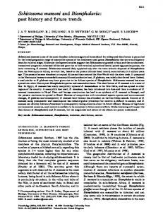
Fig. 1. Membrane immunofluorescence reactivity of 5-day-old in vitro grown schistosomula. Larvae were incubated for the last 24 h of culture at pH 7.5–7.6 and then tested by IF with control (A, IF intensity 0+), RA vaccine (B, IF intensity 1/2+, 2+ and 3+), anti-SGTP4 peptide 5 (C, D), anti-D MAP B1-C (E), or anti-D MAP D-E (F) serum, and examined under light (C) and UV (A, B, D–F) microscopy. Scale bar (A–F) = 100 µm. Table 1. Membrane immunofluorescence reactivity of 5-dayold in vitro cultured schistosomula. Serum
Total number of larvae Control 105 RA vaccine 112 Anti-Peptide 5 136 Anti-D MAP B1-C 138 Anti-D MAP D-E 128
0+ 97.1 0.0 35.2 14.5 9.3
Percent of larvae showing IF intensity of* ½+ 1+ 2+ 3+ 2.8 0.0 0.0 0.0 12.5 13.3 13.2 60.7 19.8 15.4 23.5 5.8 0.0 38.4 34.7 12.3 25.0 35.9 25.0 4.6
*Representative of three independent experiments; IF intensity as depicted in Fig. 1.
trol, serum was evident. Larvae also bound antibodies of anti-SGTP4 peptide P5 (anti-Peptide 5), anti-D MAP B1-C, and anti-D MAP D-E sera, with IF intensity of 0+ to 3+ (Fig. 1, Table 1). Larval IF reactivity diminished following absorption of anti-D MAP B1-C or D-E sera with rSG3PDH. It is of note that larvae did not bind antibodies of five individual BALB/c mouse anti-D MAP A-B sera (data not shown).
182
Membrane immunofluorescence reactivity of ex vivo lung-stage schistosomula Ex vivo lung-stage larvae emerging from lung pieces under 5% CO2 did not bind antibodies from control or test sera whether fixed with 1% paraformaldehyde or 0.1% HCHO. When larvae isolated from lung cells were then maintained for 12 h at pH 7.5–7.8 in the presence of 20% or 2% FCS, only about 36% and 44%, respectively of ex vivo schistosomula showed 1/2+ to 1+ IF reactivity with RA vaccine serum, while they were entirely negative with control, anti-SGTP4 peptide P5, or any anti-D MAP sera in three independent IF experiments. However, when ex vivo larvae were incubated for 12 h at pH 7.5–7.8 in FCS-free RPMI medium, approximately 80–100% therefrom were IF positive when tested with RA vaccine, anti-SGTP4 peptide P5, anti-DMAP B1-C, or anti-D MAP D-E sera (Figs. 2, 3). Schistosomular IF intensity was greatly diminished following absorption of anti-D MAP B1-C or anti-D MAP D-E sera with rSG3PDH (Fig. 2F), but not BSA. No reactivity was evident in repeated IF tests with five individual
Tallima, El Ridi: SG3PDH on schistosome surface membrane
Fig. 2. Membrane immunofluorescence reactivity of ex vivo lung-stage schistosomula. Ex vivo larvae incubated for 12 h at pH 7.5–7.8 in FCS-free medium were tested by IF with control (A), RA vaccine (B), anti-SGTP4 peptide 5 (C), anti-D MAP B1-C (D), anti-D MAP D-E (E), or rSG3PDH-absorbed anti-D MAP B1-C (F) serum, and examined under UV microscopy. Scale bar (A–F) = 150 µm.
BALB/c mouse anti-D MAP A-B sera (data not shown), despite the strong antisera recognition of the peptide immunogens and rSG3PDH (El Ridi et al. 2004a). It is of note that when ex vivo larvae were incubated for 12 h at pH 7.5–7.8 in FCS-free RPMI medium supplemented with 7.5, 10, or 20 µM GW4869, a specific inhibitor of tegument-bound nSMase, IF reactivity to RA vaccine, anti-D MAP B1-C, and anti-D MAP D-E sera significantly (P 150,000 Da) binding and effector activity only in developing and adult parasites suffering excessive loss of tegument integrity. Such worms are certainly poised to die without the contribution or help of antibody-dependent killing. Nevertheless, it is vital to identify the worm surface membrane antigens, as elucidation of the molecular organisation at the parasite-host interface will help in developing effective drugs and vaccine strategies. Acknowledgement. The research work was supported by the Arab Science and Technology Foundation, project No. BT 05 2 05.
REFERENCES ALVAREZ R.A., BLAYLOCK M.W., BASEMAN J.B. 2003: Surface localized glyceraldehyde-3-phosphate dehydrogenase of Mycoplasma genitalium binds mucin. Mol. Microbiol. 48: 1417– 1425. BASCH P.F. 1981: Cultivation of Schistosoma mansoni in vitro. I. Establishment of cultures from cercariae and development until pairing. J. Parasitol. 67: 179–185. BRASCHI S., CURWEN R.S., ASHTON P.D., VERJOVSKI-ALMEIDA S., WILSON A. 2006: The tegument surface membranes of the human blood parasite Schistosoma mansoni: a proteomic analysis after differential extraction. Proteomics 6: 1471– 1482. BRASCHI S., WILSON R.A. 2006: Proteins exposed at the adult schistosome surface revealed by biotinylation. Mol. Cell Proteomics 5: 347–356. CAMPANELLA M.E., CHU H., LOW P.S. 2005: Assembly and regulation of a glycolytic enzyme complex on the human erythrocyte membrane. Proc. Natl. Acad. Sci. U.S.A. 102: 2402– 2407. CHAI M., MCMANUS D.P., MCINNES R., MOERTEL L., TRAN M., LOUKAS A., JONES M.K., GOBERT G.N. 2006: Transcriptome profiling of lung schistosomula, in vitro cultured schistosomula and adult Schistosoma japonicum. Cell. Mol. Life Sci. 63: 919–929. CHIANG C.-.P., CAULFIELD J.P. 1989: Human lipoprotein binding to schistosomula of Schistosoma mansoni. Displacement by polyanions, parasite antigen masking, and persistence in young larvae. Am. J. Pathol. 135: 1015–1024. COULSON P.S. 1997: The radiation-attenuated vaccine against schistosomes in animal models: paradigm for a human vaccine? Adv. Parasitol. 39: 271–336.
DAUM G., KELLER K., LANGE K. 1988: Association of glycolytic enzymes with the cytoplasmic side of the plasma membrane of glioma cells. Biochim. Biophys. Acta 939: 277–281. DEAN D.A. 1977: Decreased binding of cytotoxic antibody by developing Schistosoma mansoni. Evidence for a surface change independent of host antigen adsorption and membrane turnover. J. Parasitol. 63: 418–426. DESSEIN A., SAMUELSON J.C., BUTTERWORTH A.E., HOGAN M., SHERRY B.A., VADAS M.A., DAVID J.R. 1981: Immune evasion by Schistosoma mansoni: loss of susceptibility to antibody or complement-dependent eosinophil attack by schistosomula cultured in medium free of macromolecules. Parasitology 82: 357–374. DHAR-CHOWDHURY P., HARRELL M.D., HAN S.Y., JANKOWSKA D., PARACHURU L., MORRISSEY A., SRIVASTAVA S., LIU W., MALESTER B., YOSHIDA H., COETZEE W.A. 2005: The glycolytic enzymes, glyceraldehyde-3-phosphate dehydrogenase, triose-phosphate isomerase, and pyruvate kinase are components of the K(ATP) channel macromolecular complex and regulate its function. J. Biol. Chem. 280: 38464–38470. EL RIDI R., MAHROUS A., AFIFI A., MONTASH M., VELEK J., JEŽEK J. 2001a: Human and murine humoral immune recognition of multiple peptides from Schistosoma mansoni glyceraldehyde 3-P dehydrogenase is associated with resistance to Schistosomiasis. Scand. J. Immunol. 54: 477–485. EL RIDI R., MOHAMED S.H., TALLIMA H. 2003: Incubation of Schistosoma mansoni lung-stage schistosomula in corn oil exposes their surface membrane antigenic specificities. J. Parasitol. 89: 1064–1067. EL RIDI R., MONTASH M., TALLIMA H. 2004a: Immunogenicity and vaccine potential of di-peptidic multiple antigen peptides
185
from Schistosoma mansoni glyceraldehyde 3-phosphate dehydrogenase. Scand. J. Immunol. 60: 392–402. EL RIDI R., SHOEMAKER C.B., FAROUK F., EL SHERIF N.H., AFIFI A. 2001b: Human T- and B-cell responses to Schistosoma mansoni recombinant glyceraldehyde 3-phosphate dehydrogenase correlate with resistance to reinfection with S. mansoni or Schistosoma haematobium after chemotherapy. Infect. Immun. 69: 237–244. EL RIDI R., TALLIMA H. 2006: Equilibrium in lung schistosomula sphingomyelin breakdown and biosynthesis allows very small molecules, but not antibody, to access proteins at the hostparasite interface. J. Parasitol. 92: 730–737. EL RIDI R., TALLIMA H., MOHAMED S.H., MONTASH M. 2004b: Depletion of Schistosoma mansoni lung-stage schistosomula cholesterol by methyl-β-cyclodextrin dramatically increases specific antibody binding to surface membrane antigens. J. Parasitol. 90: 727–732. FOLEY M., MACGREGOR A.N., KUSEL J.R., GARLAND P.B., DOWNIE T., MOORE I. 1986: The lateral diffusion of lipid probes in the surface membrane of Schistosoma mansoni. J. Cell Biol. 103: 807–818. GOUDOT-CROZEL V., CAILLOL D., DJABALI M., DESSEIN A.J. 1989: The major parasite surface antigen associated with human resistance to schistosomiasis is a 37 kDa glyceraldehyde3-phosphate dehydrogenase. J. Exp. Med. 170: 2065–2080. HARROP R., WILSON R.A. 1993: Protein synthesis and release by cultured schistosomula of Schistosoma mansoni. Parasitology 107: 265–274. HEARD K.S., DIGUETTE M., HEARD A.C., CARRUTHERS A. 1998: Membrane-bound glyceraldehyde-3-phosphate dehydrogenase and multiphasic erythrocyte sugar transport. Exp. Physiol. 83: 195–202. HOCKLEY D.J. 1973: Ultrastructure of the tegument of Schistosoma. Adv. Parasitol. 11: 233–305. HOCKLEY D.J., MCLAREN D.J. 1973: Schistosoma mansoni: changes in the outer membrane of the tegument during development from cercaria to adult worm. Int. J. Parasitol. 3: 13– 25. KOLMAKOVA A., KWITEROVICH P., VIRGIL D., ALAUPOVIC P., KNIGHT-GIBSON C., MARTIN S.F., CHATTERJEE S. 2004: Apolipoprotein C-I induces apoptosis in human aortic smooth muscle cells via recruiting neutral sphingomyelinase. Arterioscler. Thromb. Vasc. Biol. 24: 264–269. KUSEL J.R., AL-ADHAMI B.H., DOENHOFF M.J. 2007: The schistosome in the mammalian host: understanding the mechanisms of adaptation. Parasitology 134: 1477–1526. KUSEL J.R., GORDON J.F. 1989: Biophysical studies of the schistosome surface and their relevance to its properties under immune and drug attack. Parasite Immunol. 11: 431–451. LAZDINS J.K., STEIN M.J., DAVID J.R., SHER A. 1982: Schistosoma mansoni: rapid isolation and purification of schistosomula of different developmental stages by centrifugation on discontinuous density gradients of Percoll. Exp. Parasitol. 53: 39–44. LOUKAS A., TRAN M., PEARSON M.S. 2007: Schistosome membrane proteins as vaccines. Int. J. Parasitol. 37: 257–263. LUBERTO C., HASSLER D.F., SIGNORELLI P., OKAMOTO Y., SAWAI H., BOROS E., HAZEN-MARTIN D.J., OBEID L.M., HANNUN Y.A., SMITH G.K. 2002: Inhibition of tumor necrosis factor-induced cell death in MCF7 by a novel inhibitor of neutral sphingomyelinase. J. Biol. Chem. 277: 41128–41139. MARCHESINI N., LUBERTO C., HANNUN Y.A. 2003: Biochemical properties of mammalian neutral sphingomyelinase 2 and its
role in sphingolipid metabolism. J. Biol. Chem. 278: 13775– 13783. MCLAREN D.J., TERRY R.J. 1982: The protective role of acquired host antigens during schistosome maturation. Parasite Immunol. 4: 129–148. MURONETZ V.I., SHCHERBATOVA N.A., NAGRADOVA N.K. 1996: Interaction of NAD-dependent dehydrogenases with human erythrocyte membranes. Evidence that D-glyceraldehyde-3phosphate dehydrogenase and lactate dehydrogenase are catalytically active in a membrane-bound state. Appl. Biochem. Biotechnol. 61: 39–46. PEARCE E.J., BASCH P.F., SHER A. 1986: Evidence that the reduced surface antigenicity of developing Schistosoma mansoni schistosomula is due to antigen shedding rather than host molecule acquisition. Parasite Immunol. 8: 79–94. POGLAZOV B.F., LIVANOVA N.B. 1986: Interaction of actin with the enzymes of carbohydrate metabolism. Adv. Enzyme Regul. 25: 297–305. SEIFERT K.N., MCARTHUR W.P., BLEIWEIS A.S., BRADY L.J. 2003: Characterization of group B streptococcal glyceraldehyde-3-phosphate dehydrogenase: surface localization, enzymatic activity, and protein-protein interactions. Can. J. Microbiol. 49: 350–356. SKELLY P.J., TIELENS A.G.M., SHOEMAKER C.B. 1998: Glucose transport and metabolism in mammalian-stage schistosomes. Parasitol. Today 14: 402–406. STEINMANN P., KEISER J., BOS R., TANNER M., UTZINGER J. 2006: Schistosomiasis and water resources development: systematic review, meta-analysis, and estimates of people at risk. Lancet Infect. Dis. 6: 411–425. TALLIMA H., EL RIDI R. 2005: Methyl-β-cyclodextrin treatment and filipin staining reveal the role of cholesterol in surface membrane antigen sequestration of Schistosoma mansoni and S. haematobium lung-stage larvae. J. Parasitol. 91: 720–725. TALLIMA H., HAMADA M., EL RIDI R. 2007: Evaluation of cholesterol content and impact on antigen exposure in the outer lipid bilayer of adult schistosomes. Parasitology 134: 1775–1783. TALLIMA H., MONTASH M., VEPŘEK P., VELEK J., JEŽEK J., EL RIDI R. 2003: Differences in immunogenicity and vaccine potential of peptides from Schistosoma mansoni glyceraldehyde 3-phosphate dehydrogenase. Vaccine 21: 3290–3300. TALLIMA H., SALAH M., EL RIDI R. 2005: In vitro and in vivo effects of unsaturated fatty acids on Schistosoma mansoni and S. haematobium lung-stage larvae. J. Parasitol. 91: 1094– 1102. VEPŘEK P., JEŽEK J., VELEK J., TALLIMA H., MONTASH M., EL RIDI R. 2004: Peptides and multiple antigen peptides from Schistosoma mansoni glyceraldehyde 3-phosphate dehydrogenase: preparation, immunogenicity and immunoprotective capacity in C57BL/6 mice. J. Pept. Sci. 10: 350–362. WERNSTEDT C.O., ÅGREN G.K., RONQUIST G. 1975: Enzyme activities at the surface of intact Ehrlich tumor cells with albumin in the isotonic assay medium. Cancer Res. 35: 1536– 1541. WU M., HARVEY K.A., RUZMETOV N., WELCH Z.R., SECH L., JACKSON K., STILLWELL W., ZALOGA G.P., SIDDIQUI R.A. 2005: Omega-3 polyunsaturated fatty acids attenuate breast cancer growth through activation of a neutral sphingomyelinase-mediated pathway. Int. J. Cancer 117: 340–348. XU K.Y., BECKER L.C. 1998: Ultrastructural localization of glycolytic enzymes on sarcoplasmic reticulum vesicles. J. Histochem. Cytochem. 46: 419–427.
Received 12 November 2007
Accepted 7 April 2008
186