European Review for Medical and Pharmacological Sciences
2012; 16: 704-706
Hematemesis from esophageal varices associated with esophageal perforation: sclerotherapy and endoscopic clipping M. ZIPPI, G. TRAVERSA, R. PICA, G. OCCHIGROSSI Unit of Gastroenterology and Digestive Endoscopy, Sandro Pertini Hospital, Rome (Italy)
Abstract. – A 46-year-old man was referred to our Unit for hematemesis. The medical history of the patient revealed an HCV-related cirrhosis, a human immunodeficiency virus (HIV) infection and recent and persistent episodes of emesis. An urgent gastroscopy disclosed evidence of active bleeding from varices of the lower third of the esophagus and a concomitant laceration of the esophageal wall due to the emesis. These two conditions have been endoscopically diagnosed and successfully treated by sclerotherapy and endoscopic clipping. Key Words: Esophageal varices, Esophageal perforation, Endoscopic clipping.
Introduction The spontaneous perforation of the esophagus (Boerhaave’s syndrome) due to severe retching and vomiting, is an emergency situation. Its management is still controversial. This clinical condition has been traditionally treated with surgery or with a conservative approach. Endoscopic clips have been used mainly for the control of gastrointestinal bleeding. The closure of esophageal perforation by clipping has been described only in a few cases1-8, in which endoclips have been used immediately after the recognition of the leak. Case Report A 46-year-old man was referred to our Unit for hematemesis. In the preceding few months the patient suffered from episodes of emesis. The medical history of the patient included an HCVrelated cirrhosis and a human immunodeficiency virus (HIV) infection. The therapy was assumed at the moment for these diseases. 704
On admission, the physical examination revealed poor conditions. The patient appeared suffering. Laboratory findings showed anemia (Hb 9.7 g/dL) and decreased in white blood cell count (2.410/mm3) and in platelet count (52.000/mm3). An urgent gastroscopy disclosed evidence of active bleeding from varices of the lower third of the esophagus that was treated by injection sclerotherapy of polidocanole 1%, 6 cc (Atossisclerol, Kreussler). During the examination, despite the presence of a considerable amount of fresh blood, we could observe a longitudinal tear of 4 cm in the medium esophagus and suspected a laceration of the esophageal wall due to the emesis. A computed tomography (CT) of the chest was performed and it revealed bilateral pneumothoraces due to an esophageal rupture (Figure 1). The option of thoracic surgery was discussed but was declined, in consideration of the poor conditions of the patient. An endoscopic attempt of closure the perforation was proposed to the patient. After receiving the patient’s informed consent, we performed another gastroscopy under deep sedation with propofol and anestesiology assistance which confirmed the 4 cm longitudinal perforation of the medium esophagus (Figure 2a). Five clips (Resolution Clip, Boston Scientific) were applied to close the perforation (Figure 2b). The control of closure was performed with contrast agent (Gastrografin) first with an ERCP catheter and after with free flushing through the endoscope without signs of extravasation (Figure 3a). After the procedure the patient developed a small subcutaneous emphysema of the neck. Therapy was started with bowel rest, parenteral nutrition, intravenous broad-spectrum antibiotics plus proton pump inhibitors.
Corresponding Author: Maddalena Zippi, MD; e-mail:
[email protected]
Esophageal varices and perforation
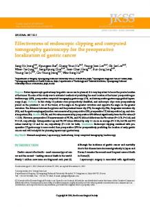
Figure 1. Chest CT on admission demonstrated a bilateral pneumothoraces.
The day after, an esophagography showed no leakage of the contrast agent (Gastrografin) from the esophagus (Figure 3b). On the fourth hospital day, a follow-up CT scan of the chest showed a significant decrease of the extraluminal air in the mediastinum and a regression of the neck emphysema. After two weeks, oral intake was resumed. The patient recovered well and was discharged a month later. Eight month later, the patient was asymptomatic and in good clinical conditions.
Discussion To our knowledge, the present report is the first description of hemostasis of bleeding esophageal varices and concomitant closure of esophageal perforation with clips in the same pa-
Figure 2. A, Esophagoscopy demonstrated 4 cm longitudinal perforation of the esophagus (white arrow). B, Endoscopic view of closure of perforation by five metallic clips.
Figure 3. A, Radiograph showing injection of contrast agent (Gastrografin) through endoscope, performed with an ERCP catheter, didn’t revealed extravasation. B, Esophagography showed no leakage of the contrast agent (Gastrografin) from the esophagus and confirmed the presence of the clip (white arrow).
tient. For both entities, early identification and treatment is mandatory. In fact, a delay of 24 hours can increase the mortality up to 50%9. Perforation of the esophagus is a relatively rare condition. The number of cases described in literature is small and it can be classified into three groups: iatrogenic, traumatic and spontaneous. Twenty to 40% of all cases of esophageal perforation are spontaneous10 and this condition is known as Boerhaave’s syndrome11. This syndrome is generally observed in patients aged 4060 years and it’s more frequently described in men with a ratio ranging from 2:1 to 5:112. In recent years, endoclip placement has been showing to be successful in the closure of spontaneous esophageal perforations (Boerhaave’s syndrome) (1), ingestion of fish bone (2), ingestion of metal hook (3), empyema (4), dilatation of esophageal stricture (5), achalasia (6) and anastomotic stricture of esophagojejunostomy (7). In addition, endoclips were showed to be successful in the closure of postoperative esophageal leakage13-15. The endoclips were reported to be successful in closing perforations from a few millimeters to 2 cm, with the larger ones requiring multiple endoclips and, in some cases, two or 3 separate sessions13. Shimizu et al16 suggest that the size limit for endoscopic clip application may be 1 to 1.5 cm in shortest diameter, because the width of an open clip is less than 1 cm. For esophageal leaks larger than 2 cm, the insertion of a covered stent should be preferred17. 705
M. Zippi, G. Traversa, R. Pica, G. Occhigrossi
Due to the small number of cases described in the literature there is no controlled trial in which surgical and conservative management are compared. Nowadays, endoscopic banding ligation is the first choice treatment in variceal esophagel bleeding. In this case we didn’t use this procedure to control variceal bleeding, firstly because the amount of fresh blood didn’t allow a clear vision and secondly because in the clinical suspect of esophageal laceration the rings cannot close a longitudinal perforation and don’t permit a second look if required. Thoracic surgery was considered only as a saving life procedure if the clinical status deteriorated due to the poor condition of the patient (HIV and cirrhosis) and the association of variceal bleeding. However, endoscopy must be executed carefully avoiding over insufflation of air that can worse pneumothorax or cause subcutaneous emphysema of the neck, as happened in our patient. We want to underline the importance of the early diagnosis, in fact, the prompt closure of the perforation certainly contributed to the good outcome of our patient. In conclusion, endoscopic clipping seems to be the most effective method for treating esophageal laceration, because of the possibility to observe directly the closure of the perforation and because avoids the necessity of the open surgery.
References 1) HURLSTONE DP. Successful endoscopic haemoclipping in Mallory-Weiss syndrome with concurrent closure of oesophageal perforation: further prospective evaluation of the technique is required. Scand J Gastroenterol 2002; 37: 866. 2) SHIMAMOTO C, HIRATA I, UMEGAKI E, KATSU K. Closure of an esophageal perforation due to fish bone ingestion by endoscopic clip application. Gastrointest Endosc 2000; 51: 736-739. 3) ABE N, SUGIYAMA M, HASHIMOTO Y, ITOH N, NAKAUR A H , I Z U M I S A T O Y, M A T S U O K A H , M A S A K I T, NAKASHIMA M, MORI T, ATOMI Y. Endoscopic nasomediastinal drainage followed by clip application for treatment of delayed esophageal perforation with mediastinitis. Gastrointest Endosc 2001; 54: 646-648.
706
4)
5) 6) 7)
8)
9) 10) 11) 12) 13) 14)
15) 16)
17)
VAN B ODEGRAVEN A, K UIPERS E, B ONENKAMP H, MEUWISSEN SG. Esophagopleural fistula treated en-
doscopically with argon beam electrocoagulation and clips. Gastrointest Endosc 1999; 50: 407410. RAYMER GS, SADANA A, CAMPBELL DB, ROWE WA. Endoscopic clip application as an adjunct to closure of mature esophageal perforation with fistulae. Clin Gastroenterol Hepatol 2003; 1: 44-50. WEWALKA FW, CLODI PH, HAIDINGER D. Endoscopic clipping of esophageal perforation after pneumatic dilation for achalasia. Endoscopy 1995; 27: 608-611. CIPOLLETTA L, BIANCO MA, ROTONDANO G, MARMO R, PISCOPO R, MEUCCI C. Endoscopic clipping of perforation following pneumatic dilation of esophagojejunal anastomotic strictures. Endoscopy 2000; 32: 720-722 MARTINEK J, KOVACOVA S, NOSEK V, VERNER T, VASICEK M, S PICAK J. Successful endoscopic treatment (clipping) of esophageal perforation during balloon dilatation in a patient with achalasia. Endoscopy 2008; 40: E61-E62. BLADERGROEN MR, LOWE JE, POSTLETHWAIT RW. Diagnosis and recommended management of esophageal perforation and rupture. Ann Thorac Surg 1986; 42: 235-239. KIM-DEOBALD J, KOZAREK RA. Esophageal perforation: an 8-year review of a mutispeciality clinic’s experience. Am J Gastroenterol 1992; 87: 1112-1119. BOERHAAVE H. Atrocis nec descripti prius morbi historia, secundum medicae artis leges conscripta. “Lugd. Bat. Boutesteniana” 1724. ZWISCHENBERGER JB, SAVAGE C, BIDANI A. Surgical aspects of esophageal disease. Am J Respir Crit Care Med 2001; 164: 1037-40. RODELLA L, LATERZA E, DE MANZONI G, KIND R, LOMBARDO F, CATALANO F, RICCI F, CORDIANO C. Endoscopic clipping of anastomotic leakages in esophagogastric surgery. Endoscopy 1998; 30: 453-456. MIZOBUCHI S, KUGE K, MAEDA H, MATSUMOTO Y, YAMAMOTO M, SASAGURI S. Endoscopic clip application for closure of an esophagomediastinaltracheal fistula after surgery for esophageal cancer. Gastrointest Endosc 2003; 57: 962-965. QADEER MA, DUMOT JA, VARGO JJ, LOPEZ AR, RICE TW. Endoscopic clips for closing esophageal perforations: case report and pooled analysis. Gastrointest Endosc 2007; 66: 605-611. SHIMIZU Y, KATO M, YAMAMOTO J, NAKAGAWA S, KOMATSU Y, TSUKAGOSHI H, FUJITA M, HOSOKAWA M, ASAKA M. Endoscopic clip application for closure of esophageal perforations caused by EMR. Gastrointest Endosc 2004; 60: 636-639. RAJU GS, THOMPSON C, ZWISCHENBERGER JB. Emerging endoscopic options in the management of esophageal leak. Gastrointest Endosc 2005; 62: 278-286.