© 2016. Published by The Company of Biologists Ltd | Development (2016) 143, 24-34 doi:10.1242/dev.124602
STEM CELLS AND REGENERATION
RESEARCH ARTICLE
Selection and dynamics of embryonic stem cell integration into early mouse embryos
ABSTRACT The process by which pluripotent cells incorporate into host embryos is of interest to investigate cell potency and cell fate decisions. Previous studies suggest that only a minority of the embryonic stem cell (ESC) inoculum contributes to the adult chimaera. How incoming cells are chosen for integration or elimination remains unclear. By comparing a heterogeneous mix of undifferentiated and differentiating ESCs (serum/LIF) with more homogeneous undifferentiated culture (2i/LIF), we examine the role of cellular heterogeneity in this process. Time-lapse ex vivo imaging revealed a drastic elimination of serum/LIF ESCs during early development in comparison with 2i/LIF ESCs. Using a fluorescent reporter for naive pluripotency (Rex1-GFP), we established that the acutely eliminated serum/LIF ESCs had started to differentiate. The rejected cells were apparently killed by apoptosis. We conclude that a selection process exists by which unwanted differentiating cells are eliminated from the embryo. However, occasional Rex1− cells were able to integrate. Upregulation of Rex1 occurred in a proportion of these cells, reflecting the potential of the embryonic environment to expedite diversion from differentiation priming to enhance the developing embryonic epiblast. KEY WORDS: Pluripotency, Embryonic stem cell, Mouse blastocyst, Chimaera, Live imaging
INTRODUCTION
Pluripotent stem cells provide a valuable system to explore intrinsic and extrinsic requirements for self-renewal in vitro. Murine embryonic stem cells (ESCs) are derived from epiblasts of late blastocysts (Boroviak et al., 2014; Brook and Gardner, 1997; Ying et al., 2008). Their potential to produce all tissues, including gametes, when injected into host embryos defines them as naive pluripotent (Nichols and Smith, 2009). ESCs can be propagated in medium containing foetal calf serum and leukaemia inhibitory factor (LIF) (Smith et al., 1988; Williams et al., 1988). In these conditions, developmentally advanced cells can be distinguished 1
Wellcome Trust-Medical Research Council Cambridge Stem Cell Institute, University of Cambridge, Tennis Court Road, Cambridge CB2 1QR, UK. 2 Department of Physiology, Development and Neuroscience, University of 3 Cambridge, Downing Street, Cambridge CB2 4BG, UK. Division of Stem Cells and Cancer, Deutsches Krebsforschungszentrum (DKFZ), Im Neuenheimer Feld 280, 4 Heidelberg 69120, Germany. Heidelberg Institute for Stem Cell Technology and Experimental Medicine (HI-STEM gGmbH), Im Neuenheimer Feld 280, Heidelberg 69120, Germany. *Author for correspondence (
[email protected]) This is an Open Access article distributed under the terms of the Creative Commons Attribution License (http://creativecommons.org/licenses/by/3.0), which permits unrestricted use, distribution and reproduction in any medium provided that the original work is properly attributed.
Received 22 March 2015; Accepted 10 November 2015
24
and cultures exhibit heterogeneous expression of markers for naive pluripotency, such as Nanog, Rex1 (Zfp42), Stella (Dppa3), Pecam1 and Klf4 (Chambers et al., 2007; Furusawa et al., 2004; Hayashi et al., 2008; Kalmar et al., 2009; Marks et al., 2012; Toyooka et al., 2008). A culture regime was subsequently developed based upon inhibition of the MEK/ERK pathway and GSK3, known as ‘2i’ (Ying et al., 2008). ESCs propagated in 2i exhibit more homogeneous expression of naive pluripotency markers (Nichols and Smith, 2009; Wray et al., 2010). Comparative profiling of ESCs propagated in serum/LIF versus 2i/LIF confirmed these differences (Marks et al., 2012). Generation of chimaeras from ESCs is used extensively to create transgenic mouse lines (Thomas and Capecchi, 1987) or to test the potency of putative pluripotent stem cells (Bradley et al., 1984). This is generally achieved by providing 8-20 ESCs to a host morula or blastocyst. An inoculum of fewer donor cells tends to produce chimaeras less efficiently (Beddington and Robertson, 1989). A probable explanation of this phenomenon is that only a proportion of the injected cells can integrate into the embryo. In support of this, a maximum of three ESCs per chimaera were observed to produce progeny contributing significantly to the adult animal (Wang and Jaenisch, 2004). Based upon experimental enrichment of ESCs expressing markers of naive pluripotency, it might be assumed that the ESCs permitted to contribute to the embryo are those residing in the naïve state (Furusawa et al., 2004; Toyooka et al., 2008). The capacity of the morula environment to alter the developmental trajectory of lineage-specified cells isolated from blastocysts was a surprising revelation (Grabarek et al., 2012). Whether the embryonic niche can exercise a similar effect on lineage-priming ESCs is currently unknown. Understanding how the environment can influence exit from pluripotency and its potential reversion is important for the design of in vitro differentiation protocols and interpretation of transplantation studies. The recent advances in transgenic reporters and live imaging open the possibility to explore how incoming ESCs incorporate into chimaeras and determine the fate of those that are rejected. In this study, we exploit two culture regimes: serum/LIF (SL) and 2i/LIF (2iL) to provide ESCs that are more (SL) or less (2iL) heterogeneous for markers of naive pluripotency. ESCs are injected into host embryos at the 8-cell stage. By tracking the process of chimaera formation, spatial and temporal trends for integration or exclusion can be uncovered. We also use a validated destabilised GFP reporter of the zinc finger protein Rex1 (Rex1-GFPd2), which correlates closely with naive pluripotency in vivo and in vitro (Pelton et al., 2002; Wray et al., 2011). This enables separation of SL-cultured ESCs into naive pluripotent (Rex1+) and developmentally advanced (Rex1−) populations prior to injection. In addition, GFP fluorescence enables assessment of the pluripotency status of integrating or excluded cells during chimaera formation.
DEVELOPMENT
Stoyana Alexandrova1,2, Tuzer Kalkan1, Peter Humphreys1, Andrew Riddell1, Roberta Scognamiglio3,4, Andreas Trumpp3,4 and Jennifer Nichols1,2,*
Our results uncover some interesting phenomena. Firstly, a large proportion of SL-cultured ESCs is dramatically eliminated by apoptosis within the first few hours after injection. Coincidentally, surviving ESCs appear to undergo compensatory proliferation. Secondly, 2iL-cultured ESCs continue to proliferate throughout the experiment, but undergo increased apoptosis during the second day of culture, in concert with the second lineage segregation event of the host embryo. Finally, although the majority of eliminated cells appear to have begun exit from pluripotency, Rex1− cells can occasionally upregulate GFP expression during development, but this is not a conditional prerequisite for integration into the epiblast. RESULTS ESCs cultured in 2iL out-perform those from SL conditions during chimaera formation
To test the hypothesis that ESCs in the state of naive pluripotency preferentially integrate into chimaeras, we used two alternative culture conditions. ESCs propagated in SL for at least four passages exhibited a substantial level of heterogeneity, both morphologically and by immunohistochemistry (Fig. 1A). Those expanded using 2iL formed more compact, rounded colonies and a higher proportion expressed pluripotency markers Sox2 and Nanog (Fig. 1B). ESCs labelled with ubiquitous tdTomato-H2B (Morgani et al., 2013) were used to facilitate tracking in chimaeras. This reporter localises to chromatin and therefore serves as an ideal identifier of nuclear fragmentation associated with cell death and separation of
Development (2016) 143, 24-34 doi:10.1242/dev.124602
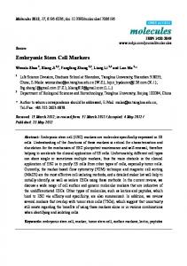
chromosomes during mitosis (Fig. S1A,B). Each pre-compacted 8cell embryo was injected with 3-7 ESCs (Table S1); pooled data from two experiments were separated into five groups based on the number of injected ESCs per embryo (3, 4, 5, 6 or 7 cells). Embryos isolated at mid-day on the third day after mating were assumed to approximate 60 h post coitum (hpc). They were tracked by live imaging from about 10 h post-flushing until the late blastocyst stage, 36-38 h later (Fig. 1C). The emergence of morphology consistent with cell death or division was recorded every 20 to 30 min. The first 10 h after flushing was not recorded to minimise fluorescence exposure and promote healthy development. However, analysis of movies revealed condensed and fragmented ESC nuclei assumed to have undergone apoptosis at the start of imaging (Fig. S1C), indicating that cell death occurred soon after injection. Apoptotic cell counts during 60-70 hpc were extrapolated by adding ESC deaths detected before imaging. To score the incidence of death, division and location of injected ESCs, chimaeras were filmed in 4D (three physical dimensions and time). Using Fiji (ImageJ) TrackMate manual tracking, a total of 46 embryos across two experiments were analysed: 18 SL-injected embryos ( producing 16 chimaeras) and 28 2iL-injected embryos (28 chimaeras). Analysis of time-lapse movies produced a dataset containing all ESC deaths and divisions scored temporally for each embryo. The numbers of viable ESCs were determined per embryo (Fig. 1D and Table S1). Injected ESCs and their progeny are referred to as ‘ESCs’ Fig. 1. Comparison of ESCs cultured in conventional versus ground-state conditions. Morphology and immunohistochemistry of ESCs cultured for 2 days in (A) conventional, serum/LIF (SL) or (B) ground-state, 2i/LIF (2iL) conditions. Left panels: bright field; second and third panels: immunoreactivity to Sox2 (green) and Nanog (white), respectively; right panels: overlay of Sox2 and Nanog. (C) Scheme for the experimental strategy: 8-cell embryos were injected with fluorescently labelled ESCs and chimaeras transferred to an immobilising grid for live imaging for 2 days. (D) Bar plot of the average numbers of ESC deaths, divisions and resulting viable ESCs by the end of captured development. (E) Plot of the average numbers of viable ESCs per embryo over time (hpc). Grey bars reflect s.e.m. between the curves of the five embryo groups ( profiles per embryo injected with 3-7 ESCs). See Table S1 for full data. Scale bars: 100 µm in A,B.
DEVELOPMENT
STEM CELLS AND REGENERATION
25
STEM CELLS AND REGENERATION
hereafter, for simplification. Comparison of the percentage increase of viable 2iL and SL ESCs across embryos at the end of culture revealed a statistically significant difference (Table S1; P=0.0265). Embryos injected with 2iL ESCs incorporated a higher number of viable ESCs (137.4±41.3%; mean±s.d.) compared with those injected with SL ESCs (34.9±20.2%; Fig. 1E). The survival rate of 2iL ESCs within the embryo remained significantly higher compared with SL ESCs for the duration of recorded development (Fig. 1E; P