Vol. 268, No. 8, Issue of March 15,pp. 5605-5614,1993 Printed in U.S.A .
THEJOURNAL OF BIOLOGICAL CHEMISTRY 0 1993 by The American Society for Biochemistry andMolecular Biology, Inc.
Sequence and Expressionof the Gene Encodingthe Corrinoid/IronSulfur Protein fromCZostridium thermoaceticum and Reconstitution of the Recombinant Protein to FullActivity* (Received for publication, October 13, 1992)
Wei-Ping Lu, Iunia Schiau, John R. Cunningham, and Stephen W. RagsdaleS From the Department of Biochemistry, Uniuersity of Nebraska, Lincoln, Nebraska 68583-0718
Thecorrinoid/iron-sulfurprotein(C/Fe-SP)from CO or CO, into cell carbon in acetogenic, methanogenic, and Clostridium thermoaceticum acts as a methyl group sulfate reducing bacteria and has been characterized in greatcarrier in the anaerobic acetyl-coA pathway ofCO est detail in Clostridium thermoaceticum. A reversal of this and COz fixation.Consisting of a small (-33 kDa) pathway appears to be responsible for conversion of acetic 1 acid to methane and COz by acetotropic methanogenic bacand a large (-55 kDa) subunit, the C/Fe-SP contains mol of cobaltin a corrinoid cofactor and 1 mol of teria or to HzS and CO, by acetate-grown sulfate reducing [4Fe-4SI2+/’+cluster/mol of crj3 dimer. Cobalt is the site bacteria. The reductive acetyl-coA pathway plays an imporof methylation, and the [4Fe-4S] center appears to tant role in the global carbon cycle since it appears to be the serve an electron transfer function. The genes encoding both subunits have been cloned previously andare major mode of CO, fixation under anaerobic conditions(1). The acetyl-coA pathway involves the conversion of 1 mol genes located within a gene cluster that includes other required for COz fixation by anaerobic bacteria.When of CO, to the methylgroup and 1 mol of CO, to the carbonyl group of acetyl-coA.Acetyl-coAthencan be utilized for the genes encoding the C/Fe-SP were expressed in Escherichia coli,the protein was found to be inactive.anabolic reactions or converted toacetic acid by coupling the We report the amino acid sequences of the large and hydrolysis of the thioester bond of acetyl-coA to the formasmall subunits of the C/Fe-SP based on the DNA se- tion of ATP via substrate-level phosphorylation. quences of the cloned genes. The [4Fe-4S] cluster was Corrinoids have been known to play an important role in found to be located in the large subunit. Although the acetate synthesis by acetogens for over two decades (2, 3); primary structural lattice cobamide for binding resides however the purificationof a corrinoidprotein tohomogeneity are required for and determination of its role in the acetyl-coA pathway is in the small subunit, both subunits formation of a stable cobamide-binding protein. Based rather recent (4, 5). The corrinoid protein was partially purion sequence comparisons with other [4Fe-4S]-contain- fied from C. thermoaceticum and found to be an (YP dimer ing proteins,3 of the 4 cysteine residues that serveas consisting of two subunits of 33 and 55 kDa and near stoichiligands to the iron sites in the cluster been have located. ometric content of corrinoid(4).Whenit was purified to The two subunits were independently overexpressed homogeneity, it was found to contain one 5-methoxybenzimin E. coli to a level of 30-5070 of cell protein; however, the resulting protein was inactive, lacked stoichiomet- idazolylcobamide and one [4Fe-4S] cluster per dimeric unit ric amounts of Fe-S cluster, and lacked cobamide. By and was named the “corrinoid/iron-sulfur protein” (C/FeSP)’ ( 5 ) . combining the recombinant subunits, unfolding them There aretwo important methyl transfer steps in acetylthe with urea, and refolding in the presenceof cobamide, iron, and inorganic sulfide, the resulting C/Fe-SP wasCoA pathway that involve the C/Fe-SP. It is the acceptor of found to contain stoichiometric amounts of cobamide the N-5 methyl group of 5-methyltetrahydrofolate (methyland [4Fe-4S] cluster and had spectroscopic and enzy- H,folate) in a reaction catalyzed by another protein, methylmatic properties similar to those of the native protein. transferase, forming enzyme-bound methylcobamide (EquaWe expect that the methods developed here may be tion l) (4). This reactionrequires reductive activation of both used for heterologous overexpression and reconstitu- the cobalt and [4Fe-4S] centers of the C/Fe-SP ( 5 , 6). The tion of other complex metalloenzymes. The C/Fe-SP important redox transitions between the 2+ and 1+states of was found to utilize with equalefficiency either vita- these centers have been found to be nearly isopotential with min BI2 or the natural cofactor 5-methoxybenzimida- formal equilibriumreduction potentials of “510 mV (6). zolylcobamide as a methyl carrier. Methylated C/Fe-SP also donates its methylgroup to carbon monoxidedehydrogenase (CODH) (Equation 2), forminga methylmetal species (7). The final steps in the assembly of Anaerobic bacteria fix CO, or CO via the reductive acetyl- acetyl-coA are catalyzed by CODH and occur via methylacetyl-CODH intermediates (1) CoA (Wood/Ljungdahl) pathway (1).This pathway converts CODH,CODH-CO,and (Equation 3). * This is publication 10160 from the Agricultural Research Division, University of Nebraska. The costs of publication of this article were defrayed in part by the payment of page charges. This article must therefore be herebymarked “aduertisement” in accordance with 18 U.S.C. Section 1734 solely to indicate thisfact. The nucleotide sequencefs) reported in thispaper has been submitted to theGenBankTM/EMBL Data Bank withaccession numberfs)LO7099 and L07100. $ T o whom correspondence should be addressed Dept. of Biochemistry, East Campus,University of Nebraska, Lincoln, NE 68583-0718. Tel.: 402-472-2943; Fax: 402-472-7842; Bitnet:
[email protected].
CH3-H4folate+ [Co+]-C/Fe-SP + CH3-[Co3’]-C/Fe-SP + Hlfolate CH3-[Co3+]-C/Fe-SP+ CODH + CH,-CODH + [Co+]-C/Fe-SP CHS-CODH + CO + COA+ CH&O-SCoA + CODH EQUATIONS 1-3 The abbreviations used are: C/Fe-SP, corrinoid/iron-sulfur protein; kb, kilobase(s); CODH,carbon monoxide dehydrogenase; IPTG, isopropyl-6-D-thiogalactopyranoside;PCR, polymerasechain reaction; PAGE, polyacrylamide gel electrophoresis; ORF, open reading frame.
5605
Protein
5606
Corrinoidllron-Sulfur
oligonucleotideprobes generated based on the N-terminal amino acid sequence of the purified large or small subunit of the C/Fe-SP (8). Nucleotide sequences were determined by the dideoxynucleotide method (14) for double-stranded DNA using Sequenase version 2.0 (United States Biochemical Corp., Cleveland, OH) and [ u - ~ ' P ] ~ A T P (ICN Biochemicals) according to the manufacturer's instructions. The formation of secondary structures in the DNA wasprecluded by 7-deaza-2'-deoxyguanosine-5'-triphosphate (7-deazathe use of dGTP) andperforming electrophoresis in acrylamide gels containing 45% formamide. In some cases, sequence information was obtained by priming the polymerase reaction with synthetic oligonucleotides. Codon translation and open reading frames were analyzed using the DNAsis program (Hitachi Software Engineering Co.). For protein and DNA homologysearches and codon probability (15) analyses, the Sequence Analysis Software package of the Genetic Computer Group was used (16). The probability plots (15) were based on the average codon usage of carbon monoxide dehydrogenase (17) and formyltetrahydrofolate synthase (18) from C. thermoaceticum. Purification and Determination of the Amino Acid Sequence and Composition of the C/Fe-SP-The C/Fe-SP was purified from C. thermoaceticum under strictly anaerobic conditions as described (5) at 16 "C. The subunits were separated and purified (8), and peptides were generated by trypsin digestion and purified by high performance liquid chromatography with a reverse phase C-18 column. The Nterminal sequences of the two subunits of the C/Fe-SP and thetryptic peptides, were done on an Applied Biosystems model 477A pulsed liquid-phase sequenator with an on-line amino acid analyzer at the Protein and Nucleic Acid Sequencing Facility, MedicalCollege of Wisconsin, Milwaukee. MATERIALS ANDMETHODS Overexpressionof the C/Fe-SP Genes-Overexpression of acsC was Strains and Cultivation-Strains and plasmids used in this study accomplished by insertion of the acsC-D region into a PET plasmid are listed in Table I. E. coli strain J M 109 was grownat 37 "C on YT behind a T7 RNA polymerase promoter as depicted in Fig. 1. Plasmid medium (9) supplemented with 0.1 mg/ml ampicillin. 5-Bromo-4- pCt946B (8) (Fig. 2) was digested with BamHI and Hind111 and chloro-3-indoyl-~-~-galactopyranoside and isopropyl-0-D-thiogalac- inserted into the BamHI/HindIII sites of pBluescript KS M13- to topyranoside (IPTG) were added for colony screening at final con- yield pBS946B.Using the polymerase chain reaction (PCR) technique centrations of0.03% and 1.6mM, respectively. c. thermoaceticum to mutate this gene, a1-kb PCR fragment was generated with pBS946B as the template. The two primers used were: a 21-base 3' was cultured at 55 "C under C02 asdescribed (10). Chemicals-Hydroxocobalamin, chymotrypsin, coenzyme A, and primer complementary to bases 1026-1047 downstream of the initia 5' mutagenesis primer, Dowex-5OW-H+ werepurchased from Sigma. (6s) CH3-H4folatewas ation codon of the large subunit, and &-base TAATCATGAAAGAAAGGAGctcACAcATATGCCTTTGAC. In obtained from Sapec S.A. Fine Chemicals (Switzerland). "CH3H4folate was purchased from Amersham Corp. Restriction enzymes the latter primer, an NdeI site (underlined) coinciding with the initiation ATG codon for the large subunit, and a Sac1 site (underand T4 DNA ligase were obtained from Promega. DNA Isolation and Sequencing-Plasmid DNA was prepared on a lined) were included. The mutated bases are shown as lowercase large scale by the alkaline lysis method (11) followedby gradient letters. A 175-base pair SacI-Not1 subfragment was inserted into pBS946B to generate the mutant, pBS946B55. The Not1 site is ultracentrifugation in CsC1-ethidium bromide (9). Sets of nested deletions were generated by exonuclease I11 digestion located 160 bases downstream of the initiation codon of the large (12) according to strategies described under "Results" with the Erase- subunit gene. The sequence of the 175-base PCR-generated insert A-Base kit following the manufacturer's instructions (Promega). Ex- was verified by DNA sequence analysis. Finally, the entire gene onuclease-treated DNA was excised from a gel, ligated, and trans- encoding the C/Fe-SP was inserted as an NdeI-Hind111fragment of formed (13) into E. coli strain JM109. Colonies were grown on YT pBS946B55 into the NdeIIHindIII sites of PET3a (19) to generate pET946B55. Plasmid pET946B55 was transformed (13) into the plates containing ampicillin, 5-bromo-4-chloro-3-indoyl-/3-~-galactopyranoside, andIPTG,and screened by colony hybridization expression strain BL21(DE1) from Novagen (19), which was grown (GenescreenPlusinstruction manual, Du Pont) with radioactive aerobically on LB-ampicillin medium at 37 "C. To induce production
The large and small subunits of the C/Fe-SP have been isolated and the genes encoding them have been cloned and found to reside in an -10-kb gene cluster which also encodes other proteins involved in conversion of CH3-H4folate, CO and CoA to acetyl-coA by C. thermoaceticum (8). We will refer to this gene cluster as the acetyl-coA synthesis (acs) gene cluster and, based on their location, the genes encoding the large and small subunits of the C/Fe-SP will be referred to as acsC and acsD. The coding regions for acsC and acsD are separated by a 2.5-kb spacer. AcsC and AcsD were previously expressed in Escherichia coli at levels from 1 to 5% of cell protein; however, the proteins were inactive and extremely labile (8), preventing further mechanistic studies via site-directed mutagenesis. We report the amino acid sequences of the two subunits, based on the DNA sequences of the cloned genes. After overexpressing acsC and acsD at levels between 30 and 50% of cell protein, the recombinant protein was reconstituted by unfolding with urea and refolding in the presence of cobamide, iron, and sulfide. The spectroscopic and enzymatic properties of the resulting C/Fe-SP were nearly identical to those of the native protein. Subunit localization of the prosthetic groups was accomplished, and 3 of the 4 cysteine ligands for the iron sites in the cluster were identified.
TABLE I Bacterial strains and plasmids Strains or vectors
Strains C. thermoaceticum E. coli K-12 HBlOl BL21 (DE3) Plasmids pCtJSA, pCt946A, pCt946B, pCt946C Bluescript KSBluescript SK+ PSK~, PSCP~, PSCP,, pET946B55 pETa3
Genotype
References
DSM 521
F' traD36 proAB lacZ-Z-M15/supE44 9 thi SupE44 hsdS20(r~-mf)recA13 lacy1 Lysogenic for XDE3; carries T7 the RNA 19 polymerase under lacUV5 control (F-ompT rB-mB-)
9
8
Strategene Strategene This work 19
5607
Corrinoidllron-Sulfur Protein
ColFe-S P
8 of Co/Fe-S
P
Sac1 81 Not1 Digest
Ligation
pBS@46B(55) T7
Hindll I
1
Ligation
FIG. 1. Strategy for construction of pET946B55. of the C/Fe-SP, 0.4 mM IPTG was added during the log phase of growth. Overexpression of acsD wasachieved by subcloning a ClaI-Hind111 fragment from pCt946B (8)into pBluescript vector KS M13-, giving pSCP, (Fig. 2) and transformed into E. coli strain HB101. Reconstitution and Purification of the Recombinant C/Fe-SP-Four hours after addition of IPTG, when the Awn,,, had reached -2.0, E.
coli strain BLZl(DE1) (pET946B55) cells were centrifuged, suspended in Buffer A (50 mM Tris-HC1, 5 mM dithiothreitol, pH 7.6), and lysed using a Heat Systems XL sonicator inside of a Coy anaerobic chamber. Centrifugation of the lysate at a setting of 4 (-1500 X g) for 30 min in an IEC Clinical centrifuge yielded a turbid cell-free supernatant. This supernatant was then loaded anaerobically into capped stainless steel centrifuge tubes and centrifuged in a type 35
5608
Corrinoidllron-Sulfur Protein
rotor in a Beckman L8-55M ultracentrifuge at 30,000 rpm for 1 h. The pellet fraction, which consists mostly of inclusion bodies, was solubilized in Buffer A containing -4 M urea. All steps of the reconstitution were performed a t 13 “C in the anaerobic chamber. The inclusion body fraction (-500 mg of protein) Then, was solubilized in 6 M urea ina final volume of-15ml. hydroxocobalamin (5 pmol) was added and, 15 min later, -250 ml of Buffer A was added to dilute the urea concentration to below 0.4 M. After incubation for a few hours, the solution was concentrated in an Amicon ultrafiltration unit with a YM30 membrane to -5 ml and centrifuged to remove precipitated protein. Unbound cobalamin was removed either by repeated buffer exchange with a Minicon B15 concentrator or by centrifugation through a Sephadex G-50 Penefsky spun column (20). To increase the level of the small subunit in the reconstitution mixture, E. coli strain HBlOl (pSCP3) cells, which overexpress acsD, weregrown toan absorbance of-2.0, centrifuged, and lysed as described above. The suspension was then centrifuged at 30,000 rpm in a type 35 rotor as described above, and about 10 ml (300 mg) of the soluble fraction was added to the above protein solution from pET946B55 containing hydroxocobalamin and 6 M urea. To maximize the amount of [4Fe-4S] cluster in the reconstituted protein, mercaptoethanol (30 pl) or dithiothreitol (300 pmol), Na2S (3 pmol), and FeCl, (3pmol) were also added. Reconstitution of the [4Fe-4S] cluster was modeled on a procedure for reconstitution of the ferredoxin from Clostridium posteurianum (21). After these additions, the final concentration of urea was 4 M. The urea was then diluted as described above. After incubation for a few hours, the reconstituted C/Fe-SP was then isolated by a modification of a published procedure to purify the native C/Fe-SP from C. thermoaceticum (5). The reconstitution mixture was applied to a 30-ml DEAE-Protein-Pak column (5 X 20 cm) at a rate of 40 ml/h and eluted with a gradient of 0-0.33 M NaCl in 50 mM Tris-HC1, pH 7.6, a t the same rate. Fractions containing the maximum amounts of C/Fe-SP were combined based on the intensity of red color of the cobamide, Western hybridization against the C/Fe-SP antibody (8), and SDS-polyacrylamide gel electrophoresis (PAGE). Ammonium sulfate was added to the combined fractions to 0.8 M, and the solution was loaded on a phenyl-Sepharose column (1 X 10) equilibrated with 0.8 M (NH&SO4 in Buffer A and eluted with a gradient of 0.6-0 M (NH4)2S04in Buffer A. Analytical Techniques-The C/Fe-SP and its subunits were identified in cell-free extracts and during purification by Western hybridization experiments which were performed with an immunoblot kit, following the instructions of the manufacturer (Bio-Rad). The membrane was reacted with polyclonal rabbit antibodies that had been previously prepared and purified against each of these proteins (8), then with an alkaline phosphatase goat anti-rabbit IgG conjugate, and finally stained with 5-bromo-4-chloro-3-indolyl phosphate and nitro blue tetrazolium. The C/Fe-SP was also detected by silver (22) or Coomassie Blue (23) staining of gels after SDS-PAGE (24). Quantitative amino analysis of each isolated subunit and of the native protein was performed on a Beckman 6300 analyzer with post column ninhydrin modification after hydrolysis in U ~ C U Oin 6 N HCl containing 0.02% mercaptoethanol for 20 h at 110 “C. Cysteine was determined as cysteic acid after performic acid oxidation (25). In a separate analysis, the subunits were separated by SDS-PAGE, transferred to a polyvinylidene difluoride Immobilon P membrane (Millipore, Bedford, MA) (26),stained with Amido Black, excised, and hydrolyzed. Protein-bound cobamide was detected essentially as described before (5). A few crystals of KCN were added to the protein solution, which was then heated at 90 “C for 2-3 min. The concentration of CN-cobamide was detected spectrophotometrically (€36, = 30.5 X lo3 M” cm”) (27). Protein concentration was measured using the Rose Bengal dye binding assay (28). Determination of the Actiuity of the C/Fe-SP-CODH (29), C/FeS P (5), and methyltransferase (30) were purified to homogeneity as previously described. The activity of the C/Fe-SP was quantitated in the total synthesis of acetyl-coA from CH,-H4folate, CoA, and CO essentially as described (31), except that CODH and methyltransferase were present at saturatinglevels and the C/Fe-SPwas in limiting amounts. The CODH preparation used was highly pure and completely devoid of the C/Fe-SP activity. Under these conditions, the specific activity of the synthesis can be expressed in terms of the C/ Fe-SP. The reaction was performed anaerobically in the dark at 55 “C in a final reaction volume of 50 pl in a 0.5-ml glass conical reaction vial (Wheaton) capped with a red rubber serum stopper. The reaction mixture contained CODH (100 pg), C/Fe-SP (5-40 pg), methyltrans-
ferase, (8 pg), CoA (50 nmol), and 14CH3-H4folate (10,000 dpm/nmol, 13 nmol of the active 6 S form) in 10 mM Tris-Maleate buffer, pH 6.5. Progress of the reaction was determined by Dowex-50W-H+ chromatography (31). Subunit Localization of the Fe-S Cluster and the Cobamide by SDSPAGE-A mild SDS-PAGE method was developed to separate the two subunits while retaining their cofactors. The composition of a 1mm SDS-polyacrylamide (10%) gel andthe running buffer were standard (24). Approximately 0.1 mg of C/Fe-SP from C. thermoaceticum was directly applied (without boiling or treatment with SDS) to a well (0.4 cm wide)of a Mighty Small I1 (SE 250) electrophoresis device (Hoeffer Scientific Instruments) and run at a constant voltage of 60 V and 16 “C for several hours. To visualize the red and brown colors associated with the different subunits in a form suitable for a black and white photograph, the gel was scanned by a Howtek color scanner using Adobe Photoshop software and converted to a gray scale file suitable for output to an LFR Lasergraphics slide printer. EPR Spectroscopy-EPR spectra were recorded on a Bruker ECS 106 spectrometer equipped with an Oxford ITC4 temperature controller and automatic frequency counter (Hewlett Packard, model 5340A). Spin concentrationswere measured by comparing the double integrals (using supplied Bruker software) of the spectra with those of a 1 mM copper perchlorate standard. Spectroscopic parameters are given in the figure legends. RESULTS
D N A Sequencing and Analysis T h e Large Subunit of the C/Fe-SP-In order to sequence acsC, the gene encoding the large subunit of the C/Fe-SP, a 1.8-kb BanHI-PstI restriction fragment frompCt946C (8) was ligated into the same sites in polylinker the of pBluescript SK+, yielding pSK, (Fig.2). Twenty separatedeletion clones, which were generated by exonuclease I11 digestion, were sequenced using the “reverse primer” to obtain thesequence of the non-coding strand. The sequence corresponding to the N terminus was determined by primer extension due to thelack of a suitable deletion plasmid. The sequence of the coding strand was obtained from the plasmid designated pKSR (Fig. 2), generated by subcloning the PstI fragment from pCt946A (8)into the PstI site of the Bluescriptvector, KS. A set of 19 overlapping deletion clones were sequenced as described above. Both DNA strands were sequenced from100 bases upstream to 327 bases downstream of the AcsC openreading frame (ORF) (Fig. 3A). Based on codon probability analysis (15), a single ORF for acsC encoding 446 amino acids is indicated. The calculated molecular mass of 49,060 daltons is within 11% of the value determined by SDS-PAGE (4). The amino acid sequences of three peptides isolated after trypsin digestion and of the N terminus ofAcsC were determined by Edman degradation analysis and are underlined in Fig. 3A. The initial methionine has apparently been removed from the mature protein since N-terminal sequence analysis gives proline as the first amino acid. The coding region ends with a single TAG stop codon. The amino acid composition of the isolatedsubunitdetermined by amino acid analysis(not shown) is in agreement within 10% with the deduced composition. A likely Shine-Dalgarno region (AGGAGTC) is located 7 base pairs upstream of the initiationcodon. Upstream of the coding region for acsC is sequence, a TTGGCCX17TAATCA,which is highly similar tothe sequence of the E. coli consensus “-35” and “-10” transcriptional control region, TTGAGAXZ1TATAAT. T h e Small Subunit of the C/Fe-SP--In order to sequence acsD, the gene encoding the small subunit, a ClaI-Hind111 fragment from pCt946B (8) was subcloned into Bluescript vector KS M13-, giving pSCP3 (Fig. 2). E. coli strain HBlOl (pSCPs) expresses acsD at levels of about 30% cell protein, approximately 10-fold greater than strains containing
5609
Corrinoidllron-Sulfur Protein s s I I
acsB
SSK P P
K
CS
II
~1
P K
PS SKP
y
11-
acsD
acsC pCt946A
I-??
KP
!
I
pCU9A
2H
pCt946B
t pCt946C
I
t
PSK,
I
I
I
acsE
PSCP,
I I
1 kb
FIG. 2. Arrangement of acsC (the gene coding for the large subunit of the C/Fe-SP) and acsD (the genecoding for the small subunit of the C/Fe-SP) in the ucs gene cluster, and the plasmids generated for overexpression and for DNA sequencing. Restriction sites indicated are: S, SucI; K , KpnI; P,PstI; C,CluI.
pCt946B. A set of 12 nested deletions was generated from pSCP3,and selected plasmids were isolated,purified, and sequenced with the “universal primer.”T o fill the gaps in the sequence left after sequencing the deletion clones, three additionalplasmids were generated (Fig. 2). A1.3-kb SmaI fragment from pCtJ9A (8) was subcloned into the Bluescript vector KS M13-, yielding pSCP,. Elimination of a Pst fragment from pSCP, yielded pPst, and deletion of a K p n fragment gave pKpn. The opposite strand was sequenced using six primers based on the sequence determination just described which hybridize to segments of the DNA approximately 150 bases apart. Both DNA strands were sequenced from381 bases upstream to 289 bases downstream of the acsD ORF (Fig. 3B). Consisting of 969 bases beginning with an ATG initiation codon and ending with a single TAA stop codon, a single ORF is predicted based on codon probability analysis. The amino acid sequences of one peptide isolated after trypsin digestion and of the N terminus of AcsD were determined by Edman degunderlined in Fig. 3B. As with AcsC, radation analysis and are theN-terminalmethionine was apparently removed since analysis of the sequence of AcsD by Edman degradationgives alanine as the first amino acid. The acsD gene encodes 323 amino acids with a calculated molecular mass of 35,530 daltons, which is within 8% of the value given by SDS-PAGE (4). Quantitative amino acid analysis of the isolated subunit (not shown) is in agreement with the composition deduced from theDNA sequence.A likely Shine-Dalgarno region (AGGAGTG) ends 8 base pairs upstream of the initiation codon and an upstream sequence, TTGATCX,TTCAAG, was found, that is similar to theE . coli consensus transcriptional control region.
Increased Expression of the C/Fe-SP The acsC-D region was placed behind a T 7 promoter by constructingtheplasmid, pET946B55, which was transformed into the expression strain BLal(DE3). More than 66% of the cell protein was found in the form of inclusion bodies, and immunological studiesindicatethatthe large subunit accountedfor over 50% of the inclusion body protein. Separate Western hybridization studieswere used to quantitate the large subunit at 30-50% and the small subunit at -1-10% of total cell protein. The level of expression of acsD was increasedto 20-30% of cell protein by construction (described above) of E. coli strain HBlOl (pSCP3). Reconstitution of and Purification of the Recombinant C/Fe-SP from pET946B55 We could not detect protein-associated cobamide either in the inclusion body or in thesoluble fraction of cells harboring
pET946B55, as found before in cells expressingacsC and acsD at low levels (8).In addition, protein-boundcobamide was not observed when cells were grown aerobically or anaerobically under a variety of different conditions,including ( a ) in media containing added cobalamins such as vitamin BI2,coenzyme BIZ, or hydroxocobalamin, ( b ) in media prepared with the soluble fraction of autoclaved cell-free extracts of C. thermoaceticum, or ( c ) using other growth media such as M9, ZB, or the standard growth medium for C. thermoaceticum. Thus, E. coli apparently cannot synthesize an active cobamide-containing C/Fe-SP. We were successfulin reconstitutingtheC/Fe-SP with cobamide by treatment of extracts of E. coli strain BL21(DE3) (pET946B55) with 6 M urea to unfold it and thenrefold it in thepresence of cobamide(see “MaterialsandMethods”). Binding of hydroxocobalamin was demonstrated by spectroscopic and activity measurements. The EPR spectrum of the unpurified protein solution revealed that -70% of the cobalamin was in the “base-off” conformation, in which benzimidazole isnotcoordinatedto cobalt. Thisconformation is characteristic of the protein-boundcobamide of the C/Fe-SP, since free hydroxocobalamin is base-off only at pH values below 3.0. Based on the UV-visible spectrum of the cyano derivative, approximately 1 nmol of cobalamin/mg of protein was found to beassociated. Most importantly, theunpurified reconstituted C/Fe-SP was found to be active in catalysis of the synthesisof acetyl-CoA fromCO, methyltetrahydrofolate, and CoA with a specific activity of -10 nmol of acetyl-coA formed/min/mg of protein. Although extracts from pET946B55 prepared as just described have C/Fe-SP activity, acsD is expressed at levels about “-fold lower than those of the large subunit. However, although the C/Fe-SP was not optimally reconstituted, generation of active C/Fe-SP containing cobamide and cluster was an achievable goal. Since low levels of acsD are expressed from pET946B55, optimization of the expression of the C/Fe-SP required addition of the small subunittothereconstitutionmixture. Since E. coli strain HBlOl harboring pSCP3 (generated for sequencing, see above) expresses acsD at levels of -20-30% of total cell protein (mostly in the soluble fraction), thesoluble fraction from these cells was added to thesolubilized inclusion body fraction from cells containing pET946B55. Then cobalamin, iron, and sulfide were added in the presence of urea and the C/Fe-SPwas refolded as described under “Materials and Methods.” The reconstitution mixture was directly applied to and chromatographed ona DEAE-ion exchange column.Furtherpurificationon a phenyl-Sepharose column resulted in a homogeneous recombinant C/Fe-SP, which migrated on an SDS-PAGE gel as the native protein from C.
5610
Corrinoidllron-Sulfur Protein a 1 66 1
ATC~CATTATGTGAGAGCCTGCACCCGGTTCCGGGGCTATCCCGGGGATTGGCCCCGGMCCGAG
65
CTGAATTMTCATGAAAGAGGAGTCAACAAATATGCC~GACGGGACTGGAGA~ACMGC
130 11
M P L T G L E I Y K Q
-
131 12
AGCTACCCAAAAAGAATTGTGGCGAGTGCGGGACACCCACCTGTCTGGCCTTCGCCATGAACCTG
196 34
A
L
P
K
K
N
C
G
E C
G
T
P
T
E
L
A
F
A
N
N
L
GCCTCCGGAAAGGCCAGCCTTGATTCCTGTCCGTATG~CAGATGCCGCCCGGGAGGCCCTGGA
S
G
K
A
S
L
D
S
S
P
Y
V
S
D
A
A
R
E
A
L
D
261 55
CGCGGCCGCGGCACCACCCATTGCCMGGTAGTCCTGGGCGCCGGGCCGACTGCCGTAG~TGG
326 77
GGGATGAGACGGMCTCTTCCGCCATGATAAACG~ACCATGAAACCGCCATTGCCATCCAG
391 99
A
A
D
A
E
A
T
P
E
P
L
I
F
A
R
K
H
V
D
V
K
L
R
G
F
A
Y
G
H
P
E
T
T
A
A
V
I
E
A
M
I
G
Q
S
D
N
L
S
S
E
E
L
K
A
K
V
E
A
I
N
G
L
N
260 54 325 76 390
90
G T T A G C G A C A A C T T G A G C A G T G M G M C T G M G G C T A A A G C T T
V
19 5 33
F
455 119
456 120
CGACCGGGTGGGCCAGCACTACACCATCCAGGCCATAGCCATCCGCCATGATGCCGATGACCCTG
521 142
CTGCTPTCAAGGCAGCGGTAGCCAGTGTAGCCGCCGCCGCTACCCAGTTAAACCTTGTCCl'l'ATGGCC 585
586 164
GATGATCCTGACGTATTAAAGGMGCCTAGCAGGAGTAGCCGACCGCMGCCCCT~ATATGC
651 185
CGCCACCGGCGCTAATTACGMGCCATGACCGCCCTGGCC~GAAAACAA~GCCCCCTGGCCG 7 15
7 16 207
TCTATGGTAACGGTCTGGAGGMCTGGCCGMCTGGTAGAT~TCGTTGCCCTGGGCCACAAG
781 229
CAGTTGGTCCTCGATCCCGGTGCCAGGGAGACCTCCAGGGCCATCGCGGA~CACCCAGATCCG
846 250
910 CCGCCTGGCCA~AAGAAACGTTTCCGTTCCGTTC~CGGTTATCCCATTATCGCC~A~ACTGCTG
A
271
911 272
CCAATCCATTAGACGAGGTACTCCAGGCAGTTMCTATGTGACCAAGTATGCTAGCTTGGTGGTT
975 293
976 294
TTACGCACCGATGCCAAAGACACCTGCTCCCCCTCPTGTCCGA
1041 3 15
CCCCCAGGTTCCCATCAGGGTAGAGGAGAAACTGAATGAAATCGGTGCCGTCMCGAGAATTCGC
1106 337
CGGTCTACGTMCCACCMCTPCTCCCCTGACCTATTACTCCGTCGAGGGCGAGATCGAGAGCACC
1171 359
MGATCCCCAGTTACCTGCTCTCGGTGGATACCGACGGACTGTCAGTCTTGACGGCCTATGCCGA
1236 380
TGGTMATTTGAAGCCGAGAAAATCGCCGCCGTTATGAAAAAGGTGGACCTGGACAATMGGl'l'A
1301 402
MCGCCACCGGATCATTATTCCCGGGGCTGTCGCCGTCCTGMGGGCAAACTGGMGA~MCT
1366 424
GGATGGGAAGTPATCGTTGGCCCCAGGGAAGCCAGCGGCATCGTGGCC~TGCCCGGGCCAACCT G W E V I V G P R E A S G I V A F A R A N L
1431 445
GGCTTCATAGGATAAACGGGAGGGGATGCCTGTGGATCAG~GCTGTCACC~ACCTGATA 1495 446 A S
1496
ATATCACCGTCCGGGTGGCGGCTGGTACCAGCATTATGGAGGCGGCCMCCAGGCCGGCCTGCCC
1560
CTGAAAAGCACCTGCGGCGGGGCCGGCACCTGTGGTCGCTGTGCCATCMGGTCCAGGAGGGGM
1625
1561
D R V G Q H Y T I Q A I A I R H D A D D P A A D
F
D
A
K
P
T
Y Q
G
N
V
L
D
K
E
L
L
L
A
V
N
D
Y
D
K
V
K
P
A
G
S
Y
H
K
V
R
V
T N
E
V
A
Y E
A
I
T
L
163
A
Q
L
I
A
L
T
N
E
L
T
N V
G
L
A
N
P
L
Q A
L
F
A
Y
L
E
D
R
E
V
N
I
G
V
K
A
W I
S
I
T
L
N
I
S E
R
I
P
V
N
E
A
L
Y
L
K
Y
N
Y
A
R
L L
Q
A
G
P
T
V
S
F
V
T
L
L
K
L
G
T
L E
S
E
A
A
A
S
L E
A
E
R
Q
A
T
R
H
F
A
M
F
V
N
A
A
E R
T
E
V
V
A
R
K I
E
G
K
E
S
E
P
A
T
A
Y
K
P
V
L
D
D V
Y
G
L
V
N
I
T
Q
V
L
A
R
P
N
P
A
A
V
L
L
D
G
L
R
A
D S
S
P
T
R
K H
F R
E I
A I
E I
K P
I G
A A
A V
V A
M V
K L
K K
V G
D K
L L
D E
N D
K L
V
650 184 206 780 228 845 250
1040 315 1105 336 1170 358 1235 380
K J P S Y L L S V D T D G L S G
520 141
K
T
1300 401 1365 423 1430 444
FIG. 3. DNA and amino acid sequences of the large and small subunits of the C/Fe-SP. The underlined amino acids show the protein sequence obtained from N-terminal sequencing of each subunit and some tryptic fragments. Heavy underlines highlight the cysteine residues in AcsC. The asterisks indicate the termination codons of the peptides.
thermoaceticum (Fig. 4). Phenyl-Sepharose could not be used as thefirst step in the purification scheme, since the proteins in the reconstitution solution precipitated upon addition of 0.7 M ammonium sulfate. Characterization of the Recombinant C/Fe-SP The EPR spectrum of the purified recombinant C/Fe-SP at 70 K (Fig. 5a) is identical to that of the native C/Fe-SP
from C. thermoaceticum ( 5 ) and is characteristic of low spin Co2+in a cobamide cofactor. Such spectra arise from an S = l / 2 system with gll 2.0 and g l 2.3 (32, 33). The splitting of the gll resonance into eight lines centered at g 2.0 results from hyperfine interaction between the unpaired electron and the cobalt nucleus which has a nuclear spin, I , of 7/2 (32). The strengthof this interaction is described by the hyperfine coupling constant, All, which was 140 G for both the recom-
-
-
-
CorrinoidfIron-Sulfur Protein
5611
b
1 66 1
G T ~ C ~ A ~ ~ C A A ~ C G T A T ~ ~ G C A G T ~ G 65C T A T C ~ C C C G A ~ C A ~ M G T T G A A A G G A G T G ~ A A A A T G G C C G ~ C A G A ~ A130 ~ ~ A ~ T A ~
M
A
V
O
I
L
R
D
R
10
S
CGAGCTGCCGTCCAGAAAGTGTCCTGCGCGCCACCAAAGACCAGGGGGGTACCCGCAGCCATAC 195
131 11
R
196 33
CATCGTCGTCGGTGGCGATGCTGCCCTGCCTTTCCACCATTTCGMGGAGAGATTGTCMCAGGC 260 I V V G G D A A L P F H H F E G E X V ~ R54 ~
261 55
CGGTAATCGGTATGGAAGTGCAGGATATCGTACCCGACTGGCCCGACGTTCTC!AMGATCCCTl'C
326 76
ACCGATGTTATTMTGAACCGGCGCTGGGCCCMAAGTGCGTAGCCGAGTATGG~C~
391 98
T A T C T A C C T G ~ ~ A C ~ ~ A C C C C ~ ~ G C ~ C C A ~ ~ 455 ~ C C A G ~ T A G
A
119
456 120
CTACTGTTAAAGAGGTCCTGCAGGCCGTGGGGGTACCCCTGGTAGTGGTAGGTTGCGGCGATGTG T V K E V L Q A V G V P L V V V G C G D V
520 140
521 141
GAAAAGGACCATGAGGTCCT~MGCAGTAGCCGAGGCTGCTGCCGGCGAGAATCTCCTCCTGGG
585 162
586 163
TAACGCTGAACAGGAAAACTATAAATCCCTMCGGCAGCCTGCATGGTCCACAAGCATAATATCA N A E Q E N Y K S L T A A C M V H K H N I I
650 184
651 185
TCGCCCGTTCGCCCC'IGGATATTAACATTTGTAAACAACTCAACATCCTGATCAA'iGAAA'iGAAC A R S P L D I N I C K Q L N I L I N B M N
715 205
716 206
~ C C C ~ G A ~ A T A T C G T C A T C G A C C C G T C C A T ~ C G G ~ G G T T A T ~ T A 780 ~GMTA~ 227 L P L D H I V I D P S I G G L G Y G I E Y S
781 228
~ ~ C G A ~ A T G G M C G C A T C C G T ~ G G ~ C C ~ C A G G ~ G A T M G A ~ C T C T C845 CA~CCGG F S I M E R I R L G A L Q G D X M L S M P V 249
846 250
TCATCTGCACCGTAGGCTATGAGGCCTGGCGCGCCAAGGAAGCCTCGGCACCGGTGAGCGMTAC 910
911 271
CCGGGCTGGGGTAAGGAAACCGAGCGTGGCATCCTCTGGGMGCCGTTACCGCCACTGCCCTGCT 975 292 P G W G K E T E R G I L W E A V T A T A L L
976 293
CCAGGCCGGCGCCCACATCCCCTCATGCGCCATCCGGAAGCCGTAGCCAGGGTGMGGAGMTA Q A G A H I L L M R H P E A V A R V X E N I
A
A
V T
I
D I
E
V
G V
Y
K
I
Q
M I
L
D
C
K
E N
K
B
T
V
V E
L
E
V
V
Q P
D
V
G
L
R G
G
L
Y
G
I R
A
E
E
A
V W
D
A
A
T
P A
P
V
W
K
D Q
E
A
R
D
W K
G
E
A
O
E C
A
A
K
G
D V
N
A
E
Y
G
A
J
D
L V
L
S
L
F
C
L
V
'
P
Q
N
P
H
D
D
E
A
S
%
V
G
S
R
L
S
A
A
T
V
H
A
E
G
L
E
G
Y
32
325 75 390 97
270
1040 314
1041 315
T C G A C C A G T T M T G G T G A G C A A C G C C A ~ ~ C C T G G T M T G G G C C T A C A T G ~ G G ~ G ' I1105 G D Q L M V S N A Y 335
1106
C T G G ~ ~ G ~ G ~ A G ~ A C ~ G G A ~ A G ~ A ~ ~ G C C C C G ~ ~ 1170 G C A G G G G A G C ~
1171
~ A ~ ~ G G ~ ~ T G C C C C ~ C G A T G ~ C ~ ~ G T G A G ~ G ~ G A A G 1235 C C ~ A T A T A T C C FIG. 8"coratinued
Localization of the (4Fe-4SJ Cluster in the Large Subunit binant and native C/Fe-SP. Double integration of the Co2+ and Its Reconstitution spectrum yielded 0.9 spins/dimeric C/Fe-SP. Quantitation of the concentrationof cobalamin by UV-visible spectroscopyof In addition to the comparison of the sequence of the cysthe cyano derivative also yielded a value of 0.9 mol of cobal- teine-rich region atthe N terminus of the large subunit amin/mol of dimer. (above), other results indicate that the [4Fe-4S] cluster is The EPR spectrum of the reduced recombinant C/Fe-SP located within this subunit. Wewere successful in separating at 10 K (Fig. 5b) reveals a n S = 1/2 rhombic signal resulting the subunitsof the C/Fe-SP without disturbing the[4Fe-4S] from the paramagnetic I+ state of a [4Fe-4S] cluster. The g cluster. The method involved performing SDS-PAGE under values are at 2.06, 1.93, and 1.85 with a very slow return of fairly mild conditions (see "MateriaIs and Methods" for deline, features characteristic of the tails). A brown and a red spot were observed on the gel. A the 1.85 feature to the base native C/Fe-SP as well ( 5 ) . Double integration of this EPR black and white image of the gel is presented in Fig. 6. By signal relative to a Cu2+ standard yielded 0.7 spins/mol of comparing the location of these spots with thatof the bands observed after staining the gel with Coomassie Blue, it was dimeric C/Fe-SP. clear that the upper andlower colored spots corresponded to The activity of the recombinant C/Fe-SPwas measured by the large and small subunitsof the C/Fe-SP, respectively. At following the rate of synthesis of acetyl-coA from CO, CoA, higher temperatures (>20 "C) or higher voltages (>I00 V), and methyltetrahydrofolate in the presence of CODH and the cobamide appearedto dissociatefrom theprotein, as methyltransferase. With limiting C/Fe-SP, there is a linear observed by the red color of the cobamide forming a diffuse relationship between the amountof the C/Fe-SP and the rate band at the top of the well. When the SDS concentration was of the synthesis. The recombinant C/Fe-SP has an activity decreased by one-third,or whenelectrophoresis was perof 200 +- 50 nmol of AcCoA formed min" (mg of C/Fe-SP)", formed at lower temperatures (-4 "C), the two subunits and comparabie to an activityof 250 & 50 for the native protein. the cofactors remained associated and appeared as a single
5612
Protein
Corrinoidllron-Sulfur
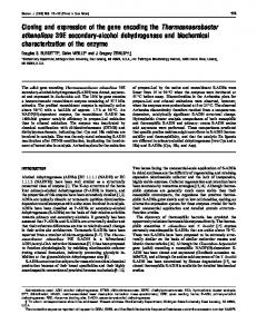
FIG.4. SDS-PAGE of the native and recombinant C/Fe-SP. 12 pg of the native protein (left lane) and 10 pg of the recombinant protein were used. The gel was stained with Coomassie Blue.
band on thegel as when nondenaturing PAGEwas used. The color of the inclusion body prepared from pET946B55, which expresses high levels of the large subunit, dissolved in urea (4-6 M) is brown, indicating the presenceof iron-sulfur clusters. After solubilizing the inclusion body fraction and reducing it with dithionite, a rhombic EPR signal with g values of 2.07, 1.93, and 1.86 wasobserved,which closely resembles that of the native C/Fe-SP (5). Spin quantitation of this signal yielded 0.3 spins permol of C/Fe-SP, assuming that the C/Fe-SP accounts for 50% of the protein in the inclusion body. This canbe increased to-0.6 spins/mol of C/ Fe-SP when the cluster is reconstituted by adding iron and 3000 34003200 3600 40003800 sulfide in the presence of urea as described under “Materials Gauss and Methods.” Thus, a significant fraction of the C/Fe-SP FIG.5. EPR spectra of the purified recombinant CFe-SP. molecules contain a [4Fe-4S] cluster before reconstitution; a, 20 mg/ml protein in 50 mM Tris-HCI buffer, pH 7.6, was used. however, reconstitution of the cluster was necessary for gen- EPR conditions were as follows: temperature, 70 K; microwave freeration of a protein with stoichiometric amounts of cluster. quency, 9.445 GHz; microwave power, 5 milliwatts; modulation amThese results supportlocalization of the iron-sulfur cluster in plitude, 9.96 G; modulation frequency, 100 kHz; receiver gain, 2 X lo‘. The A,.co is about 140 Gauss. b, protein reduced with dithionite the large subunit, since the concentration of the small subunit (0.5 mM) in the same buffer. The spectroscopic conditions were as in the inclusion body is approximately one-seventh that of describedin a except that temperaturewas 10 K and microwave the large subunit, insufficient to generate theobserved inten- power was 10 milliwatts. sity of the [4Fe-4S] EPR signal. sequenced. The sequence of the first 10 amino acids was Localization of the Binding Sitefor the Cobamide identical to thatof the small subunit. Although the above results suggest that thecobamide bindWhen E.coli cells containing pSCP3 werecultured inmedia containing 10 mM vitamin BI2, washed to remove unbound ing site islocalized in the small subunit, both subunits appear cobalamin, and lysed by sonication, thecobamide content was to be required either for optimal bindingof the corrinoid and/ elevated 3-fold relative to thatof the cells containingthe large or for stability of the protein. When the reconstitution mixture containing extracts from pET946B55 and pSCP3, urea, subunit or of a control culture lacking a plasmid. However, we were unable to furtherpurify a cobamide-containing pro- cobamide,iron, and sulfidewas chromatographedonthe tein since the small subunit from pSCP3 precipitated when- DEAE column, the large subunit eluted a t -0.1 M NaCl, the a t -0.15 M ever the temperaturewas raised above 10 “C. When the sub- holo-C/Fe-SP containing both subunits eluted units were separated by mild conditions of SDS-PAGE, one NaC1, and the small subunit eluted at -0.2 M NaCl. After can clearly observe that the red color of the cobamide comi- independently concentrating the three fractions, cobalamin was detected only in the fraction containing both subunits. grated with the small subunit (Fig. 6). When the C/Fe-SP was digested with chymotrypsin, thelarge subunit was extenDISCUSSION sively degraded before losing the small subunit. Mild conditions of SDS-PAGE resulted inloss of the brown color assoThe aminoacid sequences of the two subunits of the C/Feciated with loss of the large subunit and comigration of the SP were deduced from theDNA sequences of acsC and acsD. red color with the small subunit. T o ensure that the unde- We performed several analyses to ensure that thesequences graded small subunit wasbeingobserved and not a small are correct. 1) The sequences of both DNA strands were in proteolytic fragment of the large subunit, the red band was total agreement.2) The aminoacid sequences of interspersed transferred toa polyvinylidene difluoride membrane (26) and tryptic peptides of both subunits match those deduced from
Protein
Corrinoidllron-Sulfur
FIG. 6. Digitized scan of a nonstained gel after separation of the subunits using mild conditions of SDS-PAGE. The left lane contains molecular weight markers, and the right lane contains 100 pg of the C/Fe-SP. The two markers on either side of the two right lune have molecular subunits of the C/Fe-SP shown in the masses of 43 and 29 kDa. The large subunit retains the dark brown color of the [4Fe-4S] cluster and the small subunit retains the pink color of the cobamide.
the DNA sequences. That thededuced and determined amino acid sequences remained in alignment indicates that deletion or addition of bases (which would have changed the reading frame) did not occur during sequence analysis. 3) The molecular massesdeducedfrom the DNA sequencesagree with those determined by SDS-PAGE within experimental error (-10%). 4) The deduced andexperimentallydetermined amino acid compositions of both subunits are in agreement. 5) Codon probability plots for bothsubunitsindicatethe beginning of highly probable coding regions at the deduced N termini and endat thepredicted C termini. One of our objectives wasto increase thelevel of expression of acsC and acsD by E. coli. A low level of expression (1-5% cell protein) was found earlier and is not surprising in retrospect (B), since acsC and acsD have upstream sequences that show a high degree of similarity with the consensus E. coli Shine-Dalgarno and“-35” and “-10” regions. Methods such as primer extension analysis will be required to unambiguously define the transcriptional control signals fo? these genes. By placing the acsC-D region directly under control of the T7 promoter and transforming the genes into a strain of E. coli containing high levels of the T7RNA polymerase, AcsC was expressed at a level of 30-50% of cell protein. However, for unknown reasons, AcsD was expressed a t a 7-fold lower level. It is possible that the differential expression of these genes results from transcriptional or translational polarity effects. Another possibility is that, althoughacsC is controlled by the T 7 promoter, acsD could remain under control of C. thermoaceticum transcriptional and translational signals, as is the case with the genecloned in Bluescript or pUC (8). It is interesting that by subcloning acsD in Bluescript, generating pSCP3, the AcsD levels were -30% of cell protein, 5-6-fold higher than in the PET vector. Therefore, by using a two-
5613
vector system, levels of AcsC and AcsD were elevated to 3050% of cell protein levels. Whether acsC and acsD were expressed in E. coli a t low (8) or athigh levels, the resulting C/Fe-SPlacked cobamide and was inactive. Although E. coli can express other active cobalamin-containing proteins (34,35), it apparently cannot incorporate cobamide into the apo-C/Fe-SP and can only partly incorporate the [4Fe-4S] cluster. By unfolding and refolding thetwosubunitsinthepresence of cobamide, iron,and sodium sulfide, we were successful in reassembling the two subunits of the C/Fe-SP and reconstituting the cobamide and [4Fe-4S] centers. The resulting C/Fe-SPwas fully active as a methyl transfer protein in the synthesis of acetyl-coA from methyl-H,folate, CO, and CoA and exhibited EPR spectraof the cobamide and the [4Fe-4S] cluster that are indistinguishable from those of the protein isolated from C. thermoaceticum. Most characteristic of the cobamide in the C/Fe-SP is the existence of eightsingletscentered at gll -2.0, which unambiguously demonstrates that the benzimidazole base of the protein-boundhydroxocobalamin is uncoordinated to the cobalt. Since the recombinant protein was reconstituted with hydroxocobalamin, which possesses a dimethylbenzimidazole base instead of 5-methoxybenzimidazole, our combined results suggest that the substituentsof the benzene portion of the heterocyclic base have little influence either on activity or in controlling the propensityof the base to coordinate to cobalt. We have identified the likely residues involved in coordinating the [4Fe-4S] cluster. The arrangement of the 5 cysteines in AcsC is: C-X2-C-X4-C-Xl~-C-P-(X)ls~-C-P. Thus, the 1st 3 cysteines arelocated in a block near the N terminus in an arrangement that is reminiscent of that of bacterial ferredoxin, which has a highly conserved sequence: C-X2-CX2-,-C-X3-C-P (36,37). The cysteineresidues act as a ligands to the iron sites in the cluster. In AcsC, the arrangement is most similar to that seen in “Group 2” iron-sulfur proteins, which have the consensus sequence C-X2-C-X4-C(38). This group includes E. coli formate dehydrogenase (FdnH) (39), hydrogenase (HycB) (40), nitrate reductases (41,42), and the 21-kDa Fe-S subunit of the CODH from Rhodospirillum rubrum (38).Therefore, it ishighly likely that the1st 3 cysteine residues in the large subunit are involved in coordinating the iron in the [4Fe-4S] cluster. Further studies, including sitedirected mutagenesis, are under way to determine which of the remaining 2 cysteine residues provide the fourth coordination site in thecluster. An important goal has been to locate the cobamide binding site.This was not a straightforwardtasksinceboththe subunits and cobamide are tightly bound. In addition, independent expression of either acsC or acsD in E. coli was unsuccessful inyielding a stable cobamide-containing protein. Several results indicate that the small subunit of the C/FeSP plays a major role in binding the cobamide. First, E. coli cells that express acsD have elevated cobamide content relative to controls. Second, the red color of the cobamide was observed to comigrate withthesmallsubunit when mild separation of thesubunits by SDS-PAGE was achieved. Third, analysis of the predicted secondary structure of the region between residues 130and 280 of AcsD reveals a pattern of alternatinga-helicesandP-sheets(not shown) that is reminiscent of the region in methionine synthase predicted to contain its cobalamin binding site (35, 43). The common structural element among four cobalamin-binding proteins (BtuR, MutB, EalB, and MetE)was a pattern of alternating a-helices and P-sheets. Recently, this domain of methionine synthase was crystallized (44). Although the helix/sheet re-
5614
Protein
Corrinoidllron-Sulfur
6. Harder, S. R., Lu,W.-P.,Feinberg, B.A., and Ragsdale, S. W. (1989) gion extends over a larger area in the C/Fe-SP(-100 amino Biochemistry 28,9080-9087 7. Lu, W.-P., Harder, S. R., and Ragsdale, S. W. (1990) J. Biol. Chem. 2 6 5 , acids) than in the other BIZ-binding proteins (-50 amino 219A-RlRR ""acids),itis possible thatthedeterminants forcobamide 8. Roberts, D. L., James-Hagstrom, J. E., Smith D. K., Gorst C. M., Runquist, J. A., Baur, J. R., Haase, F. C., and Ragsdale, S. W. (i989) Proc. Natl. binding in the C/Fe-SP may differ from those of other BIZAcad. Sci. U. S. A. 86,32-36 containing proteins because benzimidazole is coordinated to 9. Sambrook, J., Fritsch, E. F., and Maniatis, T. (1989) in Molecular Cloning: A Laboratory Manual, 2nd Ed., Cold Spring Harbor Laboratory, Cold cobalt in all of these proteins except for the C/Fe-SP. Since Spring Harbor, NY the base of ATP and the benzimidazole moiety of cobamide 10. Ljungdahl, L. G., and Andreesen,d . R. (1978) Methods Enzyrnol. 53,.360have similar structures, both subunits of the C/Fe-SP were 11. 372 . e r S L , andKimmel, A. R. (1987) Guide to Molecular Cloning searched for the nucleotide binding site motif, zcflnique;, Academic Press, San Diego Henikoff. S. (1984) Gene (Amst.) 28.351-3.59 GXXGXGKXT, described for a number of ATP-binding pro- 12. 13. Hanahan, D.'(1983) J. Mol. Biol.' 166, 557-580 teins (45, 46). A similar motif, GXGKXT, was observed 14. Sanger, F., Nicklen, S., and Coulson, A. R. (1977) Proc. Natl. Acad. Sei. U. S. A. 74,5463-5467 between residues 272 and 277 of AcsD. The location of this 15. Staden, R., and McLachlan, A. D. (1982) Nucleic Acids Res. 10, 141-156 16. Devereaux, J., Haeberli, P., and Smithies, 0. (1984) Nucleic Acids Res. 12, motif supportsthe possibility that the helix/sheet region 2R7-39.5 ". ___ between residues 130 and 280 described above could be in- 17. Morton T. Runquist, J. A., Ragsdale, S. W. Shanmugasundaram, T., Wood, H: G., and Ljungdahl, L. G. (1991) J.' Biol. Chem. 266, 23824volved in binding cobamide. When thesequences of the AcsC 23838 and AcsD proteins were compared with those of several co- 18. Lovell, C. R., Przybyla, A., and Ljungdahl, L. G . (1990) Biochemistry 2 9 , 5687-5694 balamin-binding proteins, including BtuB (47), BtuR (45), 19. Studier, F. W. Rosenberg, A. H., Dunn, J. J., and Dubendorff,J. W. (1990) MutA and MutB (48), intrinsic factor (49), and two bacterial Methods E&yrnol. 1 8 5 , 60-89 Penefsky, H. S. (1977) J. Biol. Chern. 252,2891-2899 methionine synthases (35, 50), no regions of significant ho- 20. 21. Rabinowitz, J. (1972) Methods Enzymol. 24,431-446 22. Merril, C. R., Goldman, D., Sedman, S. A., and Ebert,M. H. (1981) Science mology were detected. In addition, no significantsequence 2 1 1 , 1437 homology was detected between either subunit of the C/Fe- 23. Reisner, A. H., Nemes, P., and Bucholtz,C. (1975) Anal. Biochern. 64,509516 SP and any other peptidesequence available in the National 24. Laemmli, U. K. (1971) Nature 227,680-685 Biomedical Research Foundation orSwiss Protein databases. 25. Hirs, C. H. W. (1967) Methods Enzymol. 1 1 , 59-62 Matsudaira P. (1987) J. Biol. Chem. 262,10035-10038 Our results indicate that, although cobamide binds toAcsD, 26. 27. Dawson, R.'M. C., Elliott, D. C., Elliott, W. H., and Jones, K. y.(1986) the native protein forms a morestable complex with cobamide Data for BzochemzcalResearch, 3rd Ed., Oxford Science Pubhcatlons, London than the isolated subunit. One possibility is that the cobamide 28. Elliott, J. I., and Brewer, J. M. (1978) Arch. Biochem. Biophys. 1 9 0 , 351357 binding site is shared between both subunits. Another possiRagsdale, S. W., and Wood, H. G. (1985) J. Biol. Chem. 260,3970-3977 bility is that the cobamide bindingsite is entirely on the small 29. 30. Drake, H. L., Hu, $.-I., and Wood, H.G. (1981) J. Biol. Chem. 256,1113711144 subunit, but in theabsence of the large subunit, the protein 31. Roberts, J. R., Lu, W.-P., and Ragsdale, S. W. (1992) J. Bacteriol. 1 7 4 , is unstable resultingin dissociation of cobamide. In the latter J. R. (1982) in Vitamin B,, (Dolphin, D., ed) Vol. 1, pp. 431-462, case, the large [4Fe-4S]-containing subunitwould be consid- 32. Pilbrow, John Wiley & Sons, New York ered to play an indirectrole in cobamide binding. 33. Pilbrow, J. R., and Winfield, M. E. (1973) Mol. Physiol. 2 5 , 1073-1092 34. McKie, N., Keep, N. H., Patchett, M. L., and Leadlay,P. F. (1990)Bzochem.
__"
Acknowledgments-We are grateful to Dr. Mary Graves (Dept. of Molecular Genetics, Hoffman La Roche) for synthesizing oligonucleotides for DNA sequence analysis. We thank Dr. Liane MendeMueller (Proteinand Nucleic Acid Sequencing Facility, Medical College of Wisconsin, Milwaukee, WI) for performing N-terminal amino acid sequence analysis of the two subunits of the C/Fe-SP and isolated tryptic fragments of the subunits and for performing amino acid analyses. We are grateful to the Center for Biotechnology Core Research Facilities, University of Nebraska-Lincoln, for DNA sequencing to verify the site-directedmutagenesis and synthesizing oligonucleotides for PCR. 1. 2. 3. 4. 5.
REFERENCES Ragsdale, S. W. (1991) Crit. Reu. Biochem. Mol. Biol. 2 6 , 261-300 Poston, J. M., Kuratomi, K., and Stadtman, E. R. (1964) Ann. N . Y. Acad. Sci. 112,804-806 Ljungdahl, L., Irion, E., and Wood, H. G. (1965) Biochemistry 4 , 27712780 Hu, S.-I., Pezacka, E., and Wood, H. G. (1984) J. Biol. Chern. 259,88928897 Ragsdale, S. W., Lindahl, P. A., and Munck, E. (1987) J. Biol. Chern. 2 6 2 , 14289-14297
*J. 269. 293-298 35. Baner'ee, R. V., Johnston, N. L., Sobeski, J. K., Datta, P., and Matthews, R. (1989) J. Biol. Chem. 264,13888-13895 36. George. D. G., Hunt, L. T.,Yeh, L. L.. and Barker. W. C. (1985) J. Mol. EL&' 22,20-31 37. Otaka, E., and Ooi, T. (1987) J. Mol. Euol. 2 6 , 257-267 38. Kerby R. L. Hong S. S. Ensign, S. A. Coppoc L. J., Ludden, P. W., and Roderts, d. P. (1b92) Bacteriol. li4,520415294 39, Berg, B. L., LI, J., Helder, J., and Stewart, V. (1991) J. Biol. Chem. 2 6 6 ,
d.
i.
99WL99WK """"
l l " " y
40. Bohm R. Sauter M., and Bock A. (1990) Mol. Microbiol. 4, 231-243 41. Blascd, F.: Iobbi, %.,Giordano, b.,Chippaux, M., and Bennefoy, V. (1989) Mnl 222.249-2.56 -. - - .. Opn. - .... Gonot - -. .- -. --, - .- -. . 42. Blasco F. Iobbi C. Ratoucbniak, J. Bonnefoy, V., andChippaux, M. (199b) k o l . Gek. Gknet. 222, 104-lil 43. Banerjee R. V. and Matthews, R. G. (1990) FASEB J. 4,1450-1459 44. Luchinsiy, C. Drummond, J. T., Matthews, R. G., and Ludwig, M. L. (1992) J. Mol. Biol. 222. 557-560 45. Lundrigan, M. D. and Kadner, R. J. (1989) J. Bacteriol. 171,154-161 46. Fry, D. C., Kuby,'S. A., and Mildvan, A. S. (1986) Proc. Natl. Acad. Sci. U. S. A. 83,907-911 47. Heller K., and Kadner, R. J. (1985) J. Bacteriol. 1 6 1 , 904-908 48. Marsd, E. N., McKie, N., Davis, N. K., and Leadlay, P. F. (1989) Biochem. J . 260.345-3.52 , ." 49. Diekgraefe, B. K., Seetharam B., Banaszak L Leykam, J. F., and Alpers, D. H. (1988) Proc. Natl. Acad. Sci. U. S. 85,46-50 50. Old, I. G., Margarita, D., Glass, R. E., and Girons, I. S. (1990) Gene (Amst.) 8 7 , 15-21
t.,
~
~~
A.