Tropical Medicine and Health Vol. 41 No. 1, 2013, pp. 21-25 doi:10.2149/tmh.2012-31 Copyright 2013 by The Japanese Society of Tropical Medicine
21
Short communication TMH Seroepidemiological
Study of Chagas Disease in the Southern Amazon Region of Ecuador
Angel G. Guevara1, Richard D. Atherton1, Michael A. Wauters1, Yosselin Vicuña1, Marcos Nelson2, Jose Prado3, Hirotomo Kato4, Manuel H. Calvopiña1 and Yoshihisa Hashiguchi1,5,6* ??
Received 14 December, 2012 Accepted 2 January, 2013
Published online 14 February, 2013
Abstract: To determine extent Medicine of Trypanosoma cruzi infection and/or transmission in the southern Amazon © 2013 Japanese Society ofthe Tropical region of Ecuador, three indigenous communities in the provinces of Pastaza and Morona Santiago were serosurveyed. ChagatestTM, Immunocomb®II and immunofluorescent (IF) assays were used. Among the 385 inhabitants examined, nine (2.34%) were seropositive for T. cruzi infection. Of the nine positive sera, four (44.4%) fall in the 10–19, one each in the 20–29, 30–39 and 40–49, and two in the 50–59 age groups. These results suggested the possible existence of an autochthonous active T. cruzi transmission in the region and provide the first serological evidence for T. cruzi infection in the southern province of Morona Santiago bordering Peru. Further studies are needed in these Amazonian provinces to ascertain the spread of T. cruzi infection in the area. Key words: Trypanosoma cruzi, Ecuador, Amazon, Chagas disease, seroprevalence
INTRODUCTION Chagas disease is a chronic, systemic, parasitic infection caused by infection of the protozoan parasite Trypanosoma cruzi (Kinetoplastida: Trypanosomatidae). T. cruzi was reported for the first time in the Amazon region of Brazil in 1909. The disease affects about 8 million people in Latin American countries, of whom 30–40% either have or will develop cardiomyopathy, digestive megasyndrome, or both [1, 2]. It is a neglected infectious disease in the tropics and an emerging health problem in the developed nations of Europe and the USA [3]. The parasite is transmitted by blood-sucking insects of the subfamily Triatominae (Hemiptera, Reduviidae). There is currently no effective vaccine and disease control therefore relies on domestic vector elimination through application of residual insecticides [4]. In Ecuador, cases of Chagas disease have been confirmed in eight provinces, i.e., Guayas, Manabí, El Oro, 1
Loja, Napo, Sucumbíos, Orellana and Pastaza [5–8]. It is estimated that 2.3 to 3.8 million people, out of the total Ecuadorian population of approximately 11 million, are at risk of infection by T. cruzi [5]. In 1991, sylvatic foci of Chagas disease were reported in Napo and Sucumbíos provinces, the first such cases in the Amazon region of Ecuador [9]. A further study in the Amazon region along the Napo River showed 15 of the 18 (83.3%) communities to be seropositive for T. cruzi infection, suggesting active transmission in an indigenous population in the northern Amazon region of Ecuador [6]. Moreover, another study described seroprevalence and risk factors for T. cruzi infection in the northern and central provinces of the Amazon region, including Sucumbíos, Napo, Orellana and Pastaza [7]. In 1997, Guevara et al. reported two cases of severe digestive pathology associated with chronic Chagas disease with megacolon, confirmed by polymerase chain reaction (PCR), in patients from the provinces of Loja and Morona Santiago [10]. However, the southern areas of the Amazon region of
Laboratorio de Parasitologia Molecular y Medicina Tropical, Centro de Biomedicina, Facultad de Medicina, Universidad Central del Ecuador, Quito, Ecuador 2 Hospital Vozandes Shell, Pastaza, Ecuador 3 Programa de Enfermedad de Chagas, Servicio Nacional de Erradicación de la Malaria (SNEM), Ministerio de Salud Publica, Ecuador 4 Laboratory of Parasitology, Department of Disease Control, Graduate School of Veterinary Medicine, Hokkaido University, Hokkaido, Japan 5 Programa Prometeo, Servicio Nacional de Educación Superior, Ciencia, Tecnologia e Innovación (SENESCYT), Ecuador 6 Department of Parasitology, Kochi Medical School, Kochi University, Kochi, Japan *Corresponding author: Parasitologia Molecular y Medicina Tropical, Centro de Biomedicina, Facultad de Medicina, Universidad Central del Ecuador, Quito, Ecuador Tel: +593-2-3228455 Fax: +593-2-3228454 E-mail:
[email protected]
22
Ecuador, mainly composed of Pastaza and Morona Santiago provinces, have not yet been studied in detail for the presence and prevalence of T. cruzi infection. The present seroepidemiological study, therefore, was conducted with a focus on Morona Santiago and neighboring areas in order to determine the status of the disease especially in the southern Amazon region of Ecuador. In this study, the inhabitants of three communities, i.e., Arajuno, Yuwientsa and Makuma, located in the southern provinces of Pastaza and Morona Santiago, were examined by means of three serological tests. Our positive results indicate that T. cruzi infection and/or transmission is gradually spreading from the northeast to the southeast in the Amazon region of Ecuador, especially in Pastaza and Morona Santiago provinces, very close to the northwest Amazon region of Peru.
Tropical Medicine and Health Vol.41 No.1, 2013
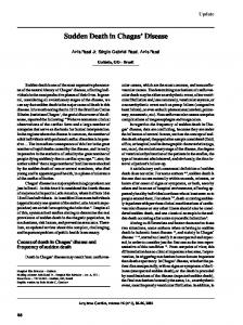
MATERIALS AND METHODS Study area The community of Arajuno is a rural settlement located in the eastern Ecuadorian province of Pastaza, while the communities of Yuwientsa and Makuma are situated in the southeastern jungle of Ecuador in the province of Morona Santiago (Fig. 1). A road was recently constructed into Arajuno, but Yuwientsa and Makuma remain accessible only on foot or by small aircraft. The entire area is a tropical lowland rainforest with a six-month rainy season (from November to May). The average temperature is 27°C (24– 31°C). The population in the study areas consists almost entirely of members of the indigenous race “Shuar” with a small number of migrants. Agriculture and hunting are the main economic activities of the inhabitants.
Fig. 1. Map of Ecuador, showing the three study areas: 1, Arajuno, Pastaza province; 2, Makuma, Morona Santiago province; 3, Yuwientsa, Morona Santiago province.
A.G. Guevara et al.
Study subjects Prior to the study, the protocol of the epidemiological and laboratory study was approved by the Bioethics Committee of Universidad Central del Ecuador, Quito, Ecuador. Specific permission was granted in writing from the community leaders, and when necessary, the study plan was explained in their primary language. In addition, the subjects as well as parents of children voluntarily consented to participate in this study after receiving a detailed explanation of the procedures by our research members (AGG & MHC). A cross-sectional study was conducted in August 2007 in Arajuno and in June 2008 in Yuwientsa and Makuma. The blood samples were obtained from the inhabitants living in the communities, irrespective of whether they were symptomatic or asymptomatic for Chagas disease. Children under the age of one year were excluded along with those who preferred not to participate. Blood sampling Blood collection was performed by venipuncture using disposable needles and vacuum 10 mL tubes (VACUETTE®, Greiner bio-one Gmbh, Austria). The samples were allowed to clot and then centrifuged for 15 minutes at 1000 g; the serum obtained was placed in a cryovial and stored at –20°C until use. Serologic analysis Blood samples were initially analyzed using an ELISA kit with T. cruzi recombinant antigens (ChagatestTM, Wiener lab, Argentina). Then, positives and suspected samples were subjected to an additional test (ImmunoComb®II, ORGENICS, Israel), as well as an immunofluorescent immunoassay (IFI) using cultured T. cruzi epimastigotes. Any sample testing positive on two or more of the individual assays was declared positive, according to WHO criteria [11]. ChagatestTM Protocol In brief, 200 μL diluent was added to each well of the supplied antigen-coated plate. This was followed by the addition of 10 μL of sample, including kit positive and negative controls to specific wells. The plate was covered and incubated at 37°C for 30 minutes. The plate was then washed five times with wash solution and incubated with 50 μL of conjugate per well for 30 minutes at 37°C. The wash step was repeated, and then 50 μL of substrate A was added to each well followed immediately by 50 μL of substrate B. The plate was then incubated for 30 minutes at room temperature and a drop of stop solution was added to each well. The plate was read at 450 nm (1° λ) and 620–650 nm (2° λ) in an ELISA reader (EIA 400 AT, Wittaker Bioproducts, Austria). According to the kit instructions, the optical density (OD) of negative controls should be below 0.150 and
23
positive controls should have an OD above 0.600. The samples were classified as positive, negative or doubtful. All the positive and doubtful samples were assayed again. Immunocomb® II, Chagas Ab Protocol Briefly, 10 μL of the sera samples was added to a comb pre-impregnated with T. cruzi antigen and allowed to react for 10 minutes at room temperature. The comb’s teeth were washed for two minutes and goat-human anti-IgG alkaline phosphatase conjugate was added and incubated for 10 minutes at room temperature. Washing solution was twice applied to the comb and a chromogenic substrate: 5-bromo-4chloride-3-indol-phosphate (BCIP) and nitroblue tetrazolium (NBT) was added for 10 minutes. Positive results were based on distinguishable color development in both the test and control areas of each comb tooth. Immunofluorescent Immunoassay (IFI) Protocol All positive sera for the previous described tests were tested using an in house IFI assay. Sera previously identified as positive or negative for T. cruzi were used as the positive and negative controls, respectively. Using standard 12-well glass slides, 20 μL of T. cruzi epimastigotes (1–2 ×10⁶ parasites per 1 mL) was fixed in each well with 4% formaldehyde solution in phosphate buffered saline (pH 7.2 PBS) and left at room temperature for 24 hours. Afterwards, 30 μL of each sample diluted 1:40 in PBS was added to the corresponding well and incubated for 30 minutes at 37°C. Each sample was tested in duplicate. After incubation, the slides were washed three times with PBS, and 30 μL of goat antihuman IgG fluorescein conjugate (Sigma, St. Louis, MO) diluted 1:100 in Evans blue was added to the specific wells and incubated for 30 minutes at 37°C. Each slide was washed a final time with PBS, and a drop of 1:1 PBS/ glycerol was added to each well before cover slide application. The slides were read with a fluorescence microscope under 100X power with oil immersion objective. Positive and negative controls were included for each assayed slide.
RESULTS Of the 385 individuals examined, nine (2.34% of the study subjects) tested positive for two or more tests in the three separate trials. This included four of the 246 in Arajuno, Pastaza province; five of the 95 in Yuwientsa, Morona Santiago province, and none of the 44 in Makuma, Morona Santiago province (Table 1). The seropositive cases were arranged by the age of subjects from Arajuno and Yuwientsa. Of the nine seropositive subjects, four fall in the 10–19, one each in the 20–29, 30–39 and 40–49, and two in the 50–59 year age groups. Thus, the subjects in the 10–19 to 50–59 year age groups showed T. cruzi seropositivity ranging from 1.64% (1/61 tested) to 7.69% (2/26 tested)
24
Tropical Medicine and Health Vol.41 No.1, 2013
Table 1. Seropositivity of inhabitants against T. cruzi infection by community Community Arajuno Yuwientsa Makuma
Province Pastaza Morona Santiago Morona Santiago
Subjects tested 246 95 44
Seropositivity (%) 4 (1.63) 5 (5.26) 0 (0.00)
Table 2. Prevalence of Trypanosoma cruzi seropositivity according to age group Age (Years) 1–9 10–19 20–29 30–39 40–49 50–59 60–69 70–79 80–89
No. examined No. positives 72 95 52 61 49 26 20 9 1
0 4 1 1 1 2 0 0 0
Seropositivity (%) — 4.21 1.92 1.64 2.04 7.69 — — —
(Table 2). Additionally, all the previously identified positive controls showed seropositivity for T. cruzi while none of the previously tested negative control sera showed T. cruzi positivity in any of the assays utilized.
DISCUSSION The present study supports and expands the findings of previous studies [6, 7], providing serological evidence for T. cruzi infection in Arajuno, Pastaza province and in Yuwientsa, Morona Santiago province, and suggesting the further spread of Chagas disease into the southern areas in the Amazon region of Ecuador. The results presented here are the first report to demonstrate the possible existence of T. cruzi infection in the province of Morona Santiago, suggesting that the disease reaches farther south in the Amazon regions than previously reported. It has been postulated that T. cruzi was introduced into the Amazon region due to the large migration of people from other provinces of the country, some of which are endemic for Chagas disease [6]. Within the last few years, Arajuno, a previously remote community accessible only by small aircraft, has been connected via roads to the larger town of Puyo, Pastaza province. The opening and development of Arajuno village has facilitated migration and trade in the area, which may be a possible cause for the introduction of T. cruzi infection. However, there is evidence of au-
tochthonous Chagas disease in humans from other provinces in the Amazon region of Ecuador [9]. The environment of Arajuno is ideal for the spread of T. cruzi, a fact that highlights the risk of possible transmission of T. cruzi in the region. Future studies involving the collection, identification, and examination of T. cruzi infection by the resident triatomine insect vectors in this area could thus yield valuable information. Our preliminary search for Triatomine bugs showed the presence of Trypanosoma positive Panstrongylus herreri (unpublished data), a significant T. cruzi vector in northern Peru [12], captured inside the house where one of the present seropositive subjects lives. The findings in Yuwientsa, Morona Santiago province, where the majority of positive cases were encountered in youths ≤19 years of age except one among five tested in the 50–59 year age group, suggest the active transmission of T. cruzi in this region. However, the identification of vector species and their habitat preference in the area are still unknown. In this study, only two communities in the entire province of Morona Santiago were tested for T. cruzi infection. Considerable future studies are necessary therefore to ascertain the true extent of the disease in the area. The present limited number of samples, i.e., only 44 subjects examined in Makuma, may account for the lack of evidence for T. cruzi existence in the area. The present seroepidemiological study indicated that human T. cruzi infection is prevalent in the studied areas and revealed the active transmission of the parasite in the southern areas of the Amazon region of Ecuador. Further investigations in the surrounding Amazon areas as well as the less accessible regions further to the southeast are necessary to ascertain the full extent of Chagas disease in the regions, from both serological and vector-entomological points of view. In addition, a detailed investigation of domestic livestock and sylvatic reservoir mammals could yield valuable information concerning the transmission and/or spread of the disease in the areas. It is important to mention that the present T. cruzi seropositive subjects were treated appropriately according to the guidelines of the World Health Organization and the Ministry of Public Health of Ecuador.
ACKNOWLEDGEMENTS We thank the US Fulbright Commission for the scholarship provided to MW. RA is a postdoctoral fellow from the London School of Tropical Medicine and Hygiene. The study was supported in part by Grants-in-aid for Scientific Research from the Ministry of Education, Culture, Sports, Science and Technology (MEXT) of Japan (Grant Nos. 2178027 and 23580424). We thank Enrique Mayorga for his help with the design of Figure 1.
A.G. Guevara et al.
25
CONFLICTS OF INTEREST The authors have no conflicts of interest connecting the work reported in this paper.
REFERENCES 1. Rassi A, Rassi A, Marin-Neto JA. Chagas disease. Lancet 2010; 375: 1388–1402. 2. Garod-Artal FJ, Gascon J. Chagas disease and stroke. Lancet Neurol 2010; 9: 533–542. 3. Basquiera AL, Sembaj A, Aguerri AM, Omelianiuk M, Guzmán S, Barral MJ, Caeiro TF, Madoery RJ, Salomone OA. Risk progression to chronic Chagas cardiomyopathy: influence of male sex and of parasitaemia detected by polymerase chain reaction. Heart 2003; 89: 1186–1190. 4. Panzera F, Dujardin JP, Nicolini P, Caraccio MN, Rose V, Tellez T, Bermúdez H, Bargues MD, Mas-Coma S, O’Connor JE, Pérez R. Genomic changes of Chagas disease vector, South America. Emerg Infect Dis 2004; 10: 438–446. 5. Aguilar MH, Abad-Franch F, Racines JV, Paucar AC. Epidemiology of Chagas disease in Ecuador. A brief review. Mem Inst Oswaldo Cruz 1999; 94: 387–393. 6. Chico MH, Sandoval C, Guevara AE, Calvopiña MH, Cooper PJ, Reed SG, Guderian RH. Chagas disease in Ecuador: Evidence for disease transmission in an indige-
7.
8.
9.
10.
11.
12.
nous population in the Amazon region. Mem Inst Oswaldo Cruz 1997; 92: 317–320. Grijalva MJ, Escalante L, Paredes RA, Costales JA, Padilla A, Rowland EC, Aguilar MH, Racines J. Seroprevalence and risk factors for Trypanosoma cruzi infection in the Amazon region of Ecuador. Am J Trop Med Hyg 2003; 69: 380–385. Ocaña-Mayorga S, Llewellyn MS, Costales JA, Miles MA, Grijalva MJ. Sex, subdivision, and domestic dispersal of Trypanosoma cruzi lineage I in southern Ecuador. PLoS Negl Trop Dis 2010; 4(12): e915. doi:10.1371/journal. pntd.0000915. Amunarriz MU, Chico ME, Guderian RH. Chagas disease in Ecuador: a sylvatic focus in the Amazon region. J Trop Med Hyg 1991; 94: 145–149. Guevara AG, Eras JW, Recarde M, Vinueza L, Cooper PJ, Ouassi A, Guderian RH. Severe digestive pathology associated with chronic Chagas disease in Ecuador: report of two cases. Rev Soc Bras Med Trop 1997; 30: 389–392. World Health Organization (WHO). Control of Chagas disease. Second report of the WHO expert committee. WHO Tech Rep Ser 2002; 905: 1–109. Cuba-Cuba CA, Abad-Franch F, Rodriguez JR, Vasquez FV, Velasquez LP, Miles MA. The triatomines of northern Peru, with emphasis on the ecology and infection by trypanosomes of Rhodnius ecuadoriensis (Triatominae). Mem Inst Oswaldo Cruz 2002; 97: 175–183.