Editorials: Cell Cycle Features
Editorials: Cell Cycle Features
Cell Cycle 11:19, 3529-3530; October 1, 2012; © 2012 Landes Bioscience
Signaling dynamics and embryonic development Aryeh Warmflash,* Eric D. Siggia and Ali H. Brivanlou Center for Studies in Physics and Biology and Laboratory of Molecular Vertebrate Embryology; The Rockefeller University, New York, NY USA
During embryonic development, signaling molecules convey positional information within the embryo and direct cells to adopt particular fates. A crucial question is how do the receiving cells of the embryo interpret these signals? While most work on this issue has focused on the biochemical and genetic dissection of the molecular circuitry of signal transduction, understanding signaling as a dynamic process is crucial to understanding development. During development, the timing and duration of signaling can play an important role in determining cell fate.1-3 The TGF-β pathway plays a crucial role during the development of organisms from fly to human and mediates events such as mesoderm induction and DV patterning in vertebrates.4 In a recent study, we focused on unraveling the dynamics of TGF-b signaling in two developmentally relevant contexts, the myoblastic C2C12 cell line and the early Xenopus embryo.5 Activation of TGF-b receptors by ligand binding leads to the phosphorylation of receptor-regulated Smads (R-Smads), binding of R-Smads to Smad4, translocation of this complex to nucleus and transcriptional activation.6 Understanding the dynamics of a signaling pathway is equivalent to answering the question: if a cell is exposed to ligand with some particular dynamics, what are the resulting dynamics of signaling activity in that cell? In our study, we focused on the simplest possible situation: cells were exposed to a step increase in ligand concentration, and we monitored signaling activity as a function of time. This situation presents two possibilities (Fig. 1). In the simplest, the output tracks the ligand so that under sustained stimulation, the output is similarly sustained. An alternative is known as an adaptive response;7,8 that is, the cells respond to the change in
ligand concentration but then “adapt” to the higher levels. Under this scenario, sustained ligand stimulation would produce a transient response. We created a clonal cell line of C2C12 cells expressing an RFP-Smad2 fusion protein (Smad2 is an R-Smad) to monitor the activity of the pathway in individual living cells. The results show that Smad2 is activated and remains localized to the cell nucleus as long as ligand is present in the medium. Thus, these results are consistent with the simple scenario outlined above. We then monitored TGF-bmediated transcriptional activity using both luciferase reporters of pathway activity and qRT-PCR measurements of representative target genes and found that both assays revealed transient pathway activity under constant stimulation. These results suggested that the pathway dynamics are adaptive, a finding contrary to a central tenant of the field, that R-Smad activity is synonymous with transcriptional activation. To begin to understand the discrepancy between R-Smad activation and transcriptional dynamics, we created a clonal cell line expressing a GFP-Smad4 fusion protein to report on the activity of this crucial R-Smad binding partner. In contrast to the results for the R-Smad, under constant ligand stimulation, Smad4 entered the nucleus transiently with dynamics that mirror the dynamics of transcription. Thus, taken together, our cell culture data suggest that R-Smad activity simply rises and falls with the level of ligand; however, the Smad4 response is adaptive, leading to a similarly adaptive transcriptional response. Next we turned to dissecting the Smad response in the Xenopus embryo. We used the future ectoderm (animal cap) as a model system, because it is a convenient
tissue for imaging and is responsive to both endogenous and exogenously provided TGF-b signals. Consistent with our cell culture data, we found that at the late blastula stage, R-Smads are homogenously localized throughout the tissue and reflect the ligand that the cells are exposed to. Surprisingly, we found that at this stage of development Smad4 localization is heterogeneous, strongly accumulated in the nuclei of some cells, while excluded from others. There was no apparent spatial pattern to the nuclear localization, and timelapse imaging revealed that it resulted from repeated, asynchronous, transient pulses of Smad4 nuclear localization. Injection into the embryo of mRNA encoding dominant-negative type I TGFb receptor Alk3 abrogated these bursts, showing that they are signal-dependent. Further studies are needed to determine whether the pulse rate varies in space or time during embryogenesis. If this is the case, it will be interesting to ask whether the number of pulses of Smad4 nuclear localization play a role in determining cell fate. It is interesting to speculate about the utility of these adaptive and pulsatile dynamics during embryonic development. The TGF-b pathway is used iteratively to guide a variety of cell-fate decisions. For example, TGF-b signaling is necessary to induce mesoderm, but then increasing levels of TGF-b specify more dorsal fates within the mesoderm.9,10 Adaptation could be a mechanism of recycling the same signaling pathway, because only changes in concentration trigger a response. Thus, with an adaptive response, there is no need to turn off the pathway at the level of R-Smads in between inductive events. More speculatively, the pulsatile response seen in the embryo perhaps evolved to maximize output from a core adaptive circuit. A drawback of adaptation is that
*Correspondence to: Aryeh Warmflash; Email:
[email protected] Submitted: 08/01/12; Accepted: 08/10/12 http://dx.doi.org/10.4161/cc.21964 Comment on: Warmflash A, et al. Proc Natl Acad Sci USA 2012; 109:E1947-56; PMID:22689943; http://dx.doi.org/10.1073/pnas.1207607109. www.landesbioscience.com
Cell Cycle
3529
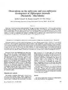
Figure 1. Schematics of simple and adaptive responses. Green lines represent ligand dynamics, and blue lines represent signaling pathway output.
the response to a step increase is limited to a fixed temporal window. It is possible that the embryo circumvents this limitation by presenting the ligand in a manner that triggers repeated bursts of transcription. Discerning whether this is the case is difficult and will require simultaneous measurements of ligand levels and transcriptional activity within the living embryo. References 1. Wacker SA, et al. Dev Biol 2004; 268:207-19; PMID:15031117; http://dx.doi.org/10.1016/j. ydbio.2003.12.022. 2. Tucker JA, et al. Dev Cell 2008; 14:108-19; PMID:18194657; http://dx.doi.org/10.1016/j.devcel.2007.11.004.
3530
3. Balaskas N, et al. Cell 2012; 148:273-84; PMID:22265416; http://dx.doi.org/10.1016/j. cell.2011.10.047. 4. Wu MY, et al. Dev Cell 2009; 16:329-43; PMID:19289080; http://dx.doi.org/10.1016/j.devcel.2009.02.012. 5. Warmflash A, et al. Proc Natl Acad Sci USA 2012; 109:E1947-56; PMID:22689943; http://dx.doi. org/10.1073/pnas.1207607109. 6. Massagué J, et al. Genes Dev 2005; 19:2783-810; PMID:16322555; http://dx.doi.org/10.1101/ gad.1350705. 7. François P, et al. Phys Biol 2008; 5:026009; PMID:18577806; http://dx.doi.org/10.1088/14783975/5/2/026009. 8. Behar M, et al. Biophys J 2007; 93:806-21; PMID:17513354; http://dx.doi.org/10.1529/biophysj.107.107516. 9. Hemmati-Brivanlou A, et al. Nature 1992; 359:609-14; PMID:1328888; http://dx.doi.org/10.1038/359609a0. 10. Green JB, et al. Cell 1992; 71:731-9; PMID:1423628; http://dx.doi.org/10.1016/0092-8674(92)90550-V.
Cell Cycle
Volume 11 Issue 19