of microbial DNA from soil for PCR. Article in Soil Biology and Biochemistry · August 1998. DOI: 10.1016/S0038-0717(98)00001-7. CITATIONS. 183. READS.
See discussions, stats, and author profiles for this publication at: https://www.researchgate.net/publication/223657380
Simple and rapid method for direct extraction of microbial DNA from soil for PCR Article in Soil Biology and Biochemistry · August 1998 DOI: 10.1016/S0038-0717(98)00001-7
CITATIONS
READS
183
228
2 authors, including: Penny R Hirsch Rothamsted Research 151 PUBLICATIONS 6,731 CITATIONS SEE PROFILE
Some of the authors of this publication are also working on these related projects:
Brazilian Microbiome Project View project
All in-text references underlined in blue are linked to publications on ResearchGate, letting you access and read them immediately.
Available from: Penny R Hirsch Retrieved on: 03 November 2016
PII:
Soil Biol. Biochem. Vol. 30, No. 8/9, pp. 983±993, 1998 # 1998 Elsevier Science Ltd. All rights reserved Printed in Great Britain S0038-0717(98)00001-7 0038-0717/98 $19.00 + 0.00
SIMPLE AND RAPID METHOD FOR DIRECT EXTRACTION OF MICROBIAL DNA FROM SOIL FOR PCR D. W. CULLEN and P. R. HIRSCH* Soil Science Department, IACR-Rothamsted, Harpenden, Herts AL5 2JQ, U.K. (Accepted 22 December 1997) SummaryÐA simple and rapid procedure for direct extraction of DNA from soils was developed to yield DNA of a high purity and quality suitable for ampli®cation using the polymerase chain reaction (PCR). Co-extracted humic material from soil was a major contaminant of DNA and methods were devised to overcome this problem. Oligonucleotide PCR primers were designed to detect and monitor a genetically-modi®ed (GM) Rhizobium leguminosarum bv. viciae strain RSM2004 (marked with Tn5) which had become established in Rothamsted ®eld soils. The key steps of the procedure were alkalineSDS buer assisted lysis of indigenous soil bacteria in a bead-beater and the puri®cation of extracted DNA by separate PVPP and Sephadex G-75 spin-column chromatography. The mean yield from Rothamsted soil was 252 1.7 mg crude DNA gÿ1 wet soil (i.e. 20 mg gÿ1 dry soil), sheared to fragment sizes of about 22±25 kb. The recovered DNA was easier to purify and of a higher quality, as veri®ed by PCR ampli®cation of a 442 bp target sequence of Tn5, than DNA extracted by a hot-SDS lysis method. The detection limit was demonstrated to be one culturable cell of RSM2004 (i.e. a single copy of Tn5) 10 mgÿ1 soil against a background of 107 diverse non-target bacteria. # 1998 Elsevier Science Ltd. All rights reserved
INTRODUCTION
However, the polymerase chain reaction (PCR), in which oligonucleotide primers with homology to speci®c DNA sequences ¯anking a target region are used to exponentially amplify the target, is much more sensitive. In theory, only one target DNA sequence per reaction is needed, although the target-to-background ratio aects the eciency of the reaction. To isolate bacterial DNA from soil (after dispersal in buer), the cells can be separated by dierential centrifugation, recovered, lysed and DNA puri®ed (Stean et al., 1988). Alternatively, the cells can be lysed in situ followed by the recovery and puri®cation of DNA (Tsai and Olson, 1991; Picard et al., 1992; Selenska and KlingmuÈller, 1992; Trevors, 1992; More et al., 1994). Lysis of bacterial cells during these direct procedures involves one or several of the following treatments: heat, detergents, enzymes (lysozyme, pronase, proteinase K), phenol, guanidine thiocyanate, EDTA, freeze±thaw cycles, sonication, microwave-heating and bead-beating. Direct extraction of microbial DNA from soils or sediments is the more commonly used approach (Trevors, 1992) because higher yields of DNA are usually recovered (Stean et al., 1988), more samples can be processed in a shorter time and the biased recovery of cells that are easily dislodged during cell separation procedures is avoided. The major disadvantage is that the lysis treatments result in the co-extraction of other organic soil com-
The application of molecular genetics to soil microbiology has great potential. Genetic material is the ultimate diagnostic tool for detecting particular microbes, an important consideration since only a small proportion (0.001 to 0.3%) of the bacteria from natural habitats can be cultivated on laboratory growth media (Amann et al., 1995). Selective media are not available for many groups of microbes and if numbers are low they may be impossible to detect by conventional plate culture, swamped by more numerous and faster-growing organisms. This can hinder studies of the survival of bacterial inoculants after addition to soil and is a particular problem for monitoring GM organisms after environmental release. Methods which can circumvent the bias and underestimation of bacterial types and numbers isolated by cultivation are valuable in microbial ecology to determine the roles and functions of microbes and study microbial community structure in natural environments. Hybridization of DNA extracted from the soil microbial community to labelled DNA probes for particular genetic markers lacked sensitivity, with a maximum detection limit equivalent to one bacterium with a single marker sequence in a background of 105 dierent strains (Holben et al., 1988). *Author for correspondence. 983
984
D. W. Cullen and P. R. Hirsch
ponents, particularly so-called humic and fulvic acids. The humic materials present in DNA extracts can inhibit restriction endonucleases (Tebbe and Vahjen, 1993), interfere with DNA±DNA hybridization experiments (Stean et al., 1988) and inhibit Taq DNA polymerase (Tsai and Olson, 1992a; Tebbe and Vahjen, 1993). Puri®cation of DNA is therefore a critical step. The original direct extraction methods and modi®ed versions used caesium chloride±ethidium bromide (CsCl±EtBr) equilibrium density gradient ultracentrifugation or hydroxyapatite chromatography puri®cation steps (Ogram et al., 1987; Stean et al., 1988). However, the more recent extraction procedures have avoided such laborious techniques by using programmes based on one or a combination of steps including: size-exclusion chromatography (gel ®ltration) with Sephadex spin-columns (Tsai and Olson, 1992b; Flemming et al., 1994) and commercial ion-exchange chromatography columns (Tebbe and Vahjen, 1993; More et al., 1994; Tas et al., 1995); glassmilk puri®cation and selective precipitation for removal of proteins and humic substances (Smalla et al., 1993) and agarose gel electrophoresis (Knaebel and Crawford, 1995). Water insoluble polyvinylpolypyrrolidone (PVPP) has been added during direct extraction to remove humic substances (Holben et al., 1988; Stean et al., 1988) and water-soluble polyvinylpyrrolidone (PVP) was incorporated in agarose gels to prevent the migration of humic materials with crude DNA (Young et al., 1993). PVPP (or PVP) removes those humic acids with phenolic groups from crude DNA extracts via hydrogen bonding and the formation of a PVPP±phenolic complex. Our objectives were to optimise the methods used for the direct extraction and puri®cation of DNA from soil together with PCR ampli®cation of the DNA, to monitor survival of RSM2004, a genetically-modi®ed rhizobial inoculant which has become established in the ®eld at Rothamsted since its release in 1987 (Hirsch and Spokes, 1994). This was achieved by the speci®c detection of a marker sequence inserted in the genome of RSM2004, a single copy of transposon Tn5. Three soil extraction methods: hot-SDS lysis (Selenska and KlingmuÈller, 1991), freeze±thaw lysis and the physical disruption of cells in a bead-beater, were compared on the basis of yield and quality of recovered DNA. Techniques to purify the DNA from contaminating humic substances were developed, leading to the development of a simple, rapid and reliable method for direct extraction of DNA from soil. MATERIALS AND METHODS
Soil characteristics and sample collection The soil at the release site, the Rothamsted ``Garden Plots'', is a silty clay loam (Batcome
series, loam over clay with ¯ints, overlying chalk; otherwise known as a ®ne loamy chromic luvisol with 32% clay, pH 7.5; J. Catt, personal communication). Ten separate soil samples of ca. 50 g were collected from the ®eld plot at IACR-Rothamsted. Samples were pooled, mixed and sieved (2 mm mesh) on return to the laboratory then used immediately for DNA extraction. To estimate soil moisture, sub-samples were dried overnight at 808C: moisture contents remained within a range of 10± 20%. Direct extraction of microbial DNA from soil Three dierent methods were tested to evaluate DNA extraction in terms of yield and purity of recovered DNA; all used 120 mM sodium phosphate buer pH 8.0 (SPB) for the initial extraction; extracts were stored at ÿ208C. Hot-SDS lysis (Selenska and KlingmuÈller, 1991) involved incubation of 1±2 g soil samples in SPB with 1% SDS at 708C, centrifugation, two subsequent extractions from the soil pellet, PEG precipitation of DNA from the supernatant and puri®cation by CsCl±ethidium bromide density gradient centrifugation. We introduced a modi®cation, adding 300 mg dry PVPP powder to each 5 ml soil suspension after the ®rst incubation, before centrifugation. For freeze±thaw lysis, soil samples (1.0 g wet wt) were suspended in 3 ml SPB, 1% SDS and mixed before they were frozen in liquid N2 (ÿ1968C, 2 min) and then thawed in a 708C water bath (5 min). After 3 successive freeze±thaw cycles, each sample was centrifuged at 2800 � g for 15 min at 108C, the supernatant was collected, kept on ice until subsequent puri®cation. Mechanical lysis using a bead-beater was performed after suspending soil samples (1.0 g wet wt.) in 3 ml SPB, 1% SDS in 4 ml capacity snap-top polypropylene tubes containing 1.0 g glass beads of various sizes (0.5, 2.0 and 3.0 mm dia, approximately equal weights of each size). Each tube was then ®tted to a bead-beater (Mikro-dismembrator II, B. Braun, Melsungen, Germany) and shaken at an amplitude of 5 mm for 5 min according to the manufacturer's instructions. Tubes were then centrifuged at 2800 � g for 15 min at 108C and the supernatant was transferred to a clean tube. EDTA (500 mM, pH 8.0) was then immediately added to each extract to 100 mM, followed by a 1/10 volume of potassium acetate (5 M, pH 5.5). Samples were incubated on ice for 20 min and then centrifuged at 10,000 � g for 5 min at 48C to remove precipitated proteins, humic substances, SDS, and RNA. The DNA in the supernatant fraction was precipitated with 1 volume of isopropanol for 1 h at room temperature. DNA was recovered by centrifugation at 10 000 � g for 5 min at 48C and pellets were resuspended in 200 ml TE buer (10 mM Tris±HCl pH
Extraction of microbial DNA from soil
985
8.0, 1 mM EDTA). Soil DNA extracts were puri®ed using PVPP and Sephadex G-75 spin-column chromatography as described in Section 2.3. During optimization, conditions were varied by extraction of 1 or 2 g samples, in SPB with 2% SDS, for 5 or 10 min periods with amplitudes of 5± 10 mm.
thaw lysis procedures. A soil suspension was made by adding 10 g soil to 30 ml SPB, 1% SDS (total volume 40 ml) and after vigorous mixing for 2 min, 4 ml aliquots of soil slurry (equivalent to 1.0 g soil) were subjected to dierent lysis procedures. Agarose gel electrophoresis (0.8%) enabled semi-quantitative, qualitative examination of DNA (Fig. 3).
DNA puri®cation by spin-column chromatography
Microscopic evaluation of cell lysis
Crude extracts of soil DNA were puri®ed using two Bio-Spin polypropylene columns (0.64 � 5 cm; Bio-Rad Laboratories, U.K.) that contained waterinsoluble polyvinylpolypyrrolidone (PVPP; Sigma, U.K.) and Sephadex G-75 (Fine Grade; Pharmacia Biotech, U.K.). For PVPP spin-columns, Bio-Spin columns were ®lled with dry PVPP powder to a height of 26± 30 mm (1 ml vol.). A sterile 1.5 ml capacity microcentrifuge tube without a cap was ®tted to the bottom of each column before they were placed into 50 ml polypropylene tubes. Columns were conditioned by sequential addition of 0.5 ml and 0.25 ml sterile distilled water (dH2O), each addition followed by 5 min centrifugation in a swing-out rotor at 2000 � g; residual water was removed by a ®nal spin. Crude DNA (up to 200 ml) was added to the top of each column and the puri®ed eluate collected in a new sterile 1.5 ml tube after two successive centrifugation steps at 2000 � g for 5 min at 108C. PVPP spin-columns could be prepared several months in advance and stored at 48C after the conditioning step. Sephadex G-75 spin-columns were used to introduce size exclusion chromatography as the ®nal step in the puri®cation of soil extracted DNA. Sephadex G-75 powder was equilibrated in TE buffer and then autoclaved. Bio-Spin columns were ®lled with ca. 1 ml G-75 slurry to obtain a column with a height of 12±15 mm. A sterile microcentrifuge tube was then ®tted to the bottom of each column before inserting into a 50 ml polypropylene tube. Residual TE buer from each column was removed by centrifugation at 1600 � g for 6 min in a swing-out rotor. Each column was conditioned by the addition of 100 ml sterile dH2O and centrifuged as above. DNA extract (200 ml) was loaded slowly onto the top of each semi-dried Sephadex column, the beads were allowed to expand for 5 min, then the columns were centrifuged at 1600 � g for 6 min at 108C on two successive occasions and the puri®ed DNA was collected in a new sterile microcentrifuge tube. Sephadex G-75 spin-columns could be prepared several weeks in advance, stored at 48C and conditioned with sterile dH2O before use.
An acridine orange direct count (AODC) was used to enumerate the total number of cells in soil samples and to determine the extent of cell lysis during direct extraction procedures (Smith and Stribley, 1994).
Variations to direct lysis procedures The yields of DNA recovered from soil samples were compared after extraction by various modi®cations to the hot-SDS, bead-beating, and freeze±
Determination of DNA quantity and purity DNA in soil extracts was estimated using methods based on ¯uorescence, absorbance and visual analysis on agarose gels. Fluorescent emission after addition of 1 mg mlÿ1 of Hoechst 33258 dye (Sigma, U.K.) in TNE buer (10 mM Tris±HCl pH 7.4, 1 mM EDTA, 100 mM NaCl) to DNA samples (5±40 ml) was measured using an MSE Spectroplus-D spectrophotometer set at the ¯uorescence mode (excitation, 365 nm; emission, 460 nm). DNA yields were estimated by comparison of replicate samples to standards of salmon sperm (SSp) DNA (Sigma). Absorbency measurements at A230, A260 and A280 were determined with a Beckman DU-62 spectrophotometer and a small-volume quartz cuvette to calculate the concentration (1 A260 unit =50 mg mlÿ1 double-stranded DNA) and the A260/A280 and A260/ A230 purity ratios of DNA samples. A high A260/ A280 high ratio (>1.7) indicates pure DNA whereas a low ratio indicates protein contamination. A high A260/A230 ratio (>2.0) indicates pure DNA and a low ratio indicates humic contamination although A230 readings are error-prone (Stean et al., 1988). For agarose gel quanti®cation, aliquots (2±5 ml) of soil-extracted DNA and SSp DNA standards (10±300 ng) were subjected to gel electrophoresis (0.8% SeaKem1 LE agarose, Flowgen, Litch®eld, U.K.) in TBE buer (89 mM Tris±HCl, 89 mM boric acid, 2 mM EDTA, pH 8.3), stained in ethidium bromide (0.4 mg lÿ1 dH2O), de-stained in dH2O and photographed with Polaroid Type 665 instant ®lm after UV illumination (312 nm) to estimate the quantity and size range of DNA fragments. PCR ampli®cation of microbial DNA PCR ampli®cation was performed in a total reaction volume of 20 ml in 0.5 ml polypropylene tubes under a layer of mineral oil (Light White; Sigma) using an automated DNA thermal cycler (TRIOThermoblock, Biometra). Assays to establish optimal conditions for PCR contained a master mix of the following compo-
986
D. W. Cullen and P. R. Hirsch
sition: 1 � PCR buer (10 mM Tris±HCl pH 8.3, 50 mM KCl, 1.5 mM MgCl2; Boehringer Mannheim GmbH, Mannheim, Germany), 200 mM each deoxynucleoside triphosphates (dNTPs), 0.3 mM each primer (A and G), 2.0 mM MgCl2 (total Mg2+=3.5 mM), 250 mg mlÿ1 BSA (Boehringer Mannheim), and 1.25% deionised formamide. Taq DNA polymerase enzyme (0.4 U, Boehringer Mannheim) followed by template DNA was added to each tube after the addition of the master mix and water, to a ®nal volume of 20 ml. For PCR ampli®cation of soil extracted DNA, usually 1±5 ml representing 25±200 ng DNA was used as template. The PCR was programmed to run for 32 cycles at the following conditions: initial denaturation, 958C for 2 min; cyclic denaturation, 958C for 1 min; annealing, 598C for 2 min; extension, 728C for 1.5 min and a ®nal primer extension at 728C for 5 min. PCR products were detected by electrophoresis in agarose gels (3% NuSeive1GTG1, Flowgen), as above. RSM2004 DNA was used as a positive control in the PCR assay; negative controls were carried out with reagents only (omission of template DNA) or non-target DNA. The control PCR reaction used in method development employed bacteriophage lambda (l) DNA template, primers and procedure supplied in the GeneAmp1 PCR Reagent Kit (PE Applied Biosystems, U.K.). Two 20-mer Tn5-speci®c forward (Primer G, bp 191±210: 5'-TCT CAT GCT GGA GTT CTT CG3') and reverse (Primer A, bp 613±632: 5'-ACC AGG TCA ACA GGC GGT AA-3') primers were chosen for the internal region of Tn5. These primers ¯ank a 442 bp region containing an XhoI restriction site, generating two fragments (100 and 345 bp) which can be used to con®rm the identity of the product. Bacterial strains, DNA probes, hybridization and detection The genetically-modi®ed R. leguminosarum biovar viciae strain RSM2004, marked by insertion of Tn5 conferring resistance to neomycin, has an additional marker gene conferring resistance to rifampicin and can be isolated from soil on media containing these two antibiotics which counterselect the indigenous bacteria (Hirsch and Spokes, 1994). The Sinorhizobium meliloti strain RCR2001 used as a source of heterologous DNA against which to assess detection of RSM2004 is from the Rothamsted collection of rhizobia. Bacterial DNA from pure rhizobial cultures was extracted using pronase and sarkosyl to lyse the cells and puri®ed with phenol and chloroform, as described by Hirsch and Skinner (1992); general molecular techniques were as described in Sambrook et al. (1989). The 442 bp PCR product of primers A and G, labelled by random priming with DIG-11-dUPT
using the DIG system (DIG System User's Guide, Boehringer Mannheim) was used to detect and verify the identities of PCR products. The ampli®ed DNA was transferred onto positively-charged nylon membranes (Boehringer Mannheim), hybridized with the appropriate DIG-labelled DNA probe under high stringency conditions and detected with chemiluminescent CSPD (Boehringer Mannheim) according to the manufacturer's instructions. Soil inoculation with RSM2004 culture To evaluate DNA recovery and purity after direct extraction procedures, ®eld soil samples were seeded with known densities of R. leguminosarum RSM2004 culture on two separate occasions. RSM2004 was grown to early log phase in TY medium (Hirsch and Skinner, 1992) and cell numbers were determined microscopically with counting chambers. The culture was diluted in SPB and 1 ml aliquots added to duplicate 6 g soil samples to give ®nal densities of 106, 104 and 102 cells gÿ1 soil. Control soil samples with no added cells were inoculated with the same volume of SPB. Each inoculated sample was vortex-mixed for 30 s and then incubated at room temperature for 30 min to allow the binding of cells to soil particles. After incubation, 12 ml SPB was added to each sample, suspensions shaken for 30 min at 48C, then triplicate 4 ml portions (equivalent to 2.0 g soil) taken. Viable plate counts on TY (Hirsch and Spokes, 1994) were performed to determine the exact number of culturable cells added to each soil. Additional SPB and SDS were added to each remaining samples to a total volume of 5 ml (1% SDS), and direct extraction of DNA in the bead-beater and by hot-SDS lysis were carried out on duplicate samples. Freeze± thaw lysis was not included in this comparison to keep the sample numbers manageable, since the DNA yield and quality using freeze±thaw had already been found to be similar to those obtained using the hot-SDS lysis. RESULTS
In¯uence of DNA purity and target-to-background ratio on successful PCR ampli®cation PCR ampli®cation of DNA extracted from pure cultures using pronase, sarkosyl, phenol and CHCl3 routinely gave Tn5-speci®c products visible on agarose gels, in reactions that contained only 10 fg (10ÿ15 g) of RSM2004 template (equivalent to about one Rhizobium genome), even when puri®ed DNA isolated from S. meliloti strain RCR2001 was included in the reaction at a ratio of 107 per RSM2004 genome. In contrast, no Tn5-speci®c PCR ampli®cation product was detected, even after hybridization to a Tn5 probe, with soil DNA extracted by the hot-SDS direct lysis method that was estimated to contain 100 RSM2004 genomes.
Extraction of microbial DNA from soil
No l PCR ampli®cation product could be detected in reactions when l DNA (200 pg) was included in reactions with up to 50 ng soil-extracted DNA with UV absorbance A260/A230 ratios below 1.6. The inhibitory eects of some DNA samples on PCR ampli®cation of l DNA were removed after a 1:10 dilution. In general, low A260/A230 ratios indicating humic contaminants in the soil-extracted DNA samples correlated with inhibition of PCR, although other factors must also play a part in the inhibition of PCR in DNA extracted using hotSDS. In addition to PCR-inhibitory substances in the DNA extracts, failure to amplify soil extracted DNA might be attributed to sub-optimal PCR conditions, insucient concentration of target DNA, or a low target-to-background DNA ratio. It had been reported that two major inhibitory components for PCR ampli®cation of soil-extracted DNA were co-extracted humic acids (Tsai and Olson, 1992a; Tebbe and Vahjen, 1993) and high concentrations of non-target DNA (Bruce et al., 1992; Picard et al., 1992). These alternative hypotheses were tested by the addition of increasing concentrations (1 pg to 1 mg) of soil-extracted DNA prepared by hot-SDS lysis and CsCl±EtBr puri®cation (A260/A230 ratios >1.7) to a constant amount of RSM2004 template DNA per reaction, to ascertain if the PCR primers could detect target Tn5 sequences over non-target DNA in conditions analogous to using soil-extracted DNA. The expected PCR product (442 bp) of Tn5 was visible, although reduced in abundance, in agarose gels when 1 pg (100 genomes) RSM2004 template DNA was included together with up to 1 ng heterologous soil DNA (a ratio of 1-to-103) in the reaction, but no signal was detected on gels when the proportion of soil extracted DNA was increased 10-fold, even after hybridization of gel blots to a Tn5 probe. Speci®c product from RSM2004 template DNA was still not detectable when included in the PCR reaction with soilextracted DNA at ratios 1-to-104 and 1-to-106 when both the primer (0.6 and 0.9 mM) and the Mg2+ ion concentrations were increased (7.0 and 10 mM). The co-precipitation of humic substances with DNA from soil extracts The combination of heat (708C) and detergent (1% SDS) in the extraction procedure of Selenska and KlingmuÈller (1991) released a large amount of brown organic matter from soil and both DNA and these humic substances were co-precipitated together by PEG 8000, as indicated by the recovered dark brown pellet of DNA. The complete removal of co-extracted humic material was dicult and it sometimes persisted even after puri®cation by CsCl±EtBr density gradient centrifugation. The humic contamination of extracted DNA was indicated by a signi®cant absorbance reading at 230 nm
987
resulting in low A260/A230 purity ratios and it was decided to examine the DNA concentration step to improve the purity of soil extracted DNA. A comparison was made between PEG 8000, isopropanol and ethanol precipitation of both hot-SDS extracted soil DNA and SSp DNA (25 mg mlÿ1) with or without the addition of 5 mg puri®ed humic acid (Aldrich, U.K.) over dierent incubation periods. The results indicated no signi®cant dierence in DNA yields between 1 h and 18 h incubations, nor between incubation at room temperature, 48C or ÿ208C (data not shown). However, ethanol precipitation resulted in a lower recovery of DNA and an increase in the concentration of humic substances in comparison to PEG 8000 or isopropanol, whereas there were no such dierences between the latter two reagents. Because of the large volumes of pooled DNA extracts recovered (ca. 14 ml) by the hot-SDS lysis procedure, concentration of samples was necessary, and the use of ethanol also had another disadvantage due to the large volumes required (2 volumes) for DNA precipitation. PEG 8000 precipitation of DNA required the smallest volume of reagent (0.5 volumes) and had the advantage of not signi®cantly increasing the sample size, although it may interfere with the PCR reaction if not removed prior to analysis: phenol extraction can remove PEG (Ogram et al., 1987), but reduces DNA yields (Torsvik, 1980). We therefore recommend the use of isopropanol to concentrate soil extracted DNA samples because there are no additional steps required for its removal after DNA precipitation. Isopropanol precipitation of plantextracted DNA was also reported to be useful for the removal of contaminating polysaccharides (Dellaporta et al., 1983), which will also be present in soil extracts. Interference of humic acids on the accurate quanti®cation of DNA The quanti®cation of soil extracted DNA and SSp DNA contaminated with puri®ed humic acids by ¯uorometry and agarose gel electrophoresis con®rmed previous reports (Stean et al., 1988; Moran et al., 1993) that without extensive puri®cation, spectrophotometric A260 determinations were not an accurate measure of the DNA concentration. This was due to overlap in the 260 nm absorbance range by co-extracted humic contaminants (Fig. 1), which produced an overestimation of the DNA content of samples by up to 10-fold. Spectrophotometric quanti®cation of DNA samples were in close agreement with ¯uorometry only when the DNA was of a high purity. Fluorometry has a higher sensitivity and speci®city for DNA than spectrophotometry. Fluorometric quanti®cation using Hoechst 33258 dye is the method most commonly used for environmental samples (Trevors, 1992). Spectrophotometric
988
D. W. Cullen and P. R. Hirsch
Fig. 1. Eect of humic acids on the absorbance spectrum of pure DNA. ., humic acid (500 ng mlÿ1); R, SSp DNA (25 mg mlÿ1); Q, humic acid (500 ng mlÿ1), mixed with SSp DNA (25 m mlÿ1) mixed; �, sum absorbance humic acid and SSp DNA (., +, R).
A260 measurements show a lack of speci®city for DNA due to interference from other UV-absorbing compounds, including proteins, RNA, nucleotides, some detergents, and other potential contaminants in the DNA solution (pers. comm., Hoefer Scienti®c Instruments, San Fransisco, U.S.A.). In contrast, ¯uorometry using Hoechst 33258 is speci®c for double-stranded DNA only, and quanti®cation is not subject to interference by the above contaminants or high salt concentrations in buers commonly used to extract DNA, although high amounts of humic material have an aect. The quenching of DNA ¯uorescence due to the addition of humic acid showed a steady increase as the concentration of humic acid was increased (Fig. 2): 15 mg of humic acid added gÿ1 SSp DNA reduced the accurate measurement of quantity by 43% in comparison to the control without added humic acid. The quanti®cation of DNA samples contaminated with humic acids by ¯uorometry was therefore reasonably accurate below humic acid concentrations of 250 mg mlÿ1. The low DNA measurements from the ¯uorometric assay in the presence of high concentrations of humic acid were probably due to hindrance of dye intercalation by DNA-bound humic acid or from complex formation with the Hoechst dye.
required to isolate puri®ed DNA from 8 soil samples, and the complete removal of co-extracted humic substances was not always possible. Microscopic total cell counts (AODC) of bacteria before and after extraction of DNA from soil by this method revealed that 54±58% of the indigenous cells had lysed (after the third extraction). In addition, the loss of DNA during the puri®cation procedure ranged from 81 to 90% (Table 1). These data indicate that only a small proportion of bacterial DNA from the total indigenous population in soil was actually sampled. Signi®cant losses of soil extracted DNA were also reported during CsCl puri®cation by Stean et al. (1988). Given that strain RSM2004 (and therefore its Tn5 marker) was present at 102 culturable cells gÿ1 soil, together with the low recovery of DNA extracted by the hot-SDS method, it was not surprising that initial attempts to amplify DNA by the PCR using conditions speci®c for the Tn5 sequence were unsuccessful. The physical disruption of bacterial cells in a bead-beater and the use of freeze±thaw cycles to induce cell lysis were evaluated in comparison to the hot-SDS procedure because they are widely used alternative methods (More et al., 1994; Le et al., 1995). Soil suspensions were subjected to each of the three lysis methods together or to various combinations. A single ¯uorescent DNA band corresponding to fragments of about 22±25 kb was observed in all extraction methods, the DNA from bead-beating migrating slightly faster than DNA extracted by the other methods indicating slightly smaller fragment sizes (Fig. 3). A qualitative examination indicated that the highest DNA yield was
Comparison of lysis methods for direct extraction of microbial DNA from soil A major disadvantage of the hot-SDS lysis procedure (Selenska and KlingmuÈller, 1991) was that it involved time-consuming extraction, centrifugation and puri®cation steps. A total time of 4 d were
Fig. 2. Humic acid interference on the quanti®cation of DNA by ¯uorometry. Equal volumes (10 ml) of SSp DNA (100 mg mlÿ1) mixed with increasing concentrations of a pure humic acid solution, ¯uorescent emission measured after the addition of Hoechst 33258 dye (1 mg mlÿ1).
Extraction of microbial DNA from soil
989
Table 1. Comparing yield and purity of soil-extracted DNA Yield of DNA (mg gÿ1 soil) Total time
crude
puri®ed
% Recovery puri®ed DNA
Hot-SDS lysis
4d
19.3±37.8, m$ 25.42 6.0
2.29±7.42, m 4.4 21.6
Bead beating
2h
22.9±27.1, m$ 25.52 1.7
7.09±15.4, m 11.5 22.7
Method
DNA purity ratios A260/A280
A260/A230
10±19.%, m 152 3
m 1.6 20.1
m 1.5 20.5
31±67%, m 462 15
m 1.7 20.3
m 1.8 20.5
PCR detection limit for Tn5 in RSM2004-seeded soil 7 � 106 CFU gÿ1 soil (i.e. per PCR reaction, 4.5 � 105 cells in 64 mg soil) 1.3 � 102 CFU gÿ1 soil (i.e. per PCR reaction, 1 cell in 9 mg soil)
$
Mean, S.D. of 10 extractions.
recovered by a single 5.0 min extraction in the bead beater. Prior to puri®cation, a concentration equivalent to 8 mg DNA mlÿ1 was obtained after beadbeating in comparison to about 4 mg DNA mlÿ1 recovered after 2 h hot-SDS extraction, three freeze±thaw cycles, or a combination of treatments. The improved yield of DNA after bead-beating was attributed to increased cell lysis: direct counts before and after extraction showed that up to 72% of the indigenous soil bacteria were lysed. More et al. (1994) similarly reported that bead-beating was more eective than freeze±thaw extraction for cell disruption and DNA yields and freeze±thaw cycles following bead-beating gave no increase in yield. We found that bead-beating following freeze±thaw or hot-SDS treatments did not increase yields, possibly due to the activity of nucleases released during
the earlier steps or to interactions between DNA and clay particles or released humic material. It is interesting to observe DNA of apparently high molecular weight trapped in the wells for all samples where hot-SDS or freeze±thaw steps were included (Fig. 3). The use of bead-beating in the cell lysis step not only increased the DNA yield, but also decreased the amount of brown humic compounds extracted in comparison to methods that involved heat (708C) treatments. A semi-quantitative estimation of the concentration of co-extracted humic acids in crude DNA extracts was possible by a comparison of the colour of these samples, by eye, with SSp DNA (100 mg mlÿ1) standards that contained increasing concentrations of pure humic acids. On this basis, there was approximately 50 mg humic acids mlÿ1 in the crude DNA extract after 5 min bead-beating, whereas there were approximately 100±125 mg humic acids mlÿ1 co-extracted with DNA after extraction by hot-SDS or freeze±thaw treatments. Puri®cation of soil DNA extracts using PVPP and sephadex G-75 spin columns
Fig. 3. Comparison of DNA yields by agarose gel electrophoresis. 1 g soil samples extracted using dierent lysis methods without subsequent puri®cation, 10 ml extract per well. Lanes: 1, molecular size marker HindIII-digested l DNA (Boehringer Mannheim); 2, 5.0-min bead-beating; 3, hot-SDS lysis at 708C for 2 h; 4, hot-SDS lysis for 1 h followed by 5.0-min bead-beating; 5, 3 freeze±thaw cycles and 6, 2 freeze±thaw cycles followed by 5.0-min bead-beating.
PVPP had been shown to remove co-extracted humic compounds from soil extracted DNA (Holben et al., 1988; Stean et al., 1988). Humic substances can form complexes with metal ions (Muller-Wegener, 1988) which also may inhibit PCR (Higuchi, 1992), therefore in theory, both these PCR-inhibitory components can be removed by PVPP. We incorporated PVPP into the hot-SDS method because it removed some co-extracted humic substances. The soil extracted DNA recovered after CsCl±EtBr gradients, subsequently treated with PVPP treatment had A260/A230 purity ratios in the range 2.05±2.63 and no loss of DNA compared with ratios of 1.43±1.72 for DNA extracts without PVPP treatment (data not shown). To replace the time-consuming and labour intensive puri®cation step of CsCl±EtBr density gradients, we designed PVPP columns for the ecient removal of co-extracted humic contaminants. Initially, acidi®ed (100 mM H2SO4) PVPP spin-columns were used because we found it to be more ecient for binding humic substances but we found signi®cant degradation of the DNA as it passed through the column
990
D. W. Cullen and P. R. Hirsch
(indicated by visible examination of DNA after gel electrophoresis). Highly-degraded DNA may not be suitable for ampli®cation by the PCR because the formation of chimeric PCR products is increased with fragmented template (Liesack et al., 1991; Thornhill et al., 1995). It was subsequently determined that non-acidi®ed PVPP columns could be used to successfully purify concentrated DNA from humic compounds when low DNA volumes (i.e. 200 ml) to PVPP volumes (1.5 ml) were used. HotSDS extracted DNA run through PVPP spin-columns was suciently pure to be digested by EcoRI and DNase I. Because humic substances remained associated with some DNA extracts, as indicated by a slight yellow colour after passage through PVPP spin-columns, samples were puri®ed through a second spincolumn that contained Sephadex G-75 to produce DNA of a higher purity for PCR analysis. Sephadex G-75 also had the advantage of removing any PVPP ®nes that were occasionally collected in the eluate and should also retain any low molecular weight compounds present the DNA extract. A rapid DNA extraction method utilising bead-beating and spin-column chromatography We combined the most eective puri®cation methods with bead-beating to produce a new procedure which was then optimized. Increasing the SDS concentration and the time and amplitude of bead-beating did not signi®cantly improve DNA recovery. Yields of 23±27 mg crude DNA gÿ1 wet soil (18±24 mg gÿ1 dry wt) with sizes of 22±25 kb were obtained (Table 1). Increasing the amplitude to 10 mm gave only a slight reduction in size, in contrast with other reported methods which used bead-beating to generate DNA sizes from 10 kb to less than 0.5 kb (Ogram et al., 1987; Le et al., 1995). Puri®cation steps were optimised to increase DNA yields and PCR detection sensitivity. EDTA was added to crude DNA extracts immediately after bead-beating to inhibit indigenous nucleases which otherwise caused signi®cant degradation, followed by potassium acetate to precipitate SDS; it should also selectively precipitate RNA, proteins, polysaccharides and other organic materials (Ogram et al., 1987; Smalla et al., 1993). A proportion of the DNA was trapped in a large pellet of insoluble SDS and brown organic matter: examination of resuspended pellets by agarose gel electrophoresis indicated that this precipitation step resulted in the greatest loss of DNA during the whole extraction procedure. The supernatant DNA was concentrated by isopropanol precipitation before puri®cation through successive PVPP and Sephadex G-75 spincolumns without signi®cant losses of DNA in the 20±25 kb size range (Fig. 4). However, low molecular weight degraded DNA, and very large DNA
Fig. 4. Soil DNA yield and fragment size. Soil DNA after extraction by bead-beating and puri®cation via PVPP and Sephadex G-75 spin-column chromatography, 10 ml per well. Lanes: 1, molecular size marker HindIII-digested l DNA (Boehringer Mannheim); 2, Crude DNA extract after 5.0-min bead-beating; 3, crude DNA after isopropanol precipitation; 4, 5, isopropanol-concentrated DNA after passage through one and two successive PVPP spin columns, respectively and 6, DNA after passage through one Sephadex G-75 and one PVPP spin column.
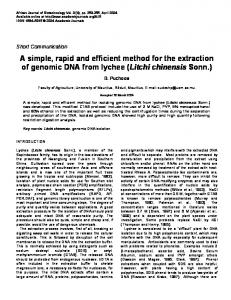
complexes seen trapped in the wells (Fig. 4, track 3), appear to be removed by spin-column puri®cation. The UV absorbance A260/A230 purity ratio increased from 0.75±0.83 for crude DNA extracts to 0.96±2.21 for puri®ed DNA samples. Although a component absorbing at 230 nm persisted in some of these samples, they were suciently pure for subsequent PCR ampli®cation. Total processing time for the extraction of soil in the bead-beater to puri®ed DNA for 8 samples was at most 2.5 h when using ready-prepared PVPP and Sephadex G-75 spin-columns, an improvement over the hot-SDS lysis procedure, with increased DNA yield and quality and a much shorter puri®cation time (Table 1). Application of the optimised extraction method to detect RSM2004 in ®eld soil Following extraction (bead-beating) and puri®cation (PVPP and Sephadex G75 spin-columns) of DNA from the release site of RSM2004, containing around 100 culturable cells gÿ1, the expected PCR product of 442 bp was faint but visible in agarose gels, con®rmed by probing gel blots. Signals of similar intensity were observed from soil inoculated with a further 102 RSM2004 cells and stronger signals corresponded to an increase in the amount of RSM2004 added to 104 and 106 cells (Fig. 5). Therefore, the sensitivity of detection was determined as 100 cells gÿ1 soil which was the equivalent of ca. 1 cell (in 50 ng DNA) from 10 mg soil per
Extraction of microbial DNA from soil
991
Fig. 5. PCR ampli®cation and detection of Tn5 in soil-extracted DNA. DNA extracted from soils inoculated with broth cultures of RSM2004, each lane (excluding 1) represents the total (20 ml) volume of PCR ampli®ed mix.: (A) EtBr-stained gel (3%); (B) gel blot after hybridisation to Tn5-speci®c probe generated by PCR. Lanes: 1, molecular size marker pBR328 DNA BglI/HinfI digest (Boehringer Mannheim); 2, 10 pg RSM2004 positive-control; 3, no DNA negative-control; 4±7, uninoculated ®eld soil (1.25 � 102 CFU RSM2004 gÿ1 soil) with 50, 100, 200, 300 ng DNA, respectively; 8±11, soil seeded with 102 RSM2004 gÿ1 (6.4 � 102 CFU RSM2004 gÿ1 soil) with 50, 100, 200, 300 ng DNA, respectively; 12±15, soil seeded with 104 RSM2004 gÿ1 (6.7 � 104 CFU RSM2004 gÿ1 soil) with 50, 100, 200, 300 ng DNA, respectively; 16±19, soil seeded with 106 RSM2004 gÿ1 (6.6 � 106 CFU RSM2004 gÿ1 soil) with 50, 100, 200, 300 ng DNA, respectively.
20 ml reaction. In contrast, in DNA extracted from soil using the hot-SDS lysis method, signal could be detected only after supplementing the numbers of RSM2004 with cultured cells to 106 gÿ1 soil.
DISCUSSION
The bead-beating plus spin-column procedure was developed to overcome the problems in obtaining PCR signals with soil DNA isolated by hot-
992
D. W. Cullen and P. R. Hirsch
SDS extraction, especially the diculty of removing co-extracted humic contaminants, and we demonstrated the importance of accurate qualitative and quantitative estimation of extracted DNA. The ®eld soil contained 102 gÿ1 RSM2004 culturable cells (i.e. copies of Tn5) against the background indigenous community of 109 bacterial cells: sensitivity is limited by the amount of soil-extracted DNA which it is practicable to use in a PCR reaction, normally equivalent to 1% of the yield from 1 g soil. With our optimised method it was possible to amplify and detect a single copy of Tn5 corresponding to one genome of RSM2004 in ®eld soil against a background of 107 diverse non-target organisms, which would not have been possible with hot-SDS extracted DNA. This PCR detection limit for Tn5marked R. leguminosarum RSM2004 was also 10 to 1000-fold greater than those limits previously reported for other organisms added to soil or sediment samples before extraction (Picard et al., 1992; Erb and Wagner-DoÈbler, 1993; Herrick et al., 1993; Smalla et al., 1993; Tebbe and Vahjen, 1993). Yields obtained by our method were comparable to the range 4±50 mg DNA gÿ1 dry soil reported from similar studies (Selenska and KlingmuÈller, 1991; Tsai and Olson, 1991; Smalla et al., 1993; Tebbe and Vahjen, 1993), although these latter values may have been overestimated because of coextracted humic material interfering with spectroscopic quanti®cation. Total direct counts of bacterial numbers in the agricultural soil extracted indicated there were up to 1.6 � 109 cells gÿ1 wet soil and assuming a cellular DNA content of 5±8 fg (McCoy and Olson, 1985), yields of 8±13 mg DNA gÿ1 soil are predicted. The DNA yield recovered by our new direct extraction method was greater than this theoretical total, indicating an overall high extraction eciency. Nevertheless, sample variability and the possible contribution of extracellular bacterial DNA and eucaryotic DNA may also be factors. The method which we have developed and optimised is simple and inexpensive with a small number of ecient lysis and puri®cation steps to maximize the yields and quality of recovered DNA and allow the processing of many samples simultaneously in a short time. It does not require the use of enzymes (lysozyme, proteinase K, pronase, RNase), phenol and chloroform, or time-consuming CsCl density gradient puri®cation. We veri®ed the method by successfully detecting the Tn5 marker in an established community of GM rhizobia in ®eld soil, but there will be many other applications. The method has been scaled-up to extract DNA from heavier soil samples (5±10 g) by using larger tubes for bead-beating. The procedure for removing coextracted humic substances from soil DNA extracts could also be scaled-down and speeded up by using microcentrifuge tubes ®tted with ®lters and PVPP.
The PVPP spin-columns which we have developed should be useful for removing phenolic contaminants in other extraction procedures. We have applied the new method to detect a Tn5 marker in bacteria in several paddy ®eld soils with high organic matter contents, and to obtain signals (using dierent PCR primers) from sandy soils and from bacteria surviving in 150-year old air-dried soils from the Rothamsted Soil Archive. Thus, in addition to the sensitive monitoring of individual strains of bacteria, our rapid and simple method can be used with wide range of dierent soil types to provide high quality DNA from the soil microbial community for PCR ampli®cation, a prerequisite of modern techniques for investigating microbial ecology. AcknowledgementsÐIACR-Rothamsted receives grantaided support from the Biotechnology and Biological Sciences Research Council of the U.K. We would like to acknowledge ®nancial support for this work from the U.K. Ministry of Agriculture, Fisheries and Food.
REFERENCES
Amann R. I., Ludwig W. and Schleifer K. H. (1995) Phylogenetic identi®cation and in situ detection of individual microbial cells without cultivation. Microbiological Reviews 59, 143±169. Bruce K. D., Hiorns W. D., Hobman J. L., Osborn A. M., Strike P. and Ritchie D. A. (1992) Ampli®cation of DNA from native populations of soil bacteria by using the polymerase chain reaction. Applied and Environmental Microbiology 58, 3413±3416. Dellaporta S. L., Wood J. and Hicks J. B. (1983) A plant DNA minipreparation: Version II. Plant Molecular Biology Reporter 1, 19±21. Erb R. W. and Wagner-DoÈbler I. (1993) Detection of polychlorinated biphenyl degradation genes in polluted sediments by direct DNA extraction and polymerase chain reaction. Applied and Environmental Microbiology 59, 4065±4073. Flemming C. A., Leung K. T., Lee H., Trevors J. T. and Greer C. W. (1994) Survival of Lux-lac-marked biosurfactant-producing Pseudomonas aeruginosa UG2L in soil monitored by; non-selective plating and PCR. Applied and Environmental Microbiology 60, 1606±1613. Herrick J. B., Madsen E. L., Batt C. A. and Ghiorse W. C. (1993) Polymerase chain reaction ampli®cation of naphthalene catabolic and 16S rRNA gene sequences from indigenous sediment bacteria. Applied and Environmental Microbiology 59, 687±694. Higuchi R. (1992) Simple and rapid preparation of samples for PCR. In PCR Technology, ed. H. A. Erlich, pp. 31±38. W.H. Freeman, New York. Hirsch P. R. and Skinner F. A. (1992) The identi®cation and classi®cation of Rhizobium and Bradyrhizobium. In Identi®cation Methods in Applied and Environmental Microbiology, eds R. G. Board, D. Jones and F. A. Skinner, pp. 45±65. Blackwell Scienti®c Publications, Oxford. Hirsch P. R. and Spokes J. R. (1994) Survival and dispersal of genetically modi®ed rhizobia in the ®eld and genetic interactions with native strains. FEMS Microbiology Ecology 15, 147±160. Holben W. E., Jansson J. K., Chelm B. K. and Tiedje J. M. (1988) DNA probe method for the detection of
Extraction of microbial DNA from soil speci®c microorganisms in the soil bacterial community. Applied and Environmental Microbiology 54, 703±711. Knaebel D. B. and Crawford R. L. (1995) Extraction and puri®cation of microbial DNA from petroleum-contaminated soils and detection of low numbers of toluene, octane and pesticide degraders by multiplex polymerase chain reaction and Southern analysis. Molecular Ecology 4, 579±591. Le L. G., Dana J. R., McArthur J. V. and Shimkets L. J. (1995) Detection of Tn5-like sequences in kanamycinresistant stream bacteria and environmental DNA. Applied and Environmental Microbiology 59, 417±421. Liesack W., Weyland H. and Stackenbrandt E. (1991) Potential risks of gene ampli®cation by PCR as determined by 16S rDNA analysis of a mixed culture of strict barophilic bacteria. Microbial Ecology 21, 191± 198. McCoy W. F. and Olson B. H. (1985) Fluorimetric determination of the DNA concentration in municipal drinking water. Applied and Environmental Microbiology 49, 811±817. Moran M. A., Torsvik V. L., Torsvik T. and Hodson R. E. (1993) Direct extraction and puri®cation of rRNA for ecological studies. Applied and Environmental Microbiology 59, 915±918. More M. I., Herrick J. B., Silva M. C., Ghiorse W. C. and Madsen E. L. (1994) Quantitative cell lysis of indigenous microorganisms and rapid extraction of microbial DNA from sediment. Applied and Environmental Microbiology 60, 1572±1580. Muller-Wegener U. (1988) Interaction of humic substances with biota. In Humic Substances and Their Role in the Environment, eds. F. H. Frimmel and R. F. Christman, pp. 179±192. John Wiley, New York. Ogram A., Sayler G. S. and Barkay T. (1987) The extraction and puri®cation of microbial DNA from sediments. Journal of Microbiological Methods 7, 57±66. Picard C., Ponsonnet C., Paget E., Nesme X. and Simonet P. (1992) Detection and enumeration of bacteria in soil by direct DNA extraction and polymerase chain reaction. Applied and Environmental Microbiology 58, 2717± 2722. Sambrook J., Fritsch E. F. and Maniatis T. (1989) Molecular Cloning: A Laboratory Manual. Cold Spring Harbor Press, Cold Spring Harbor. Selenska S. and KlingmuÈller W. (1991) DNA recovery and direct detection of Tn5 sequences from soil. Letters in Applied Microbiology 13, 21±24. Selenska S. and KlingmuÈller W. (1992) Direct recovery and molecular analysis of DNA and RNA from soil. Microbial Releases 1, 41±46.
993
Smalla K., Cresswell N., Mendonca-Hagler L. C., Wolters A. and van Elsas J. D. (1993) Rapid DNA extraction protocol from soil for polymerase chain reaction mediated ampli®cation. Journal of Applied Bacteriology 74, 78±85. Smith N. C. and Stribley D. P. (1994) A new approach to direct extraction of microorganisms from soil. In Beyond the Biomass, eds. K. Ritz, J. Dighton and K. E. Giller, pp. 49±55. John Wiley, Chichester. Stean R. J., Goksùyr J., Bej A. K. and Atlas R. M. (1988) Recovery of DNA from soils and sediments. Applied and Environmental Microbiology 54, 2908±2915. Tas E., Saano A., Leinonen P. and LindstroÈm K. (1995) Identi®cation of Rhizobium spp. in peat based inoculants by DNA hybridization and PCR and its application in inoculant quality control. Applied and Environmental Microbiology 61, 1822±1827. Tebbe C. C. and Vahjen W. (1993) Interference of humic acids and DNA extracted directly from soil in detection and transformation of recombinant DNA from bacteria and a yeast. Applied and Environmental Microbiology 59, 2657±2665. Thornhill R. H., Burgess J. G. and Matsunaga T. (1995) PCR for direct detection of indigenous uncultured magnetic cocci in sediment and phylogenetic analysis of ampli®ed 16S ribosomal DNA. Applied Environmental Microbiology 61, 495±500. Torsvik V. L. (1980) Isolation of bacterial DNA from soil. Soil Biology & Biochemistry 12, 15±21. Trevors J. T. (1992) DNA extraction from soil. Microbial Releases 1, 3±9. Tsai Y.-L. and Olson B. H. (1991) Rapid method for direct extraction of DNA from soil and sediments. Applied and Environmental Microbiology 57, 1070±1074. Tsai Y.-L. and Olson B. H. (1992a) Detection of low numbers of bacterial cells in soils and sediments by polymerase chain reaction. Applied and Environmental Microbiology 58, 754±757. Tsai Y.-L. and Olson B. H. (1992b) Rapid method for separation of bacterial DNA from humic substances in sediments for polymerase chain reaction. Applied and Environmental Microbiology 58, 2292±2295. Young C. C., Burgho R. L., Keim L. G., Minak-Berners V., Lute J. R. and Hinton S. M. (1993) Polyvinylpyrrolidone-agarose gel electrophoresis puri®cation of polymerase chain reaction ampli®able DNA from soils. Applied and Environmental Microbiology 59, 1972±1974.