JOURNAL OF CLINICAL MICROBIOLOGY, July 1998, p. 2093–2095 0095-1137/98/$04.0010 Copyright © 1998, American Society for Microbiology. All Rights Reserved.
Vol. 36, No. 7
Simple, Inexpensive, Reliable Method for Differentiation of Candida dubliniensis from Candida albicans EMMANUELLE PINJON,1 DEREK SULLIVAN,1 IRA SALKIN,2* DIARMUID SHANLEY,1 AND DAVID COLEMAN1 Department of Oral Medicine and Pathology, School of Dental Science and Dublin Dental Hospital, Trinity College, University of Dublin, Dublin 2, Republic of Ireland,1 and Wadsworth Center, New York State Department of Health, Albany, New York2 Received 13 February 1998/Accepted 21 April 1998
Candida dubliniensis is a recently described pathogenic species which shares many phenotypic features with Candida albicans, including the ability to form germ tubes and chlamydospores. These similarities have caused significant problems in the identification of C. dubliniensis by the average clinical mycology laboratory. To facilitate the differentiation of these species, we investigated the growth of 120 isolates of C. dubliniensis and 98 C. albicans isolates at 42 and 45°C on Emmons’ modified Sabouraud glucose agar (SGA) and 10 isolates of each species in yeast-peptone-dextrose broth. None of the C. dubliniensis isolates grew on the agar or in the broth medium at 45°C, while 11 isolates were capable of growing on SGA at 42°C. In contrast, all of the C. albicans isolates but one grew at 45°C on or in either medium. These reproducible results clearly demonstrate that the incubation of isolates suspected to be C. dubliniensis or C. albicans at 45°C provides a simple, reliable, and inexpensive method for the differentiation of the two species. of C. dubliniensis isolates were able to grow, albeit poorly, at this elevated temperature (9). Furthermore, another recent study reported that 8 of 12 C. dubliniensis isolates examined grew at 42°C (7). These findings motivated us to investigate whether incubation at an even higher temperature would provide a simple, inexpensive, and reliable means of differentiating the two species. Descriptions of the 120 C. dubliniensis isolates and 98 isolates of C. albicans used in these studies are presented in Table 1. Conventional morphological and physiological methods as well as molecular techniques were employed to identify isolates of both species (11). All yeasts studied were initially grown for 48 h at 37°C individually on 25 ml of Emmons’ modified Sabouraud glucose agar (SGA) contained in 85-mmdiameter petri dishes. A small portion of a single colony of each isolate was then aseptically removed and streaked over the surface of two plates of SGA, one of which was incubated at 42°C and the second of which was placed at 45°C. Growth, if any, on both plates was visually assessed after 24 and 48 h of incubation. In addition to these experiments, 10 isolates of each species were selected for examination of their growth in yeast-peptone-dextrose (YPD) broth at 37, 42, and 45°C. The 10 C. dubliniensis isolates included 3 which grew poorly at 42°C on SGA medium and 7 that did not grow at all on SGA at that temperature. Nine of the C. albicans isolates were randomly selected, while the 10th isolate was chosen because it failed to grow at 45°C on SGA (see below). By using a hemocytometer, a standard inoculum suspension, containing 105 CFU in sterile distilled water, was prepared from colonies of each isolate grown on SGA for 24 h at 37°C. The inoculum was then aseptically transferred to YPD broth (to a final volume of 50 ml) in 250-ml conical flasks and incubated at the three temperatures with shaking at 150 rpm. At specific time points, aliquots of each isolate were removed to spectrophotometrically measure their optical densities at 600 nm. These values were then used to plot a growth curve for each isolate at each temperature. No growth was found for any of the 120 C. dubliniensis isolates at 24 and 48 h on SGA at 45°C, although 11 isolates
Candida dubliniensis is a newly described species of the most protean genus of pathogenic yeasts (9, 11). The vast majority of C. dubliniensis isolates identified to date have been recovered from the oral cavities of human immunodeficiency virus (HIV)infected individuals, particularly those suffering from recurrent episodes of oral candidiasis (3, 8–11). However, a small number of isolates associated with other anatomical sites, including the vagina and the lung, have been reported (5, 9, 11). To gain a more complete understanding of the precise epidemiological role that this species plays in human infections, it is essential that rapid and reliable tests for the identification of C. dubliniensis in the routine clinical microbiology laboratory be available. However, the introduction of such tests has been complicated by the fact that C. dubliniensis shares many phenotypic characteristics with Candida albicans (9). The high degree of similarity between these two species has, in all likelihood, contributed to the misidentification of some C. dubliniensis isolates as C. albicans (3). The most reliable tests currently used for discriminating between the two species are based on molecular techniques such as DNA fingerprinting with repetitive-sequencecontaining DNA probes, pulsed-field gel electrophoresis, and others (2, 9, 11). Although they are very effective, these techniques are not readily applicable to the identification of large numbers of isolates, nor can they presently be conducted in most standard mycology laboratories. While the use of a number of phenotypic characteristics, e.g., chlamydospore production, carbohydrate assimilation, colonial coloration on differential media such as CHROMagar Candida and methyl blueSabouraud agar, has been investigated, none has provided a completely reliable method for the differentiation of C. albicans from C. dubliniensis (7, 9). It has previously been suggested that the two species could be distinguished by the inhibition of the growth of C. dubliniensis at 42°C (2, 9, 11). However, one recent study reported that approximately 10%
* Corresponding author. Mailing address: Wadsworth Center, New York State Department of Health, Albany, NY 12201-0509. Phone: (518) 485-5386. Fax: (518) 485 5414. E-mail:
[email protected]. 2093
2094
NOTES
J. CLIN. MICROBIOL.
TABLE 1. C. dubliniensis and C. albicans isolates used in these studies Species and country of isolation
C. dubliniensis Argentina Australia Belgium Canada Finland Germany Greece Ireland Ireland Ireland Spain Switzerland United Kingdom United Kingdom United Kingdom C. albicans Australia Hong Kong Ireland
United Kingdom United Kingdom
No. of isolatesa
Specimen source(s)
HIV status of subject(s) sampledb
1 4 5 6 1 4 1 52 8 5 9 6 5 7 6
Oral cavity Oral cavity Oral cavity Oral cavity Oral cavity Oral cavity Oral cavity Oral cavity Oral cavity Vaginal Oral cavity Oral cavity Oral cavity Oral cavity Miscellaneousc
1 1 1 1 1 1 1 1 2 2 1 1 1 2 2
10 4, 10, 11 This study This study This study This study This study This study; 2 This study; This study 1, 10 This study; This study This study;
1 6 32 5 20 21 8 1 4
Oral cavity Oral cavity Oral cavity Oral cavity Vaginal Vaginal Blood Oral cavity Oral cavity
1 1 1 2 1 2 2 1 2
11 This This This This This 6 This This
Source or reference(s)
11 5 10 8
study study study study study study study
a
Each isolate was recovered from a separate individual. 1, positive; 2, negative. c These included two fecal isolates and one each from a blood culture, tracheal aspirate, sputum, and postmortem lung specimens. b
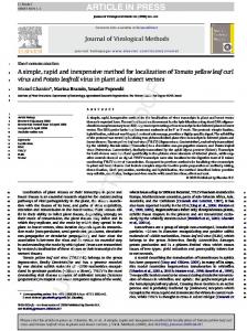
showed limited growth after 48 h of incubation at 42°C. All 98 of the C. albicans isolates showed good growth at 42°C on SGA at 48 h, and all but one exhibited substantial growth after the same period of time at 45°C. The growth of more than half of the isolates was examined on at least two occasions, and identical results were obtained in these confirmatory studies. Representative growth curves for isolates of each species grown in YPD broth at 37 and 45°C are presented in Fig. 1. Although all
10 selected isolates of C. dubliniensis grew at 37°C, 7 failed to grow at 42°C, while the 3 that showed limited growth on SGA at this temperature also showed limited growth in YPD broth. None of the 10 isolates examined showed any appreciable growth in YPD broth at 45°C. All but 1 of the 10 C. albicans isolates grew well in YPD broth at all three temperatures. The single isolate which failed to grow in the broth at 45°C (isolate CA58.1) also failed to grow when incubated on SGA at the same temperature. Preliminary studies with a single isolate of a closely allied taxon, Candida stellatoidea type I (ATCC 11006), demonstrated that it, too, like the majority of C. dubliniensis isolates, did not grow at 42 or 45°C. However, C. stellatoidea type I may be easily differentiated from C. dubliniensis on the basis of sucrose assimilation, production of b-glucosidase, and serotype (1, 11). The data from these studies clearly indicate that C. dubliniensis can be readily distinguished from C. albicans by the incubation of isolates on SGA at 45°C. This test is simple, reliable, inexpensive, reproducible, and readily applicable to large numbers of isolates in either a clinical or academic mycology laboratory. This simple procedure can be employed to retrospectively evaluate the identification of stored cultures of C. albicans held in stock collections. In a previous retrospective analysis of the authors’ collection of C. albicans isolates, 2 of 110 (1.8%) isolates recovered from the oral cavities of asymptomatic, normal, healthy individuals and 13 of 79 (16.5%) isolates which had been obtained from the oral cavities of HIV-infected individuals and had been identified as C. albicans were found to be C. dubliniensis (3). Accurate identification of C. dubliniensis isolates in archival collections and in clinical specimens should provide invaluable information concerning the epidemiology of this species and help to establish its clinical significance. In addition, data from such investigations may also help to explain the rapid emergence of C. dubliniensis as a potentially significant pathogen during the last decade. We thank our colleagues who sent us isolates of C. dubliniensis and C. albicans, including Aristea Velegraki, Department of Microbiology, National University of Athens Medical School, Athens, Greece; Markus Ruhnke, Virchow Klinikum der Humboldt Universita¨t, Berlin, Germany; Luc Giasson, School of Dentistry, Laval University, Quebec, Quebec, Canada; Jose Ponton, Departamento de Inmunologı´a, Microbiologı´a y Parasitologı´a, Universidad del Paı´s Vasco, Bilbao, Vizcaya, Spain; Elizabeth Johnson, Public Health Laboratory Service, Mycology Reference Laboratory, Bristol, United Kingdom; Frank Odds, Department of Bacteriology and Mycology, Janssen Research Foundation, Beerse, Belgium; Lakshman Samaranayake, Oral Biology Unit, Faculty of Dentistry, University of Hong Kong, Hong Kong; and Fiona Mulcahy, Department of Genitourinary Medicine, St. James’s Hospital, Dublin, Ireland. This work was supported by Irish Health Research Board grants 41/96 and 4/97. REFERENCES
FIG. 1. Growth curves of the oral isolates C. albicans 132A and CA58.1 and C. dubliniensis CD36 and CD43 in YPD broth medium at 37°C (solid lines) and 45°C (dashed lines). O.D. 600, optical density at 600 nm.
1. Boerlin, P., F. Boerlin-Petzold, C. Durussel, M. Addo, J.-L. Pagani, J.-P. Chave, and J. Bille. 1995. Cluster of oral atypical Candida isolates in a group of human immunodeficiency virus-positive drug users. J. Clin. Microbiol. 33: 1129–1135. 2. Coleman, D., D. Sullivan, K. Haynes, M. Henman, D. Shanley, D. Bennett, G. Moran, C. McCreary, L. O’Neill, and B. Harrington. 1997. Molecular and phenotypic analysis of Candida dubliniensis: a recently identified species linked with oral candidosis in HIV-infected and AIDS patients. Oral Dis. 3(Suppl. 1):S96–S101. 3. Coleman, D. C., D. J. Sullivan, D. E. Bennett, G. P. Moran, H. J. Barry, and D. B. Shanley. 1997. Candidiasis: the emergence of a novel species, Candida dubliniensis. AIDS 11:557–567. 4. McCullough, M. J., B. C. Ross, B. D. Dwyer, and P. C. Reade. 1994. Genotype and phenotype of oral Candida albicans from patients infected with the human immunodeficiency virus. Microbiology 140:1195–1202. 5. Moran, G. P., D. J. Sullivan, M. C. Henman, C. E. McCreary, B. J. Harrington, D. B. Shanley, and D. C. Coleman. 1997. Antifungal drug suscep-
VOL. 36, 1998 tibility of oral Candida dubliniensis isolates from human immunodeficiency virus (HIV)-infected and non-HIV-infected subjects and generation of stable fluconazole-resistant derivatives in vitro. Antimicrob. Agents Chemother. 41:617–623. 6. O’Connell, B., D. C. Coleman, D. Bennett, D. Sullivan, S. R. McCann, and C. T. Keane. 1995. An epidemiological study of Candida species infection in cancer patients using genetic fingerprinting and morphotyping. J. Hosp. Infect. 31:211–217. 7. Schoofs, A., F. C. Odds, R. Colebunders, M. Ieven, and H. Goosens. 1997. Use of specialised isolation media for recognition and identification of Candida dubliniensis isolates from HIV-infected patients. Eur. J. Clin. Infect. Dis. 16:296–300. 8. Sullivan, D., D. Bennett, M. Henman, P. Harwood, S. Flint, F. Mulcahy, D. Shanley, and D. Coleman. 1993. Oligonucleotide fingerprinting of isolates of
NOTES
2095
Candida species other than C. albicans and of atypical Candida species from human immunodeficiency virus-positive and AIDS patients. J. Clin. Microbiol. 31:2124–2133. 9. Sullivan, D., and D. Coleman. 1998. Candida dubliniensis: characteristics and identification. J. Clin. Microbiol. 36:329–334. 10. Sullivan, D. J., K. Haynes, J. Bille, P. Boerlin, L. Rodero, S. Lloyd, M. Henman, and D. Coleman. 1997. Widespread geographic distribution of oral Candida dubliniensis strains in human immunodeficiency virus-infected individuals. J. Clin. Microbiol. 35:960–964. 11. Sullivan, D. J., T. J. Westerneng, K. A. Haynes, D. E. Bennett, and D. C. Coleman. 1995. Candida dubliniensis sp. nov.: phenotypic and molecular characterisation of a novel species associated with oral candidosis in HIV-infected individuals. Microbiology 141:1507–1521.