0013-7227/02/$15.00/0 Printed in U.S.A.
The Journal of Clinical Endocrinology & Metabolism 87(12):5649 –5657 Copyright © 2002 by The Endocrine Society doi: 10.1210/jc.2002-020098
Single and Combined Effects of Growth Hormone and Testosterone Administration on Measures of Body Composition, Physical Performance, Mood, Sexual Function, Bone Turnover, and Muscle Gene Expression in Healthy Older Men KIMBERLY T. BRILL, ARTHUR L. WELTMAN, ANGELA GENTILI, JAMES T. PATRIE, DAVID A. FRYBURG, JOHN B. HANKS, RANDALL J. URBAN, AND JOHANNES D. VELDHUIS Departments of Internal Medicine (K.T.B., A.L.W., D.A.F., J.D.V.), Human Services (K.T.B., A.L.W.), Health Evaluation Sciences (J.T.P.), and Surgery (J.B.H.), General Clinical Research Center (K.T.B., A.L.W., J.T.P., J.D.V.), and Center for Biomathematical Technology (J.D.V.), University of Virginia, Charlottesville, Virginia 22908; Department of Geriatrics (A.G.), Medical College of Virginia, Virginia Commonwealth University, Richmond, Virginia 23298; and Department of Internal Medicine (R.J.U.), Division of Endocrinology, University of Texas Medical Branch, Galveston, Texas 77555 We examined the effects of GH and/or testosterone (T) administration on body composition, performance, mood, sexual function, bone turnover, and muscle-gene expression in healthy older men. Ten men [mean (SEM) age, 68 (2.5) yr; height, 171.5 (2.4) cm; and weight, 80 (3.0) kg] completed each of the following 1-month, double-blind interventions after a baseline (B) study in randomized order with an intervening 3-month washout: transdermal T patch (5.0 mg/daily); recombinant human GH (6.25 g/kg sc daily); and combined hormones (GHT). ANOVA with repeated measures was used to evaluate interventional effects. Integrated serum GH concentrations [mean (SEM)] were elevated comparably by GH and GHT: [B ⴝ 363 (55), GH ⴝ 1107 (120), T ⴝ 459 (131), and GHT ⴝ 1189 (46) g/ liter䡠min; P < 0.0001]. Serum IGF-I concentrations also increased commensurately after GH and GHT: [B ⴝ 168 (14), GH ⴝ 285 (16), T ⴝ 192 (25), and GHT ⴝ 294 (25) g/liter; P < 0.0001]. GHT administration increased total estradiol: [B ⴝ 110 (20), GH ⴝ 106 (13), T ⴝ 129 (13), and GHT ⴝ 153 (17) pmol/liter; P < 0.02], and both T and GHT elevated free T: [B ⴝ 12 (2.1), GH ⴝ 11 (1.5), T ⴝ 22 (2.8), and GHT ⴝ 24 (2.5) pg/ml; P < 0.0001]. No significant changes occurred in strength, flexibility, per-
H
EALTHY AGING IN men is marked by a progressive reduction in the daily production of GH (1–3) and testosterone (T) (4 –12). Daily mean serum GH concentrations decline exponentially, with a half-time of approximately 7 yr beginning in the young adult (age 18 –25 yr; Refs. 1–3). The hyposomatotropism of aging is associated with increased fatigue, decreased physical performance, reduced lean body mass, and an accumulation of abdominal visceral fat mass (1–3). T bioavailability falls in parallel, such that T production is reduced by nearly one third by age 70 and one half by age 80. Hypogonadal-like features may emerge concurrently in the aging male, e.g. loss of bone and muscle mass, diminished Abbreviations: AR, Androgen receptor; B, baseline; BMI, body mass index; CV, coefficient(s) of variation; E2, estradiol; GHT, combined GH and T; HDL, high-density lipoprotein; PSA, prostate-specific antigen; rh, recombinant human; T, testosterone; VTG, thoracic gas volume.
centage body fat, or sexual function and mood. However, fatfree mass increased under combined GHT exposure: [B ⴝ 55 (1.3), GH ⴝ 56 (1.1), T ⴝ 55 (1.5), GHT ⴝ 57 (1.7) kg; P < 0.03]. Balance improved in response to GH intervention (P < 0.05), as did 30-m walk time during T and GHT interventions [B ⴝ 6.6 (0.3), GH ⴝ 6.2 (0.7), T ⴝ 5.9 (0.3), GHT ⴝ 5.5 (0.3) sec; P ⴝ 0.04] and stair climb time for all three interventions [B ⴝ 32.2 (1.4), GH ⴝ 29.8 (1.2), T ⴝ 30.5 (1.4), and GHT ⴝ 29.9 (1.2) sec (P ⴝ 0.0034), wherein the effects of GH, T, and GHT were different from that of B]. Muscle IGF-I gene expression increased by 1.9-fold during GH administration and by 2.3-fold during GHT administration (P < 0.05, compared with B). Myostatin and androgen receptor gene expression were not affected. Serum osteocalcin increased in response to the GH and GHT interventions: [B ⴝ 4.8 (0.52), GH ⴝ 5.7 (0.54), T ⴝ 4.7 (0.33), and GHT ⴝ 5.5 (0.39); P < 0.009]. There were no significant adverse events during 30 patient-months of intervention. We conclude that 1 month of GH and/or T administration improves certain measures of balance and physical performance in older men and increases muscle IGF-I gene expression. (J Clin Endocrinol Metab 87: 5649 –5657, 2002)
libido and potency, impaired psychological well-being, and variable reduction in red cell mass (5, 6, 10, 11). Albeit unproven, an emergent hypothesis is that impoverished anabolic drive by GH and T in the aging male may contribute to loss of well-being, energy, strength, libido, and skeletal and muscle mass and to the accumulation of visceral fat (1–12). The resultant relative physical frailty, potential loss of independent activity status, evident decline in exercise capacity, and higher risk of falls and fractures can seriously impair quality of life (13, 14). Recent limited interventional studies of either GH or T supplementation indicate that important anabolic effects can be elicited in GHdeficient, hypogonadal, and healthy older men, such as enhanced lean body mass, greater strength and muscle protein synthesis, increased bone mineral content, and an improved sense of well-being (13–32). However, higher doses of androgen may cause polycythemia, diminish plasma high-
5649
5650
J Clin Endocrinol Metab, December 2002, 87(12):5649 –5657
density lipoprotein (HDL) concentrations, worsen sleep apnea, and stimulate prostate growth (23, 33–36). Likewise, GH supplementation may induce peripheral edema, arthralgias, carpal tunnel syndrome, gynecomastia, and mild glucose intolerance (17–19, 37–39). One approach to addressing the foregoing concerns would be to limit the amounts of T or GH administered. A second consideration is to combine GH and T repletion at nearly physiological doses. In the latter regard, few interventional studies in older individuals have implemented combined GH and T supplementation. On the basis of the physiological anabolic synergy expected between GH and androgen in normal puberty, the present clinical investigation explores the impact of 2-fold T and GH supplementation in healthy older men. This population is relatively hypoandrogenemic and hyposomatotropic by young adult standards. We hypothesized that combined near-physiological supplementation with GH and T would enhance selected endpoints of trophic hormone action, such as muscle strength, functional performance, skeletal mass, mood, sexual function, and body composition and elicit few side effects. We tested this postulate via a prospectively randomized, double-blind, within-subject crossover interventional design comprising the single and combined administration of recombinant human (rh)GH sc (6.25 g/kg䡠d) and T transdermally (5.0 mg/d) for 1 month each. We monitored biochemical indices of GH, muscle IGF-I, myostatin and androgen receptor (AR) gene expression, strength, balance, physical performance, percentage body fat, mood and sexual function, and any adverse impact on serum HDL concentrations, hematocrit, fasting plasma insulin/glucose ratios and glycosylated hemoglobin. Subjects and Methods Human subjects The protocol was approved by the Human Investigation Committees of the University of Virginia Health Sciences Center and the McGuire Hunter Holmes Veterans Affairs Medical Center. Inpatient studies were carried out in the General Clinical Research Center (GCRC) at the University of Virginia. All subjects were healthy older (age, 60 –78 yr) men, whose baseline physical examination and screening biochemical tests of renal, hepatic, hematological, and metabolic function (thyroid function and fasting plasma glucose) were unremarkable. A screening preintervention prostate-specific antigen (PSA) and digital prostatic exam were normal. To determine gonadal status, morning serum total T concentrations were measured on two separate occasions. Volunteers were considered eligible for the study if the latter mean exceeded 200 ng/dl (6.9 nmol/liter) but was less than 450 ng/dl (15.6 nmol/liter). In addition, serum concentration of prolactin needed to be below 25 g/liter, LH and FSH below 20 IU/liter, and IGF-I below 200 g/liter. To obviate possible confounding between T and/or GH interventions and exercise, volunteers were not allowed to enter an active exercise program during the 1-yr study period, including the 3-month washout intervals between consecutive interventions. Subjects were admitted to the GCRC at 1700 h for overnight testing on four different occasions, once at baseline and subsequently after each of three interventions. Each subject served as his own control. After the baseline study, the following 1-month treatments were each assigned in randomized order: GH/placebo (GH), testosterone/placebo (T), and GH/testosterone (GHT). A 3-month washout period intervened between hormone exposure. Interventions included daily application of transdermal placebo or T patches (Androderm, two 2.5-mg patches applied at bedtime) and evening sc injections of saline or rhGH (Genotropin, 6.25 g/kg䡠d).
Brill et al. • GH, T, and GHT Effects in Healthy Older Men
GCRC admissions. After admission to the GCRC in the evening, subjects received a eucaloric standardized meal (12 kcal/kg) with a macronutrient composition of 55% carbohydrate, 15% protein, and 30% fat. A catheter was placed in a forearm vein at 1800 h. Subjects received the last sc injection (above) and replaced T or placebo patches at 1900 h. Beginning at 2000 h, blood samples were withdrawn every 10 min for 12 h for later analysis of GH and T. At 0800 h the next morning, serum was collected for later measurements of bone turnover markers including PTH, bone specific alkaline phosphatase, and osteocalcin. A fasting timed urine specimen was obtained to measure urinary calcium and creatinine on each admission. Subjects also provided fasting serum samples on d 1, 7, 14, 21, and 28 during each interventional month. The latter was used to monitor serum concentrations of GH, IGF-I, LH, prolactin, estradiol (E2), total T, and free T. Assays. Serum GH concentrations (2000 h to 0800 h) were measured in duplicate by an ultrasensitive (0.005 g/liter threshold) chemiluminescence-based assay (Nichols Institute Diagnostics, San Juan Capistrano, CA). All samples from an individual subject were analyzed in the same assay. The assay standard was 22-kDa rhGH. The mean intra-assay coefficient of variation (CV) was 4.8%, and the interassay CV was 8.6%. Serum total T and free T concentrations (2000 h to 0800 h) were measured by solid-phase RIA (Coat-a-Count, Diagnostic Products, Los Angeles, CA), with respective intra-assay CV of 6.1% and 3.8% and interassay CV of 7.9% and 4.2%, respectively. E2 (pooled sample) was assayed by a chemiluminescence assay (Automated Chemiluminescence Systems, Bayer Corp., Diagnostics Division, Norwood, MA), with respective intra-assay CV of 5.3% and interassay CV of 6.4%. SHBG (pooled sample) was measured using an assay from Diagnostic Systems Laboratories, Inc. (Webster, TX). The intra-assay CV ranged from 2.9 –3.0%, and the interassay CV ranged from 8.9 –10%. Serum IGF-I concentrations were assayed by RIA after acid ethanol extraction (Nichols Institute Diagnostics). The intra-assay CV was 2.7%, and the interassay CV was 6.8%. Integrated serum GH concentrations over the 12-h interval 2000 h to 0800 h were calculated by the trapezoidal rule (40). Serum PTH and osteocalcin were quantitated by two-site immunoradiometric assay (Nichols Institute Diagnostics). Serum bone alkaline phosphatase was measured by immunoradiometric assay using the Tandem-R-Ostase kit from Hybritech (San Diego, CA). CV for the latter assays were 4 –10% (intraassay) and 7–15% (interassay). Muscle biopsy. Muscle biopsies were performed at 0930 h. The dominant vastus lateralis was anesthetized locally using lidocaine. A 6 ⫻ 8-mm tissue sample was removed using a Bergstrom needle and snap-frozen in liquid nitrogen. Skeletal muscle mRNA measured by RT-PCR. For RNA extraction, the frozen muscle sample was pulverized in liquid nitrogen using a mortar and pestle and homogenized for 30 sec at 8000 rpm. Total RNA was extracted using RNA STAT-60 (Tel-Test, Friendswood, TX) and quantified by duplicate absorbance determinations at 260 nm (26, 28). cDNA was reverse transcribed from 0.5 g total RNA using 1 m random hexamers and 100 U Superscript II reverse transcriptase (Life Technologies, Inc., Rockville, MD) in a final volume of 20 l at 42 C for 50 min, followed by 5 min of heat inactivation at 99 C. The cDNA was amplified from the 20-l reverse transcription reaction mixture in the same tube in a final concentration of 1⫻ PCR buffer [(Perkin-Elmer Corp., Norwalk, CT), 25 mm Tris-HCl, 50 mm KCl2, 1.5 mm MgCl2, 1.0 mm deoxy-NTP, forward and reverse primers (gene of interest, 10 pmol; glyceraldehyde-phosphodehydrogenase, 1.5 pmol]; 5 U AmpliTaq DNA Polymerase (Perkin-Elmer Corp.), and 0.0225 Ci䡠liter⫺1 [32P] deoxy-CTP (Amersham Pharmacia Biotech, Arlington Heights, IL) in a final volume of 50 l. The linear portion of the amplification curve for each transcript was defined and then used to determine the appropriate number of PCR amplification cycles in the RT-PCR analyses. Curves for IGF-I, AR, and myostatin were generated independently (data not shown). PCR for AR started with an initial denaturation at 94 C for 3 min, followed by 26 cycles of denaturation at 94 C for 35 sec, annealing at 58 C for 45 sec, extension at 72 C for 80 sec, with a final extension cycle at 72 C for 7 min. IGF-I was amplified for 28 cycles, and myostatin for 30 cycles. The PCR products (10 l from each sample tube) were then isolated by electrophoresis using precast 6% polyacrylamide-Tris-borate EDTA gels (Novex, San Diego, CA) at 90 W for 90 min. Gels were subsequently
Brill et al. • GH, T, and GHT Effects in Healthy Older Men
dried, and [32P] incorporation was determined by quantitative autoradiography using a PhosphorImager from Molecular Dynamics, Inc. (Sunnyvale, CA). For final analyses, the OD of the transcript of interest was determined in duplicate using SigmaGel 1.05 software (SPSS, Inc. Richmond, CA; see Statistical analysis). For subsequent sequencing of the IGF-I, AR, and myostatin DNA fragments, PCR products were isolated using a 5% polyacrylamide (1:25 bis to acrylamide ratio) gel with 25% glycerol. Samples were run for 3 h at 250 V in 0.5⫻ Tris-borate EDTA buffer. The gel was then stained with ethidium bromide, and the bands were excised under UV illumination. The gel slices were electroeluted for 30 min (Bio-Rad Electro-Eluter 442, Bio-Rad Laboratories, Inc., Hercules, CA). The samples were extracted with phenol-chloroform (2⫻) and ethanol precipitated. Samples were sequenced at the University of Texas Medical Branch Molecular Core Laboratory with the specific forward and reverse primers from the 3⬘ to 5⬘ ends using an ABI Prism (Model 310) Sequencer (PE Applied Biosystems, Foster City, CA). All amplified DNA fragment sequences were compared to confirm identity. Muscle RNA extracts (for samples being measured) were reversetranscribed in duplicate and subjected to PCR. Duplicate samples were electrophoresed, and OD was determined from [32P] incorporation into the relevant bands. The arbitrary value for OD was then averaged between duplicate samples. Analysis of duplicate samples minimizes the potential variability in reverse transcription, PCR cycle efficiency, and gel loading. The intra-assay variance for the entire set of duplicate samples determined (IGF-I, AR, and myostatin) was 9.7% ⫾ 0.1 (mean ⫾ sd; n ⫽ 40 pairs). Therefore, mRNA data are expressed as the average OD (arbitrary units) of the duplicate samples measured as a unitless ratio to a housekeeping (glyceraldehyde-phosphodehydrogenase) gene measured simultaneously.
Outpatient testing Body composition. Body volume and density were estimated by hydrostatic weighing (43) and air-displacement plethysmography (44). For hydrostatic weighing, each subject was weighed in air on an AccuWeigh beam scale accurate to 0.1 kg and subsequently weighed underwater on a Chatillon autopsy scale accurate to 10 g. Residual lung volume was measured using an oxygen dilution technique (45). Body density was also determined using the Bod Pod body composition system (Life Measurement Instruments, Concord, CA). Subjects were instructed to wear close-fitting swimwear, including a swim cap, and to remove all jewelry. Volunteers were first weighed on a calibrated scale to the resolution of 5 g, and then asked to enter the chamber and sit quietly during the initial measurement of body volume. The body volume trial was repeated, and the thoracic gas volume (VTG) was determined while the subject was in the chamber. The system estimates body volume based on the following equation: body volume(raw) ⫺ surface area artifact ⫹ 40% VTG (44). The computational procedure of Brozek et al. (46) was used to determine the percentage of total body fat from the body density measurements. Strength. Eccentric and concentric strength of the quadriceps femoris and biceps femoris muscle groups was assessed using the Kin-Com II isokinetic dynamometer (Chattex Corp., Hixson, TN). Tests were performed at 60°/sec. To ensure testing of the dominant leg, subjects were asked to kick a ball, and the preferred leg was used in further testing. Each subject was seated on the Kin-Com, with the lateral epicondyle of the knee aligned with the axis of the dynamometer and the inferior edge of the force pad aligned directly superior to the medial malleolus. Velcro straps were placed across the hips, thigh, and ankle of each subject for stabilization. Each subject performed three eccentric and three concentric contractions at the velocity of 60°/sec. The eccentric test contraction was followed by the concentric test without any rest. Gravity correction was performed according to the manufacturer’s protocol with the knee at 0° flexion. The dynamometer’s preload and minimal force values were set at 50 and 20 N, respectively. Balance. One-legged stance was used to assess balance. This assessment recorded the amount of time the subject remained standing on one leg with his eyes open and eyes closed. Subjects were instructed to pick a point on the wall ahead, steady themselves and lift the nonsupporting leg when ready. Total time on one leg was recorded. The clock was
J Clin Endocrinol Metab, December 2002, 87(12):5649 –5657 5651
stopped if the subject put his nonsupporting foot down or moved the supporting leg from the initial location. Left and right legs were alternated. Each test was repeated three times, beginning with the eyes-open test. Function. Functional ability was assessed during a four-flight stair climb and a 30-m walk. In the stair climb, subjects were asked to descend four flights of stairs and return to the top of the staircase as quickly as possible. Subjects were instructed to use the railing for balance only. Two trials were made, with a 2-min rest between each trial. For the 30-m walk, subjects were instructed to walk as quickly as possible for three separate trials. Flexibility. Hamstring/lower back flexibility was assessed using a sitand-reach apparatus. Subjects were asked to sit with their feet against the apparatus, place their hands one on top of the other, and reach as far forward as possible, with knees extended. Positive scores indicated the number of inches the subject could reach past his toes. Negative scores indicated that the subject was a distance away from touching his toes. Three trials were performed. Sexual function and mood questionnaire. Mood and sexual function were assessed 1 wk before baseline admission and during the last 7 d of each intervention using a self-report questionnaire. Volunteers ranked the level of sexual desire, enjoyment, and performance and assessed mood. Mood assessment included positive mood attributes (alert, friendly, full of pep, and well/good feelings) and negative mood attributes (angry, irritable, nervous, sad or blue, and tired). The ranking of sexual function and mood used a Likert-type scale, as previously described (42, 43). Adverse events. Subjects were examined on d 1, 7, 14, 21, and 28 of each interventional period and asked to report anything unusual, such as skin irritations, itching, sneezing, or fatigue. Subjects were asked to rank the severity of the symptoms they were experiencing as mild, moderate, or severe. If the symptoms were mild to moderate, subjects were provided appropriate treatment and asked whether they wanted to continue participation in the study. Subjects with severe symptoms would be treated and withdrawn from the study. Documentation was maintained in the subject’s chart. Statistical analysis. All values are expressed as the mean (sem). ANOVA with repeated measures was used to determine mean differences between baseline and each of the three interventions. The primary endpoints were 12-h integrated GH, serum IGF-I, T, free T, E2, and SHBG concentrations; percentage body fat; fat free mass; strength; flexibility; function and muscle gene expression; bone turnover; sexual function; and mood. Sexual function and mood scores were averaged for each 7-d period (25, 40, 41). Whenever a significant interventional effect was observed, mean comparisons were examined by post hoc examination. Bonferroni correction was performed to adjust for multiple comparisons. Significance was set at P ⬍ 0.05.
Results Subject characteristics
The mean age of the subjects was 68.1 ⫾ 2.4 yr; height, 171.5 ⫾ 2.4 cm; weight, 79.9 ⫾ 3.0 kg; body mass index (BMI), 26.71 ⫾ 1.05 kg/m2; and percentage body fat, 30.5 ⫾ 1.4%. GH release
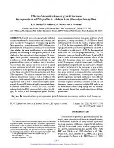
Figure 1 shows the overnight serum GH concentration profiles in one subject based on blood sampling at 10-min intervals over 12 h (2000 h to 0800 h) at baseline (B) and during randomly ordered T, GH, and GHT supplementation. Figure 2 summarizes 12-h integrated (area under the curve) serum GH concentrations at B and during the T, GH, and GHT interventions. ANOVA revealed that GH was greater than B (P ⬍ 0.001) and GHT was greater than B (P ⬍ 0.001). GH and GHT effects were not significantly different (P ⫽ 0.877), but exceeded the effect of T: GH was greater than
5652
J Clin Endocrinol Metab, December 2002, 87(12):5649 –5657
Brill et al. • GH, T, and GHT Effects in Healthy Older Men
B (P ⫽ 0.005). The responses to T and GHT were comparable (P ⫽ 0.477). E2
Administration of GHT increased serum E2 concentrations by 38% over B. In addition, we found that GH increased E2 above B (P ⫽ 0.05) and GHT increased E2 more than GH (P ⫽ 0.02; Fig. 4). T and GHT had a greater effect than GH on E2 (P ⫽ 0.05 and P ⫽ 0.02, respectively). SHBG
GH, GHT, and T interventions resulted in a nonsignificant reduction in SHBG [B ⫽ 80.4 ⫾ 12.4 nmol/liter; GH ⫽ 66.1 ⫾ 6.8 nmol/liter; GHT ⫽ 77.2 ⫾ 16.2 nmol/liter; and T ⫽ 71 ⫾ 9.4 nmol/liter (P ⫽ 0.33)]. Muscle gene expression
FIG. 1. Illustrative effects of 1 month of GH, T, and GHT supplementation on the overnight serum GH concentration profile in 1 subject (see Subjects and Methods).
Figure 5 shows semiquantitative estimates of muscle IGF-I gene expression, which was stimulated equally by GH (P ⫽ 0.05) and GHT (P ⫽ 0.05). T alone had no effect on muscle IGF-I gene expression. AR gene expression was invariant of intervention (P ⬎ 0.05; Fig. 5). Myostatin gene expression was also unaffected by T or GH supplementation, although the effect of GHT approached statistical significance (P ⫽ 0.09). Strength, flexibility, and body composition
T (P ⫽ 0.001) and GHT was greater than T (P ⬍ 0.001). T administration did not have a significant effect on 12-h integrated serum GH concentration (P ⫽ 0.468). IGF-I
Figure 2 also shows the changes in serum IGF-I concentrations, which paralleled those of GH. GH and GHT produced an approximately 70% increase in IGF-I: GH was greater than B (P ⫽ 0.01) and GHT was greater than B (P ⫽ 0.002). Mean IGF-I during the GH and GHT interventions did not differ (P ⫽ 0.498), but exceeded that during T: GH was greater than T (P ⫽ 0.04) and GHT was greater than T (P ⫽ 0.008). T did not have a significant effect on IGF-I concentrations (P ⫽ 0.508). Total T
Administration of T increased total T by 62% above B (P ⫽ 0.004), whereas GHT elicited a 75% increase in total T above B (P ⫽ 0.016). Both GHT and T significantly increased total T over GH administration: GHT was greater than GH (P ⫽ 0.003) and T was greater than GH (P ⫽ 0.011; Fig. 3). The response to GH administration was not significantly different from B (P ⬎ 0.05).
Table 1 summarizes quantitation of eccentric and concentric knee extension and knee flexion strength (P ⬎ 0.05), hamstring flexibility (P ⬎ 0.05), and percentage body fat (P ⬎ 0.05). The Bod Pod (see Subjects and Methods) but not hydrostatic weighing estimates identified significant overall increases in fat-free mass (P ⫽ 0.03). There was no statistical difference in BMI during the interventional periods compared with B or each other (P ⬎ 0.05). Function
T and GHT significantly reduced the time required to complete the 30-m walk compared with B: T was less than B (P ⫽ 0.045) and GHT was less than B (P ⫽ 0.009) and from each other, and GHT was less than T (P ⫽ 0.004). GH alone had no effect (Table 2). The stair-climb time improved significantly over B: GH was less than B (P ⫽ 0.022); T was less than B (P ⫽ 0.025); and GHT was less than B (P ⫽ 0.012). There were no differences among the interventions (P ⬎ 0.05). Balance
GH, but not T, enhanced performance in the eyes-closed, nondominant-leg stance (P ⫽ 0.047; Fig. 6). The GH effect was significantly different from that of GHT (P ⫽ 0.0294).
Free T
Figure 3 also shows the changes in free T concentrations, which are similar to changes in total T concentrations. T and GHT elicited an approximate 2-fold increase in free T above B: T was greater than B (P ⫽ 0.020) and GHT was greater than
Markers of bone remodeling
Table 3 shows no significant effects of any of the interventions on serum 25-hydroxyvitamin D or bone alkaline phosphatase concentrations, or fasting urinary calcium or
Brill et al. • GH, T, and GHT Effects in Healthy Older Men
J Clin Endocrinol Metab, December 2002, 87(12):5649 –5657 5653
FIG. 2. The effects of 1 month of GH, T, and GHT supplementation on 12-h integrated serum GH and serum IGF-I concentrations. Data are the mean (SEM). Baseline condition is referred to as B. ANOVA revealed GH ⬎ B (P ⬍ 0.0001); GH ⬎ T (P ⫽ 0.001); GHT ⬎ T and B (P ⬍ 0.001) for GH area under the curve. ANOVA revealed GH ⬎ B (P ⬍ 0.01); GH ⬎ T (P ⫽ 0.002); GHT ⬎ T and B (P ⬍ 0.008) for IGF-I.
creatinine excretion. There was a significant interventional effect on osteocalcin (P ⬍ 0.0095) for all three treatments. Sexual function and mood
Table 4 shows the values for the mood questionnaires (n ⫽ 7; three subjects did not complete the questionnaire). There were no significant changes in positive mood attributes (alertness, energy, feelings of well/good), negative mood attributes (irritable, lethargic, sad, angry), or frequency of intercourse. Adverse events
There were no significant changes in fasting blood glucose, glycosylated hemoglobin, total cholesterol, HDL cholesterol, or PSA. No subject presented with polycythemia, sleep apnea, edema, arthralgias, carpal tunnel syndrome, or gynecomastia. One volunteer presented with a mild skin irritation associated with the use of the T transdermal patch. The individual was treated with a topical steroid and continued in the study.
Discussion
An aging-associated reduction in the combined anabolic drive achieved by GH and/or T may contribute to diminished well-being, energy, strength, libido, muscle and skeletal mass, and increased abdominal visceral fat accumulation in elderly men (2–12). This inference is supported by many but not all analyses of the individual effects of T or GH supplementation in healthy older men. Some studies report that T supplementation has a salutary effect on total body mass (22), muscle mass (22, 26), strength (28 –30), and protein synthesis (28, 30). Likewise, GH administration may exert beneficial effects on body composition (15–18), protein synthesis (19), muscle strength (15, 27), and function (14, 27) in some older individuals. We reasoned that the combined administration of GH and T would exert synergistic anabolic effects on selected biochemical measures, balance, strength, performance, and/or muscle gene expression, and impose few adverse events at near-physiological doses of each. Indeed, supplementation with T and GH, single and combined, induced mid-adult serum concentrations of GH, IGF-I, T, and E2 (reference levels for the mid-adult hormones levels are
5654
J Clin Endocrinol Metab, December 2002, 87(12):5649 –5657
Brill et al. • GH, T, and GHT Effects in Healthy Older Men
FIG. 5. The effects of 1 month of GH, T, and GHT supplementation on muscle IGF-I gene expression. Data are the mean (SEM). ANOVA revealed GH ⬎ B (P ⬍ 0.05); GHT ⬎ B (P ⬍ 0.05).
FIG. 3. The effects of 1 month of GH, T, and GHT supplementation on serum total T and free T concentrations. Data are the mean (SEM). Baseline condition is referred to as B. ANOVA revealed T ⬎ GH (P ⫽ 0.011); GHT ⬎ GH (P ⫽ 0.003); T ⬎ B (P ⫽ 0.016); and GHT ⬎ B (P ⫽ 0.004) for total T. ANOVA revealed T ⬎ GH (P ⫽ 0.005); GHT ⬎ GH (P ⫽ 0.001); and GHT ⬎ B (P ⫽ 0.005) for free T.
FIG. 4. The effects of 1 month of GH, T, and GHT supplementation on serum E2 concentrations. Data are the mean (SEM). Baseline condition is referred to as B. ANOVA revealed T ⬎ GH (P ⫽ 0.05); GHT ⬎ GH (P ⫽ 0.02); and GHT ⬎ B (P ⫽ 0.05) for E2.
250-1000 g/liter䡠min, 90 –360 g/liter, 16 –33 pg/ml, and 0 – 44 pmol/liter for GH, IGF-I, T, and E2, respectively). Longer interventional intervals of 3 or more months and/or higher doses of GH (14, 15, 17, 18) and T (20 –26, 47) alone can decrease total body fat and increase lean body mass in older men (14, 15, 17, 18, 20 –26, 47). A recent 6-month trial of combined GH and injected androgen revealed a significant decrease in sc but not visceral fat (48). The present data indicate that combining T and GH administration does not evidently achieve a reduction in total body fat within 1 month.
Increased muscle strength after androgen or GH supplementation in older men has been demonstrated inconsistently. Wang et al. (25) administered sublingual testosterone cyclodextrin in hypogonadal middle-aged men, and Urban et al. (28) and Tenover (23) injected im testosterone enanthate (100 mg/wk) in older men and reported significantly increased leg strength without exercise. Welle et al. (15) administered rhGH (30 g/kg three times a week for 3 months) to older (⬎60 yr) men and observed an increase in muscle strength, but a more extended intervention in men aged 70 yr and older did not improve strength and induced adverse events that required dose reduction in 25% of subjects (16). The present analysis of a total of 30 patient-months of supplementation with T, GH, and GHT revealed no significant adverse events or drug-related dropout. This outcome may reflect the small cohort size and/or the dose of androgen (T, 5.0 mg transdermally) and GH (6.25 g/kg䡠d sc) selected here. In particular, no subject developed headache, peripheral edema, lethargy, joint swelling or pain, abdominal bloating, hypertension, arthralgia, carpal tunnel syndrome, gynecomastia, glucose intolerance, polycythemia, an HDL reduction, worsening of sleep apnea, a PSA elevation or prostate growth, all of which have been reported otherwise (23, 33, 35–39). The interventional goal was to restore hormone levels to the normal ranges of young adults and, more importantly, alleviate the symptoms of hormone deficiency. The general androgen supplementation to elderly men is a biweekly injection of 200 mg of testosterone enanthate. Although we administered 5.0 mg transdermal T, we restored T to levels similar to most authors (6, 20, 23, 25). However, it must be kept in mind that guidelines for plasma T levels used to determine androgen deficiency are not completely defined, thus making it more difficult to assess biological parameters of androgen action when existing clinical criteria are somewhat arbitrary (35). Various dose regimens for GH have been reported in GH intervention protocols (13, 15, 17, 18), thus providing a challenge when comparing results. However, the GH dosing regimen used in the present study was effective, as indicated by the achievement of IGF-I concentrations within the mid-adult range without serious adverse events.
Brill et al. • GH, T, and GHT Effects in Healthy Older Men
J Clin Endocrinol Metab, December 2002, 87(12):5649 –5657 5655
TABLE 1. Intervention effects on strength, flexibility, and body composition Measurement
B
Flexibility (in.) Knee extension concentric average (newton m/sec) Knee extension eccentric average (newton m/sec) Knee flexion concentric average (newton m/sec) Knee flexion eccentric average (newton m/sec) % Body fat (UWW) Fat-free mass (kg) (Bod Pod)a BMI (kg/m2)
T
GH
GHT
⫺2.6 (1.7) 310.4 (15.5)
⫺1.8 (1.7) 298.7 (19.2)
⫺2.5 (1.9) 311.0 (16.8)
⫺2.0 (1.4) 303.2 (26.0)
415.7 (24.0)
410.4 (35.9)
409.4 (27.6)
407.3 (32.0)
155.7 (14.2)
172.6 (8.7)
176.8 (18.8)
171.5 (11.9)
256.4 (15.7)
264.4 (21.8)
272.7 (28.9)
260.6 (18.5)
29.10 (1.66) 54.79 (1.30) 26.7 (1.05)
29.05 (1.48) 54.94 (1.46) 26.7 (1.01)
27.72 (1.43) 56.39 (1.07) 26.9 (1.01)
26.98 (1.27) 57.41 (1.74) 26.9 (0.91)
Data are presented as the mean (SEM); n ⫽ 10. No evident interventional effects; ANOVA yielded P ⬎ 0.05 for all outcomes. UWW, Underwater weighing. a ANOVA P ⫽ 0.03; no statistical differences among interventions as determined by post hoc testing. TABLE 2. Interventional effects on functional status Measurement
B
T
GH
GHT
30-m walk (sec)a Stair climb (sec)b
6.65 (0.34) 32.17 (1.39)
5.89 (0.28) 30.44 (1.39)
6.21 (0.69) 29.76 (1.24)
5.49 (0.26) 29.88 (1.19)
Data are presented as the mean (SEM); n ⫽ 10. T ⬍ B (P ⫽ 0.045); GHT ⬍ B (P ⫽ 0.009); GHT ⬍ T (P ⫽ 0.004). GH ⬍ B (P ⫽ 0.022); T ⬍ B (P ⫽ 0.0025); GHT ⬍ B (P ⫽ 0.012).
a b
FIG. 6. The effects of 1 month of GH, T, and GHT administration on effects on the one-legged stance, eyes closed (nondominant). Data are the mean (SEM). ANOVA revealed GH ⬎ GHT (P ⬍ 0.03); GH ⬎ B (P ⬍ 0.03).
Quality of life, as measured by reported mood changes and a sexual-function scale, did not change. This outcome may reflect limited statistical power for this secondary endpoint. However, it is possible that quality of life did not change in this short treatment period, because the subjects in the present study were active, healthy older men and likely had a good healthrelated quality of life. It has been shown that quality of life in the GHD subjects improves most in the individuals who had the worst initial scores (13). In support of the above, Snyder et al. (21) also observed no psychosocial benefits during 2 yr of transcrotal T administration in older men. In contrast, Wang et al. (41) reported an increase in positive affect and a decrease in negative affect in middle-aged hypogonadal men after 3 months of treatment with sublingual T. A recent 2-yr GH replacement study found no effect of GH on psychological well-
being (49). Thus, further clinical studies will be required to clarify this issue in the older male. The current data affirm the a priori hypothesis that combined intervention with GH and T for 1 month can enhance certain measures of balance and physical performance, such as a more stable stance (one-legged stance) and a faster stair climb and 30-m walk. In an older individual, such improvements in functional outcomes may be important to quality of life. Whether these effects persist or increase during longerterm dual-hormone replacement is not known, and whether fewer falls and fractures would result from improved balance remains to be determined. We reasoned that the relatively hypogonadal and hyposomatotropic state in the aging male contributes to diminished bone mineral density, because GH and T both maintain bone mass and influence bone remodeling (1–12, 50 –56). The present interventional data support (but do not prove) this notion, because short-term administration of GH with or without T significantly increased serum osteocalcin concentrations. On the other hand, serum alkaline phosphatase levels and urinary calcium loss did not change. Moreover, plasma concentrations of GH and IGF-I in older men do not always correlate with bone mineral density or markers of bone turnover (57) particularly over short intervals. In a recent study in the pan-hypopituitary elderly adult, Drake et al. (58) reported that rhGH supplementation increased bone mineral density and serum bone-specific alkaline phosphatase after 6 months. In addition, in other analyses, bone mass improved without major changes in boneturnover markers (59). Thus, GHT should not be discounted as a possible interventional strategy in aging men until more extended trials are performed. As complementary direct measures of target-tissue responses to GH and/or T, we examined specific muscle gene expression. Both GH alone and GHT produced an increase in skeletal-muscle mRNA IGF-I content. T slightly aug-
5656
J Clin Endocrinol Metab, December 2002, 87(12):5649 –5657
Brill et al. • GH, T, and GHT Effects in Healthy Older Men
TABLE 3. Interventional effects on markers of bone remodeling Measure
B
T
GH
GHT
25-Hydroxyvitamin D (ng/ml) Bone alkaline phosphatase (IU/liter) Osteocalcin (ng/ml)a Urinary creatinine (g/12 h) Urinary calcium (mg/dl)
22 (0.92) 10 (1.77) 4.8 (0.52) 0.80 (0.07) 12 (3.4)
23 (1.0) 9.8 (0.87) 4.7 (0.33) 0.78 (0.06) 10 (3.0)
23 (0.92) 9.5 (0.80) 5.7 (0.54) 0.80 (0.07) 12 (2.2)
23 (0.64) 9.4 (0.81) 5.5 (0.40) 0.84 (0.07) 12 (2.4)
Values reported are mean (SEM) (n ⫽ 10). a GH and GHT ⬎ B and T, (P ⫽ 0.0095). TABLE 4. Descriptive characterization of interventional effects on mood and sexual function Variable
B
T
GH
GHT
Alert Energetic Friendly Well/Good Angry Irritable Sad Nervous Tired Frequency of intercourse per week
5.41 (0.98) 4.62 (0.86) 5.56 (1.31) 5.56 (1.30) 0.50 (0.86) 0.78 (1.29) 1.07 (1.36) 0.56 (1.38) 2.07 (1.4) 1.10 (0.80)
5.6 (1.34) 4.51 (1.50) 5.59 (1.27) 5.48 (1.42) 0.46 (0.59) 0.96 (1.06) 0.84 (1.06) 0.46 (0.93) 2.64 (1.63) 1.78 (1.72)
6.06 (0.57) 5.24 (0.90) 6.10 (0.74) 5.98 (0.85) 0.79 (1.12) 1.63 (1.82) 0.84 (1.14) 0.32 (0.46) 2.77 (1.29) 1.75 (1.78)
5.75 (0.78) 5.36 (0.92) 5.73 (1.15) 5.87 (0.86) 0.65 (0.94) 1.05 (1.08) 0.86 (1.02) 0.38 (0.45) 2.4 (2.03) 2.0 (2.0)
n ⫽ 7. Values are mean (SD) on a Likert-type scale (see Subjects and Methods).
mented the effect of GH (Fig. 6). Thus, the present data do not exclude longer-term synergy. No treatment effect was observed for the AR gene at 1 month, although parenteral T repletion did alter this endpoint (28). Expression of the myostatin gene further determines skeletal muscle mass (28, 60). Because heightened myostatin levels are associated with muscle wasting (60), we did not predict a tendency for GH and T administration to elevate myostatin mRNA concentrations in thigh muscle (P ⫽ 0.09). An analogous response was reported in preliminary studies by Blackman et al. (61) and could indicate increased cellular turnover of this muscle protein. It should be realized that in the present study a placebo patch and placebo injection condition was not implemented. Rather, all subjects completed B before administration of GH, T, or GHT. However, because the treatment was short (1 month), administered in random order, and the washout between treatment was relatively long (3 months), it is unlikely that either an order effect or a persistent anabolic effect of repeated treatment occurred. In summary, a short-term, nonpharmacological dose of combined GH and T supplementation in older men elevates serum concentrations of GH, IGF-I, T, and E2, improves selected facets of physical performance, and increases muscle IGF-I gene expression without measurably changing body composition or muscle strength or inducing clinically adverse events. These preliminary outcomes suggest the utility of evaluating the impact and safety of longer-term, midphysiological bihormonal supplementation in older men. Acknowledgments We are grateful to Sandra Jackson and the nursing staff at the General Clinical Research Center (GCRC) for their expert clinical care; Pharmacia & Upjohn, Inc. and SmithKline Beecham for the donation of rhGH and Androderm; Ginger Bauer and the Core Laboratory for running the hormone assays; Judy Weltman and Laurie Wideman in the GCRC Exercise Physiology Laboratory for assisting with the body composition and physical performance testing; and Rachael Joyner for her editorial assistance. We thank Taylor Marcel for expert analysis of the muscle biopsy samples.
Received January 24, 2002. Accepted September 7, 2002. Address all correspondence and requests for reprints to: Arthur Weltman, Ph.D., Exercise Physiology Laboratory/Memorial Gym, University of Virginia, Charlottesville, Virginia 22904. E-mail:
[email protected]. This work was supported in part by NIH Grants RO3 AG14873, RO1 AG14799 and AG19695, and GCRC Grant RR MO100847. Results from this work were presented in part at the 82nd Annual Meeting of The Endocrine Society, Toronto, Canada, 2000.
References 1. Iranmanesh A, Lizzarlde G, Veldhuis JD 1991 Age and relative adiposity are specific negative determinants of the frequency and amplitude of growth hormone (GH) secretory burst and half-life of endogenous GH in healthy men. J Clin Endocrinol Metab 73:1081–1088 2. Veldhuis JD, Iranmanesh A, Weltman A 1997 Elements in the pathophysiology of diminished growth hormone (GH) secretion in aging humans. Endocrine 7:41– 48 3. Veldhuis JD, Liem AY, South S, Weltman A, Weltman J, Clemmons DA, Abbott R, Mulligan T, Johnson ML 1995 Differential impact of age, sex steroid hormones, and obesity on basal vs. pulsatile growth hormone secretion in men assessed in an ultrasensitive chemiluminescence assay. J Clin Endocrinol Metab 80:3209 –3222 4. Baker HW, Burger HG, de Krester DM, Hudson B, O’Connor S, Wang C, Mirovics A, Court J, Dunlop M, Rennie GC 1967 Changes in the pituitary testicular system with age. Clin Endocrinol (Oxf) 5:349 –372 5. Snyder PJ 2001 Effects of age on testicular function and consequences of testosterone treatment. J Clin Endocrinol Metab 86:2369 –2372 6. Harman SM, Metter EJ, Tobin JD, Pearson J, Blackman MR 2001 Longitudinal effects of aging on serum total and free testosterone levels in healthy men. J Clin Endocrinol Metab 86:724 –731 7. Davidson J, Chen JJ, Crapo L, Gary GD, Greenleaf WJ, Catania JA 1983 Hormonal changes and sexual function in aging men. J Clin Endocrinol Metab 57:71–77 8. Desylpere JP, Vermuelen A 1984 Leydig cell function in normal men: effect of age, lifestyle, residency, diet and activity. J Clin Endocrinol Metab 59: 955–962 9. Kaiser FE, Viosca SP, Morley JE, Mooradian AD, Davis SS, Korenman SG 1988 Impotence and aging: clinical and hormonal factors. J Am Geriatr Soc 36:511–519 10. Nankin HR, Calkin JG 1986 Decreased bioavailable testosterone in aging normal and impotent men. J Clin Endocrinol Metab 63:1418 –1420 11. Stearns EL, MacDonnel JA, Kaufman BL, Padua R, Lucman TS, Winter JS, Faiman C 1974 Declining testicular function with age: hormonal and clinical correlates. Am J Med 57:761–766 12. Veldhuis JD, Urban RJ, Lizzarlde G, Johnson ML, Iranmanesh A 1995 Attenuation of luteinizing hormone secretory burst amplitude is a proximal basis for hypoandrogenism of healthy aging in men. J Clin Endocrinol Metab 80: 3025–3031
Brill et al. • GH, T, and GHT Effects in Healthy Older Men
13. Wallymahed ME, Foy P, Shaw D, Hutcheon R, Edwards RH, MacFarlane IA 1997 Quality of life, body composition and muscle strength in adult growth hormone deficiency: the influence of growth hormone replacement therapy for up to 3 years. Clin Endocrinol (Oxf) 47:439 – 446 14. Woodhouse LJ, Asa SL, Thomas SG, Ezzat S 1999 Measures of submaximal aerobic performance evaluate and predict functional response to growth hormone (GH) treatment in GH-deficient adults. J Clin Endocrinol Metab 84:4570 – 4577 15. Welle S, Thornton C, Statt M, McHenry B 1996 Growth hormone increases muscle mass and strength but does not rejuvenate myofibrillar protein in healthy subjects over 60 years old. J Clin Endocrinol Metab 81:3239 –3243 16. Papadakis MA, Grady D, Black D, Tierney MJ, Gooding GA, Schambelan M, Grunfeld C 1996 Growth hormone replacement in healthy older men improves body composition but not functional ability. Ann Intern Med 124:708 –716 17. Rudman D, Feller AG, Nagraj HS, Gergans GA, Lalitha PY, Goldberg AF, Schlenker RA, Cohn L, Rudman IW, Mattson DE 1990 Effects of human growth hormone in men over 60 years old. N Engl J Med 323:1– 6 18. Rudman D, Feller AG, Nagraj HS, Cohn L, Shetty KR, Rudman IW, Draper MW 1991 Effects of growth hormone on body composition in elderly men. Horm Res 36(Suppl 1):73– 81 19. Marcus R, Butterfield G, Holloway L, Gilliland L, Baylink DJ, Hintz RL, Sherman BM 1990 Effects of short term administration of recombinant human growth hormone to elderly people. J Clin Endocrinol Metab 70:519 –527 20. Snyder PJ, Peachey H, Hannoush P, Berlin JA, Loh L, Lenrow DA, Holmes JH, Diewati A, Santanna J, Rosen CJ, Strom BL 1999 Effect of testosterone treatment on body composition and muscle strength in men over 65 years of age. J Clin Endocrinol Metab 84:2647–2653 21. Snyder PJ, Peachey H, Berlin JA, Hannoush P, Haddad G, Diewati A, Santanna J, Loh L, Lenrow DA, Holmes JH, Kapoor SC, Atkinson LE, Strom BL 2000 Effects of testosterone replacement in hypogonadal men. J Clin Endocrinol Metab 85:2670 –2677 22. Bhasin S, Storer TW, Berman N, Yarasheski KE, Clevenger B, Phillips J, Lee WP, Bunnell TJ, Casaburi R 1997 Testosterone replacement increases fat-free mass and muscle size in hypogonadal men. J Clin Endocrinol Metab 82: 407– 413 23. Tenover JC 1992 Effects of testosterone supplementation in the aging male. J Clin Endocrinol Metab 75:1092–1098 24. Wang C, Eyre DR, Clark R, Kleinberg D, Newman C, Iranmanesh A, Veldhuis J, Dudley RE, Berman N, Davidson T, Barstow TJ, Sinow R, Alexander G, Swerdloff RS 1996 Sublingual testosterone replacement improves muscle mass and strength, decreases bone resorption and increases bone formation markers in hypogonadal men. J Clin Endocrinol Metab 8:3654 –3662 25. Wang C, Swerdloff RS, Iranmanesh A, Dobs A, Snyder PJ, Cunningham G, Matsumoto AM, Weber T, Berman N 2000 Transdermal testosterone gel improves sexual function, mood, muscle strength, and body composition parameters in hypogonadal men. J Clin Endocrinol Metab 85:2839 –2853 26. Griggs RC, Kingston W, Jozefowicz RF, Herr BE, Forbes G, Halliday D 1989 Effect of testosterone on muscle mass and muscle protein synthesis. J Appl Physiol 66:498 –503 27. Johannsson G, Grimby G, Sunnerhagen KS, Bengston BA 1997 Two years of growth hormone treatment increase isometric and isokinetic muscle strength in GH deficient adults. J Clin Endocrinol Metab 82:2877–2884 28. Urban RJ, Bodenburg YH, Gilkison C, Foxworth J, Coggan AR, Wolfe RR, Ferando A 1995 Testosterone administration to elderly men increases skeletal muscle strength and protein synthesis. Am J Physiol 269:820 – 826 29. Sih R, Morley JE, Kaiser FE, Perry 3rd HM, Patrick P, Ross C 1997 Testosterone replacement in older hypogonadal men: a 12-month randomized controlled trial. J Clin Endocrinol Metab 82:1661–1667 30. Urban RJ, Gilkison C, Jiang J, Marcell T, Tipton K, Sheffield-Moore M, Yeckel CW, Lieberman S, Ferrando AA, Testosterone administration to older men for six months increases skeletal muscle strength, net muscle protein balance, and the expression of intramuscular IGF-I transcripts. Program of the 82nd Annual Meeting of The Endocrine Society, Toronto, Canada, 2000, p 393 (Abstract 1628) 31. Morley JE, Perry 3rd HM, Kaiser FE, Kraenzle D, Jensen J, Houston K, Mattammal M, Perry Jr HM 1993 Effects of testosterone replacement therapy in old hypogonadal males: a preliminary study. J Am Geriatr Soc 41:149 –152 32. Behre HM, Kleish S, Leife E, Link TM, Nieschlag E 1997 Long-term effect of testosterone therapy on bone mineral density in hypogonadal men. J Clin Endocrinol Metab 82:2386 –2390 33. Hajjar RR, Kaiser FE, Morley JE 1997 Outcomes of long-term testosterone replacement in older hypogonadal men: a retrospective analysis. J Clin Endocrinol Metab 82:3793–3796 34. Salehian B, Wang C, Alexander G, Davidson T, McDonald V, Berman N, Dudley RE, Ziel F, Swerdloff RS 1995 Pharmacokinetics, bioefficacy, and safety of sublingual testosterone cyclodextrin in hypogonadal men: comparison to testosterone enanthate. J Clin Endocrinol Metab 80:3567–3575 35. Vermeulen A 2001 Androgen replacement therapy in the aging male–a critical evaluation. J Clin Endocrinol Metab 86:2380 –2390 36. Borst SE, Millard WJ, Lowenthal DT 1994 Growth hormone, exercise and aging: the future of therapy of the frail elderly. J Am Geriatr Soc 42:528 –535 37. Bengtsson BA, Abs R, Monson J, Wuster C, Wilton P, No increased mortality in growth-hormone treated adults. Experience from KIMS. Program of the 82nd Annual Meeting of The Endocrine Society, Toronto, Canada, 2000, p 472 (Abstract 1951)
J Clin Endocrinol Metab, December 2002, 87(12):5649 –5657 5657
38. Hartman M, Crowe B, Melmed S, Kleinberg D, Chipman JJ, Safety of GH replacement therapy for adult GH deficiency (GHD): comparison of adverse event profiles in treated and untreated patients. Program of the 82nd Annual Meeting of The Endocrine Society, Toronto, Canada, 2000, p 471 (Abstract 1950) 39. Harman SM, Pabst KM, Munzer T, Christmas C, O’Connor KG, Bellantoni MF, Busby-Whitehead MJ, Stevens TM, Sorkin JD, Blackman MR, Adverse effects observed in healthy women and men over 65 years of age treated with GH and sex-steroid hormone replacement. Program of the 82nd Annual Meeting of The Endocrine Society, Toronto, Canada, 2000, p 395 (Abstract 1635) 40. Roch GC, Landis JR, Freeman JL, Freeman DH, Lehnen RG 1977 A general methodology for the analysis of experiments with repeated measurements of chronological data. Biometrics 33:133–158 41. Wang C, Alexander G, Berman N, Salehian B, Davidson T, McDonald V, Steiner B, Hull L, Callegari C, Swerdloff RS 1996 Testosterone replacement therapy improves mood in hypogonadal men–a clinical research center study. J Clin Endocrinol Metab 81:3578 –3583 42. Veldhuis JD, Johnson ML 1986 Cluster analysis: a simple, versatile and robust algorithm for endocrine pulse detection. Am J Physiol 250:E486 –E493 43. Katch FI, Micheal ED, Horvath SM 1967 Estimation of body volume by underwater weighing: description of a simple method. J Appl Physiol 23: 811– 813 44. Dempster P, Aitkens S 1995 A new air displacement method for the determination of human body composition. Med Sci Sport Exerc 27:1692–1697 45. Wilmore JH 1969 A simple method for the determination of residual lung volume. J Appl Physiol 27:96 –100 46. Brozek J, Grande F, Anderson JT, Keys A 1963 Densitometric analysis of body composition of men from girth measurements: revisions of some quantitative assumptions. Ann N Y Acad Sci 110:113–140 47. Lam P, Jimenez M, Zhaung TN, Celermajer DS, Conway AJ, Handelsman DJ 2001 A double-blind, placebo controlled, randomized clinical trial of transdermal dihydrotestosterone gel on muscular strength, mobility, and quality of life in older men with partial androgen deficiency. J Clin Endocrinol Metab 86:4078 – 4088 48. Munzer T, Harman SM, Hees P, Shapiro E, Christmas C, Bellantoni MF, Stevens TE, O’Connor KG, Pabst KM, St Clair C, Sorkin JD, Blackman MR 2001 Effects of GH and/or sex steroid administration on abdominal subcutaneous and visceral fat in healthy aged women and men. J Clin Endocrinol Metab 86:3604 –3610 49. Deijen JB, de Boer H, van der Veen EA 1998 Cognitive changes during growth hormone replacement in adult men. Psychoneuroendocrinology 23:45–55 50. Vermeulen A 1991 Androgens in the aging male. J Clin Endocrinol Metab 73:221–224 51. Hayes VY, Urban RJ, Jiang J, Marcell TJ, Helgeson K, Mauras N 2001 Recombinant human growth hormone and recombinant human insulin-like growth factor-I diminish the catabolic effects of hypogonadism in man: metabolic and molecular effects. J Clin Endocrinol Metab 86:2211–2219 52. Kenny AM, Prestwood, KM, Gruman CA, Marcello KM, Raisz LG 2001 Effects of transdermal testosterone on bone and muscle in older men with low bioavailable testosterone levels. J Gerontol A Biol Sci Med Sci 56:M266 –M272 53. Falahati-Nini A, Riggs BL, Atkinson EJ, O’Fallon WM, Eastell R, Khosla S 2000 Relative contributions of testosterone and estrogen in regulating bone resorption and formation in normal elderly men. J Clin Invest 106:1553–1560 54. De Rosa M, Paesano L, Nuzzo V, Zarrilli S, Del Puente A, Oriente P, Lupoli G 2001 Bone mineral density and bone markers in hypogonadotrophic hypogonadal men after prolonged testosterone treatment. J Endocrinol Invest 24:246 –252 55. Wuster C, Abs R, Bengsston BA, Bennmarker H, Feldt-Rasmussen U, Hernberg-Stahl E, Monson JP, Westberg B, Wilton P; The KIMS Study Group and the KIMS International Board. Pharmacia & Upjohn International Metabolic Database 2001 The influence of growth hormone deficiency, growth hormone replacement therapy, and other aspects of hypopituitarism on fracture rate and bone mineral density. J Bone Miner Res 16:398 – 405 56. Nilsson AG 2000 Effects of growth hormone replacement therapy on bone markers and bone mineral density in growth hormone deficient adults. Horm Res 54:52–57 57. Gurlek A, Gedik O 2001 Endogenous sex steroid, GH, and IGF-I levels in normal elderly men: relationship with bone mineral density and markers of bone turnover. J Endocrinol Invest 24:408 – 414 58. Drake WM, Rodriguez-Arnao J, James IT, James IT, Coyte D, Spector TD, Besser GM, Monson JP 2001 The influence of gender on the short and long-term effects of growth hormone replacement on bone metabolism and bone mineral density in hypopituitary adults: a 5-year study. Clin Endocrinol (Oxf) 54:525–532 59. Sartorio A, Ortolani S, Galbaiti E, Conte G, Vangeli V, Arosia M, Poretti S, Faglia G 2001 Effects of 12-month GH treatment on bone metabolism and bone mineral density in adults with adult-onset GH deficiency. J Endocrinol Invest 24:224 –230 60. Gonzalez-Cadavid NF, Taylor WE, Yarasheski K, Sinha-Hikim I, Ma K, Ezzat S, Shen R, Lalani R, Asa S, Mamita M, Nair G, Arver S, Bhasin S 1998 Organization of the human myostatin gene and expression in healthy men and HIVinfected men with muscle wasting. Proc Natl Acad Sci USA 95:14938 –14943 61. Blackman MR, Bellantoni MF, Busby-Whitehead J, Stevens T, O’Connor KG, Jayme J, Christmas C, Munzer T, Edmond J, Marcell TJ, Cottrell E, Stewart K, Tobin JD, Roy TA, Shapiro E, Hees P, Hurley B, Ivey FM, St Clair C, Pabst K, Sorkin JD, Harman SM, Growth hormone and sex steroid treatment of the elderly. Program of the 82nd Annual Meeting of The Endocrine Society, Toronto, Canada, 2000, p 47 (Abstract 215)