SIRT6 Promotes DNA Repair Under Stress by Activating PARP1 Zhiyong Mao, et al. Science 332, 1443 (2011); DOI: 10.1126/science.1202723
This copy is for your personal, non-commercial use only.
If you wish to distribute this article to others, you can order high-quality copies for your colleagues, clients, or customers by clicking here.
The following resources related to this article are available online at www.sciencemag.org (this infomation is current as of September 14, 2011 ): Updated information and services, including high-resolution figures, can be found in the online version of this article at: http://www.sciencemag.org/content/332/6036/1443.full.html Supporting Online Material can be found at: http://www.sciencemag.org/content/suppl/2011/06/15/332.6036.1443.DC1.html A list of selected additional articles on the Science Web sites related to this article can be found at: http://www.sciencemag.org/content/332/6036/1443.full.html#related This article cites 25 articles, 12 of which can be accessed free: http://www.sciencemag.org/content/332/6036/1443.full.html#ref-list-1 This article appears in the following subject collections: Molecular Biology http://www.sciencemag.org/cgi/collection/molec_biol
Science (print ISSN 0036-8075; online ISSN 1095-9203) is published weekly, except the last week in December, by the American Association for the Advancement of Science, 1200 New York Avenue NW, Washington, DC 20005. Copyright 2011 by the American Association for the Advancement of Science; all rights reserved. The title Science is a registered trademark of AAAS.
Downloaded from www.sciencemag.org on September 14, 2011
Permission to republish or repurpose articles or portions of articles can be obtained by following the guidelines here.
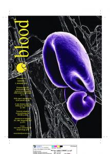
gevity, and mutations in DSB repair genes lead to premature aging phenotypes (8). Due to the involvement of sirtuins in stress response and DNA repair, we hypothesized that members of the Sir2 family may promote longevity by integrating stress signaling and DNA DSBs repair pathways. To examine the ability of SIRT proteins to promote DSB repair under stress, we used two diploid human fibroblast cell lines containing chromosomally integrated green fluorescent protein– based reporter constructs (9), which allow for the separate analysis of HR and nonhomologous end joining (NHEJ) (fig. S1). We overexpressed the four nuclear-localized human sirtuins (SIRT1, SIRT2, SIRT6, or SIRT7) in the reporter cell lines (Fig. 1A) and measured the efficiency of DSB repair. Overexpression of SIRT1 and SIRT2 had no effect on DSB repair, whereas overexpression of SIRT6 improved the efficiency of NHEJ by 3.3-fold and HR by 3.4-fold (Fig. 1B). SIRT7 overexpression increased the efficiency of NHEJ by 1.5-fold and HR by 2.8-fold (Fig. 1B). We pretreated the cells with paraquat before induction of DSBs to test the effect of oxidative stress in the DNA repair assay. Only SIRT6 overexpression under stress led to a significant stimulation of
SIRT6 Promotes DNA Repair Under Stress by Activating PARP1 Zhiyong Mao, Christopher Hine, Xiao Tian, Michael Van Meter, Matthew Au, Amita Vaidya, Andrei Seluanov,* Vera Gorbunova* Sirtuin 6 (SIRT6) is a mammalian homolog of the yeast Sir2 deacetylase. Mice deficient for SIRT6 exhibit genome instability. Here, we show that in mammalian cells subjected to oxidative stress SIRT6 is recruited to the sites of DNA double-strand breaks (DSBs) and stimulates DSB repair, through both nonhomologous end joining and homologous recombination. Our results indicate that SIRT6 physically associates with poly[adenosine diphosphate (ADP)–ribose] polymerase 1 (PARP1) and mono-ADP-ribosylates PARP1 on lysine residue 521, thereby stimulating PARP1 poly-ADP-ribosylase activity and enhancing DSB repair under oxidative stress.
SIRT
SIRT
pControl
D
SIRT6
Tubulin
Tubulin
SIRT2
SIRT7
Tubulin
Tubulin
SIRT6 Tubulin
P=0.02
2
SI
on
R
tr
R T7
0
pC 0h
2h
25 25
-H2AX nucleus g-H2AXfoci fociper per nucleus
SIRT6
Control
C
0.5
SI
T6 SI R
T2 R SI
ol
SI R T1
0
1 P=0.0001
SI R T7
4
P=0.02
P=0.001
1.5
T6
6
P=0.006
T1
P=0.02
P=0.001
2
ol
HR efficiency (GFP+/DsRed+)
P=0.0002
P=0.0009
8
0 0.25 1
SIRT1
B
Paraquat (mM)
Days 1 2 3 4 1 2 3 4
1 2 3 4
SI R
Days 1 2 3 4
SI R T2
pControl
A
tr
Fig. 1. SIRT6 stimulates DSB repair. (A) Overexpression of SIRT1, -2, -6, and -7 in human fibroblasts. Immunoblotting with sirtuin-specific antibodies after transfection with a sirtuin-expressing vector or a control vector encoding hypoxanthineguanine phosphoribosyltransferase (pControl). (B) Effect of sirtuin overexpression on the efficiency of NHEJ and HR, measured as described in (27) and fig. S1. The efficiency of DSB repair was scored in untreated cells (open bars), cells pretreated with 1 mM paraquat for 16 hours (black bars), or cells treated with paraquat and 5 mM nicotinamide for 16 hours (red bars). Error bars indicate SD; n = 8 experiments (control and SIRT6); n = 3 (other sirtuins). P values were calculated by two-tailed Student’s t test. GFP, green fluorescent protein. (C) SIRT6 overexpression accelerates the disappearance of gH2AX foci after treatment with 1 mM paraquat for 16 hours. Data represents an average of at least 50 nuclei. Error bars indicate SEM. (D) Immunoblot showing induction of endogenous SIRT6 protein levels by oxidative stress. Human fibroblasts were treated with paraquat for 16 hours.
on
*To whom correspondence should be addressed. E-mail:
[email protected] (V.G.); andrei.seluanov@ rochester.edu (A.S.)
NHEJ efficiency (GFP+/DsRed+)
Department of Biology, University of Rochester, Rochester, NY 14627, USA.
nance (1–3). SIRT1- and SIRT6-deficient cells have a reduced ability to repair double-strand breaks (DSBs) (4, 5), and SIRT6 knockout mice exhibit a premature aging phenotype associated with impaired base excision repair (BER) (6). Furthermore, SIRT6 participates in homologous recombination (HR) by deacetylating C-terminal binding protein (CtBP) interacting protein (CtIP) (7). Repair of DNA DSBs is essential for lon-
pC
M
ammalian sirtuin (SIRT) proteins function in multiple pathways, including stress response and genome mainte-
Downloaded from www.sciencemag.org on September 14, 2011
REPORTS
15 15 10 10
55
00 Paraquat 1Time after treatment (h)
www.sciencemag.org
SCIENCE
VOL 332
pControl SIRT6
20 20
17 JUNE 2011
+2
+3
0
2
+4
+5
+6
+7
4
8
16
24
Time, h
1443
REPORTS SIRT6 bound to Alu, Sirt6 bound to Alu chromatin, % %ofof input input
77 66 55
44 33 22 11 00 Time post IR (h)
Paraquat
*
B
SIRT6 bound to DSB, % of input Sirt6 bound to DSB, % of input
Untreated
A
* * *
* 0
0
0.5
2
0.5 2
8
8
14
24
14 24
0
0
0.5
2
0.5
2
* 8
14
8
*
Paraquat
22
* *
1.5 1.5
* *
11
*
0.5
0.5
00 0 Time post I-SceI (h) 0
24
14 24
Untreated
2.5 2.5
2
2
4
4
8
10
16
24
0
8 10 16 24 0
2
2
4
4
8
10
16
24
8 10 16 24
SIRT6 ChIP
SIRT6 ChIP IgG ChIP Input
IgG ChIP
H3
SIRT6
C
wt S56Y G60A R65A
B
Coomassie 32P
NAD+
SIRT6
Fig. 3. Deacetylation and mono-ADP-ribosylation activities of SIRT6 are required to stimulate DSB repair. (A) Immunoblot showing that S56Y and R65A mutations abolish the H3K9 deacetylation activity of Sirt6 and appear to exert a dominant-negative effect. (B) In vitro assay showing that S56Y and G60A mutations abolish mono-ADP-ribosylation activity of SIRT6. NAD+, nicotinamide adenine dinucleotide. (C) SIRT6 mutants for deacetylation and/or ribosylation activities have reduced ability to stimulate NHEJ and HR. Untreated cells (open bars) or cells treated with paraquat (black bars) were transfected with SIRT6-expressing vectors or pControl. Error bars indicate SD; n = 4.
NHEJ by 6.7-fold and HR by 6-fold relative to control paraquat-treated cells or 16-fold relative to untreated controls (Fig. 1B). Addition of a sirtuin inhibitor, nicotinamide, which inhibits both deacetylase and mono–adenosine diphosphate (ADP)–ribosylase activities, abolished the stimulatory effect of SIRT6 overexpression (Fig. 1B). We also observed stimulation of DSB repair by SIRT6 when a different inducer of oxidative stress, H2O2 was used (fig. S2). Furthermore, overexpression of SIRT6 accelerated clearance of gH2AX foci in paraquat treated cells (Fig. 1C) and accelerated repair of DSBs induced by neocarzinostatin, measured by a neutral comet assay (fig. S3). Additional experiments demonstrating the stimulatory effect of SIRT6 on DSB repair are shown in fig. S4. Oxidative stress elevated endogenous SIRT6 levels (Fig. 1D), indicating that ectopic overexpression enhances this physiological response. We also showed that depletion of SIRT6 compromises DSB repair, especially under stress
1444
HR HR efficiency (GFP+/DsRed+) efficiency (GFP+/DsRed+)
H3K9Ac
SIRT6 wt S56Y G60A R65A
pControl
A
NHEJ efficiency (GFP+/DsRed+) NHEJ efficiency (GFP+/DsRed+)
Fig. 2. Oxidative stress results in early recruitment of SIRT6 to DNA breaks. ChIP analysis showing kinetics of SIRT6 recruitment to Alu sequences after 8 Gy of g-irradiation (IR) (A) and sequences flanking I-SceI–induced DSB after transfection
with I-SceI expression vector (B). Asterisks indicate values significantly different from corresponding zero time points (P < 0.05). Error bars indicate SD; n = 5. Control ChIP with SIRT6−/− cells is shown in fig. S7. IgG, immunoglobulin G.
10 10
Untreated Paraquat
8 6 4 2 0 Control wt pControl wt
S56Y G60A G60A R65A R65A S56Y
1.5 1.5
Untreated Paraquat
11
0.5 0.5
0 Control wt pControl wt
S56Y G60A G60A R65A S56Y R65A
(fig. S5). Together, these observations suggest that SIRT6 plays an important regulatory role in the DNA damage response by stimulating DSB repair under oxidative stress. SIRT6 is a chromatin-associated protein (6, 10) that binds to sites of DNA DSBs (5). SIRT6 distribution in untreated cells showed distinct aggregates within the nucleus that colocalized with heterochromatin protein HP1b (fig. S6). Next, we used chromatin immunoprecipitation (ChIP) to analyze SIRT6 recruitment to damaged DNA after oxidative stress. SIRT6 was recruited to Alu elements after g-irradiation (Fig. 2A) and to the site-specific DSB generated by I-SceI (Fig. 2B and fig. S7). In the absence of oxidative stress, SIRT6 was recruited after 8 and 10 hours of g-irradiation and I-SceI transfection, respectively. When the cells were pretreated with paraquat, we observed an additional, early wave of SIRT6 recruitment, within 30 min after induction of DSBs. Thus, under normal conditions SIRT6 is recruited
17 JUNE 2011
VOL 332
SCIENCE
to DSBs relatively late, whereas preexisting oxidative stress results in early mobilization of SIRT6. Sir2 family members have two enzymatic activities, deacetylase and mono-ADP-ribosyltransferase (11–13). To determine which enzymatic activity is important for the stimulation of DSB repair, we introduced point mutations in conserved residues of SIRT6: Ser 56→Tyr 56 (S56Y) (14) lacks both deacetylase and mono-ADP-ribosyltransferase activities, G60A lacks mono-ADP-ribosyltransferase activity, and R65A lacks deacetylase activity (Fig. 3, A and B). All three mutations reduced the ability of SIRT6 to stimulate DSB repair, suggesting that both activities are important for this function (Fig. 3C). To identify the substrate of SIRT6 monoADP-ribosylation, we introduced biotin-labeled nicotinamide adenine dinucleotide (NAD), a substrate for mono-ADP-ribosyltransferase, into wildtype (WT) and SIRT6 −/− mouse embryo fibroblasts (MEFs), which were pretreated with paraquat. Poly-ADP-ribosylated proteins were depleted from the cell extracts using poly-ADP-ribose antibodies. The remaining mono-ADP-ribosylated proteins were isolated using avidin-coated beads. We observed two bands corresponding to proteins of approximately 120 and 70 kD (Fig. 4A). The 120-kD band was present at higher levels in the WT than in the SIRT6 −/− cells, suggesting that ribosylation of this protein is mediated by SIRT6. The level of the 70-kD band was independent of the SIRT6 status. We focused on the 120-kD protein hypothesizing that the band corresponded to poly(ADP-ribose) polymerase 1 (PARP1). This was confirmed by Western blotting with PARP1 antibodies (Fig. 4B). Coimmunoprecipitation showed that SIRT6 physically associates with PARP1, and the interaction between SIRT6 and PARP1 was resistant to the addition of ethidium bromide (Fig. 4C), indicating that the two proteins interact directly rather than through binding to DNA. The molecular weight of PARP1 immunoprecipitated with SIRT6 is higher than of the input PARP1, suggesting that PARP1 bound by SIRT6
www.sciencemag.org
Downloaded from www.sciencemag.org on September 14, 2011
Input
SIRT6 + - + + + + PARP1 - + + + + + kDa 250
*
*
150
22
44 22 00
1 pControl
22 1.5 1.5
11
2 SIRT6
Control 3-ABA PJ34 Para Para + 3-ABA Para + PJ34
0.5 0.5
00 1 pControl
2 SIRT6
00 2
3
4
5
w
6
1
t S5 6Y G 60 A R 65 A
SIRT6 - + + PARP1 + + + 32P NAD+
0.2 0.2
rt
E
*
0.4 0.4
T6
+8
R
+7
0.6 0.6
66
Control 3-ABA PJ34 Para Para + 3-ABA Para + PJ34
SI
+6
Relative PARP1 activity
+5
*
0.8
Si
+4
*
11 0.8
o
+3
C PA ont R rol P C 1w on t P P A tr PA AR RP ol R P1 1 w P Y t PA 1 D 88 R EE 9C P PA 1 KK R DE K P1 EK K K 52 1A
2 -
1.2 1.2
N
11
Relative PARP1 activity
100
88
Coomassie
Fig. 4. SIRT6 interacts with PARP1 and stimulates its poly-ADP-ribosylation activity. (A) Analysis of mono-ADP-ribosylated proteins in the WT and SIRT6−/− MEFs stressed with paraquat for 16 hours. Cells were transfected with biotinylated NAD, and poly-ADP-ribosylated proteins were cleared away with PAR antibodies. Immunoprecipitation (IP) of the mono-ADP-ribosylated proteins revealed bands of 120 and 70 kD. KO, knockout; WB, Western blot. (B) Immunoblotting of the extracts in (A) with PARP1 antibodies indicated that the 120-kD band is Parp1. (C) SIRT6 interacts with PARP1. Human fibroblasts were treated with 1 mM paraquat. Cell lysates were immunoprecipitated with SIRT6 antibodies in the presence of 50 mg/ml ethidium bromide (EtBr) followed by Western blotting with PARP1 antibodies. (D) PARP1 K521 is essential for activation of NHEJ by SIRT6. An NHEJ assay was performed in PARP1−/− MEFs containing an integrated NHEJ reporter. Cells were transfected with SIRT6 and/or PARP1 was mono-ADP-ribosylated (Fig. 4C). The amount of SIRT6-PARP1 complexes increased after DNA damage (fig. S8). We introduced mutations into the six known PARP1 ribosylation sites (15, 16)—D387A, E488A, E491A, K498A, K521A, and K524A—and used PARP1 knockout mouse fibroblasts containing integrated NHEJ reporter to test whether these mutants can mediate the effect of SIRT6 on repair. SIRT6 overexpression did not stimulate NHEJ in PARP1 knockout cells (Fig. 4D). However, when SIRT6 was cotransfected with the WT PARP1, but not with catalytically inactive PARP1 Y889C, it led to stimulation of NHEJ, indicating that PARP1 is required to mediate the effect of SIRT6 on repair. The PARP1 K521A mutant was sufficient to abolish the stimulation of repair by SIRT6, whereas the mutations in the other five
or PARP1 mutants. Both SIRT6 and PARP1 are required for the stimulation of repair. PARP1 Y889C is a catalytically inactive PARP1. PARP1 DEEKKK contains mutations in all six poly-ADP-ribosylation sites. PARP1 DEEKK contains the same mutations, except at K521. Error bars show SD; asterisks indicate values significantly different from control (P < 0.01). (E) PARP1 lacking the catalytic domain is mono-ADP-ribosylated by SIRT6 in vitro, whereas K521A is not. (F) In vitro assay of PARP1 poly-ADP-ribosylation activity showing that PARP1 is stimulated only by SIRT6 mono-ADPribosylation activity. The graph shows quantification of six independent experiments. Error bars show SD; asterisks indicate values significantly different from control (P < 0.01). (G) Stimulation of NHEJ and HR by SIRT6 is abolished by PARP1 inhibitors 3-ABA (5 mM) or PJ34 (20 mM). Error bars indicate SD; n = 4.
ribosylation sites had no effect, either alone or in combination (Fig. 4D), indicating that SIRT6 ribosylates PARP1 on K521. This was confirmed by showing that purified PARP1 was mono-ADPribosylated by SIRT6, whereas K521A mutant was not (Fig. 4E). Neither was K521A modified by SIRT6 in vivo (fig. S9). PARP1 binds to DNA damage sites and activates itself by automodification. It also polyADP-ribosylates other proteins around DNA damage sites facilitating recruitment of DNA repair factors (17–19). To test whether PARP1 activation is enhanced by mono-ADP-ribosylation by SIRT6, we analyzed PARP1 activity in vitro in the presence of SIRT6. WT SIRT6 protein and the R65A mutant, which has only mono-ADPribosylation activity, strongly stimulated PARP1 (Fig. 4F). The SIRT6 G60A mutant, which has
www.sciencemag.org
SCIENCE
VOL 332
Downloaded from www.sciencemag.org on September 14, 2011
F
G
(GFP+/DsRed+) NHEJ NHEJefficiency efficiency (GFP+/DsRed+)
In
PARP1 WB
PA PARP 1 K RP wt 52 1 1A wt
Relative NHEJ efficiency Relative NHEJ efficiency
wt
Input
33
00 SIRT6 wt 1-
100
-EtBr +EtBr
Input PARP1 WB
D
150
PARP1 WB
HR efficiency (GFP+/DsRed+) HR efficiency (GFP+/DsRed+)
50 37
kDa
SI R T6 w SI t R S5 T6 6 w G Y t 60 R A 65 A
100 75
IgG IP
KO
Avidin IP wt
150
C KO
KO
B Streptavidin WB
wt
kDa
IgG IP
KO
Avidin IP wt
A
pu Ig t G SI IP R Ig T6 G IP SI IP R T6 IP
REPORTS
only deacetylation activity, failed to stimulate PARP1, whereas the enzymatically inactive S56Y mutant was inhibitory (Fig. 4F). We did not detect changes in PARP1 acetylation status after incubation with SIRT6 (fig. S10). To test the effect of SIRT6 on PARP1 activity in vivo, we transfected human fibroblasts with the WT SIRT6 and SIRT6 mutants. WT SIRT6 and the R65A mutant enhanced PARP1 poly-ADP-ribosylation, whereas S56Y and G60A mutants failed to stimulate PARP1 (fig. S11). Furthermore, PARP1 inhibitors 3-ABA and PJ34 suppressed SIRT6-mediated activation of NHEJ and HR (Fig. 4G). From this result, we conclude that SIRT6 promotes DSB repair by stimulating PARP1. Multiple lines of evidence (20, 21) support the role of PARP1 in DSB repair and in suppressing aberrant recombination events by stabilizing
17 JUNE 2011
1445
broken DNA ends (22, 23). Furthermore, PARP1 is required for an alternative, DNA–dependent protein kinase catalytic subunit (PKcs) independent pathway of NHEJ (24, 25). Overexpression of SIRT6 in DNA-PKcs null MEFs up-regulated NHEJ by 1.7-fold, and in the WT MEFs by 2.3-fold (fig. S12), suggesting that SIRT6 stimulates the alternative NHEJ pathway. In this study, we identify PARP1 as the in vivo target of SIRT6 ribosylation. As PARP1 is involved in both BER and DSB repair (17, 26), the role of SIRT6 as an activator of PARP1 explains the phenotype of the SIRT6 knockout mice, which are characterized by deficient BER and genomic instability probably stemming from a defect in DSB repair (6). In the absence of oxidative stress, SIRT6 overexpression mildly induced repair, whereas under stress DNA repair was stimulated up to 16-fold. This observation suggests that SIRT6 plays a regulatory function in DNA repair by integrating DNA repair and stress signaling pathways.
3. 4. 5. 6. 7. 8. 9. 10. 11. 12. 13. 14.
15. 16.
17.
References and Notes
18.
1. L. R. Saunders, E. Verdin, Science 323, 1021 (2009). 2. S. D. Westerheide, J. Anckar, S. M. Stevens Jr., L. Sistonen, R. I. Morimoto, Science 323, 1063 (2009).
19.
E. M. Dioum et al., Science 324, 1289 (2009). P. Oberdoerffer et al., Cell 135, 907 (2008). R. A. McCord et al., Aging 1, 109 (2009). R. Mostoslavsky et al., Cell 124, 315 (2006). A. Kaidi, B. T. Weinert, C. Choudhary, S. P. Jackson, Science 329, 1348 (2010). V. Gorbunova, A. Seluanov, Mech. Ageing Dev. 126, 621 (2005). Z. Mao, A. Seluanov, Y. Jiang, V. Gorbunova, Proc. Natl. Acad. Sci. U.S.A. 104, 13068 (2007). E. Michishita, J. Y. Park, J. M. Burneskis, J. C. Barrett, I. Horikawa, Mol. Biol. Cell 16, 4623 (2005). S. Imai, C. M. Armstrong, M. Kaeberlein, L. Guarente, Nature 403, 795 (2000). J. Landry et al., Proc. Natl. Acad. Sci. U.S.A. 97, 5807 (2000). R. A. Frye, Biochem. Biophys. Res. Commun. 260, 273 (1999). Single-letter abbreviations for the amino acid residues are as follows: A, Ala; C, Cys; D, Asp; E, Glu; F, Phe; G, Gly; H, His; I, Ile; K, Lys; L, Leu; M, Met; N, Asn; P, Pro; Q, Gln; R, Arg; S, Ser; T, Thr; V, Val; W, Trp; and Y, Tyr. Z. Tao, P. Gao, H. W. Liu, J. Am. Chem. Soc. 131, 14258 (2009). M. Altmeyer, S. Messner, P. O. Hassa, M. Fey, M. O. Hottiger, Nucleic Acids Res. 37, 3723 (2009). P. O. Hassa, S. S. Haenni, M. Elser, M. O. Hottiger, Microbiol. Mol. Biol. Rev. 70, 789 (2006). D. Ahel et al., Science 325, 1240 (2009); 10.1126/ science.1177321. S. F. El-Khamisy, M. Masutani, H. Suzuki, K. W. Caldecott, Nucleic Acids Res. 31, 5526 (2003).
The Visual Impact of Gossip Eric Anderson,1* Erika H. Siegel,1* Eliza Bliss-Moreau,2 Lisa Feldman Barrett1,3† Gossip is a form of affective information about who is friend and who is foe. We show that gossip does not influence only how a face is evaluated—it affects whether a face is seen in the first place. In two experiments, neutral faces were paired with negative, positive, or neutral gossip and were then presented alone in a binocular rivalry paradigm (faces were presented to one eye, houses to the other). In both studies, faces previously paired with negative (but not positive or neutral) gossip dominated longer in visual consciousness. These findings demonstrate that gossip, as a potent form of social affective learning, can influence vision in a completely top-down manner, independent of the basic structural features of a face. ossip is a vital thread in human social interaction. As a type of instructed learning, gossip is a way to learn socially relevant information about other people’s character or personality without having to experience directly their triumphs and misadventures (1). Whether delicious or destructive, gossip is functional. It provides human beings with information about others in the absence of direct experience, allowing us to live in very large groups. It is believed that gossip was important for social cohesion during the course of human evolution (2). Scientists speculate that instead of establishing
G
1 Department of Psychology, Northeastern University, Boston, MA 02115, USA. 2Department of Psychiatry and Behavioral Science, University of California, Davis, Davis, CA 95616, USA. 3 Department of Psychiatry and the Martinos Center for Biomedical Imaging, Massachusetts General Hospital, Harvard Medical School, Charlestown, MA 02129, USA.
*These authors contributed equally to this work. †To whom correspondence should be addressed. E-mail:
[email protected]
1446
and maintaining relationships by plucking fleas off of each other, we exchange and digest juicy tidbits of chit-chat, hearsay, and rumor. Gossip allows human beings not only to transcend oneto-one interaction for getting along and getting ahead, but also to know the “value” of people we
20. H. Hochegger et al., EMBO J. 25, 1305 (2006). 21. M. N. Paddock, B. D. Buelow, S. Takeda, A. M. Scharenberg, PLoS Biol. 8, e1000428 (2010). 22. P. A. Jeggo, Curr. Biol. 8, R49 (1998). 23. F. d’Adda di Fagagna et al., Nat. Genet. 23, 76 (1999). 24. M. Wang et al., Nucleic Acids Res. 34, 6170 (2006). 25. M. Audebert, B. Salles, M. Weinfeld, P. Calsou, J. Mol. Biol. 356, 257 (2006). 26. A. Bürkle, Biogerontology 1, 41 (2000). 27. Materials and methods are available as supporting material on Science Online. Acknowledgments: This work was supported by grants from the NIH (to V.G.) and the Ellison Medical Foundation (to V.G. and A.S.). Z.M. performed DNA repair assays, immunoblots, coimmunoprecipitations, in situ staining, and biochemical assays; C.H. and A.V. performed ChIP; X.T. analyzed PARP1 mutants; M.V.M. analyzed DNA damage foci; M.A. performed comet assays; and Z.M., C.H., A.S., and V.G. designed the study, analyzed data, and wrote the manuscript.
Supporting Online Material www.sciencemag.org/cgi/content/full/332/6036/1443/DC1 Materials and Methods SOM Text Figs. S1 to S12 References 11 January 2011; accepted 9 May 2011 10.1126/science.1202723
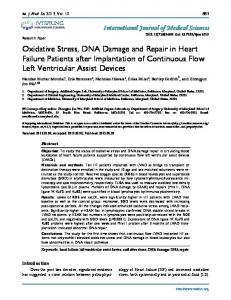
have never met. For instance, perceivers evaluate a structurally neutral face (presented alone) as “negative” for as long as 2 days after that face was paired only four times with a sentence describing a negative behavior (e.g., “threw a chair at a classmate”) (3). Gossip, when understood as a type of instructed affective learning, is a powerful way to learn whom to befriend and, even more important, whom to avoid—all without the costly and time-consuming process of learning from firsthand experience. To assess how gossip might influence conscious visual experience for other people, we capitalized on a phenomenon known as binocular rivalry (4). Binocular rivalry occurs when perceptually dissimilar images are presented to different eyes (e.g., a face to one eye and a house to the other eye) and the two percepts compete for perceptual dominance. Visual input from one eye is consciously experienced (and seen) while
Fig. 1. Example of gossip stimuli. Examples of structurally neutral faces paired with one of the following: (A) negative gossip; (B) positive gossip; (C) neutral gossip; (D) negative nonsocial information; (E) positive nonsocial information; (F) neutral nonsocial information. For a complete list of sentences, see (26).
17 JUNE 2011
VOL 332
SCIENCE
www.sciencemag.org
Downloaded from www.sciencemag.org on September 14, 2011
REPORTS